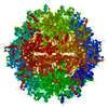
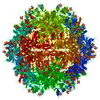
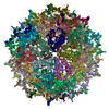
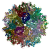
#200 - Aug 2016 Quasisymmetry in Icosahedral Viruses similarity (1)
-
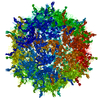
Assembly
Deposited unit
A: Capsid protein VP1 B: Capsid protein VP1 C: Capsid protein VP1 D: Capsid protein VP1 E: Capsid protein VP1 F: Capsid protein VP1 G: Capsid protein VP1 H: Capsid protein VP1 I: Capsid protein VP1 J: Capsid protein VP1 K: Capsid protein VP1 L: Capsid protein VP1 M: Capsid protein VP1 N: Capsid protein VP1 O: Capsid protein VP1 P: Capsid protein VP1 Q: Capsid protein VP1 R: Capsid protein VP1 S: Capsid protein VP1 T: Capsid protein VP1 hetero molecules
A: Capsid protein VP1 B: Capsid protein VP1 C: Capsid protein VP1 D: Capsid protein VP1 E: Capsid protein VP1 F: Capsid protein VP1 G: Capsid protein VP1 H: Capsid protein VP1 I: Capsid protein VP1 J: Capsid protein VP1 K: Capsid protein VP1 L: Capsid protein VP1 M: Capsid protein VP1 N: Capsid protein VP1 O: Capsid protein VP1 P: Capsid protein VP1 Q: Capsid protein VP1 R: Capsid protein VP1 S: Capsid protein VP1 T: Capsid protein VP1 hetero molecules
A: Capsid protein VP1 B: Capsid protein VP1 C: Capsid protein VP1 D: Capsid protein VP1 E: Capsid protein VP1 F: Capsid protein VP1 G: Capsid protein VP1 H: Capsid protein VP1 I: Capsid protein VP1 J: Capsid protein VP1 K: Capsid protein VP1 L: Capsid protein VP1 M: Capsid protein VP1 N: Capsid protein VP1 O: Capsid protein VP1 P: Capsid protein VP1 Q: Capsid protein VP1 R: Capsid protein VP1 S: Capsid protein VP1 T: Capsid protein VP1 hetero molecules
A: Capsid protein VP1 B: Capsid protein VP1 C: Capsid protein VP1 D: Capsid protein VP1 E: Capsid protein VP1 F: Capsid protein VP1 G: Capsid protein VP1 H: Capsid protein VP1 I: Capsid protein VP1 J: Capsid protein VP1 K: Capsid protein VP1 L: Capsid protein VP1 M: Capsid protein VP1 N: Capsid protein VP1 O: Capsid protein VP1 P: Capsid protein VP1 Q: Capsid protein VP1 R: Capsid protein VP1 S: Capsid protein VP1 T: Capsid protein VP1 hetero molecules
Idetical with deposited unit in distinct coordinate
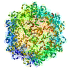
point asymmetric unit, std point frame
Type
Name
Symmetry operation
Number
transform to point frame
1
4
A: Capsid protein VP1 B: Capsid protein VP1 C: Capsid protein VP1 D: Capsid protein VP1 E: Capsid protein VP1 F: Capsid protein VP1 G: Capsid protein VP1 H: Capsid protein VP1 I: Capsid protein VP1 J: Capsid protein VP1 K: Capsid protein VP1 L: Capsid protein VP1 M: Capsid protein VP1 N: Capsid protein VP1 O: Capsid protein VP1 P: Capsid protein VP1 Q: Capsid protein VP1 R: Capsid protein VP1 S: Capsid protein VP1 T: Capsid protein VP1 hetero molecules
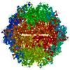
crystal asymmetric unit, crystal frame
1.65 MDa, 20 polymers
Theoretical mass
Number of molelcules
Total (without water)
1,646,369
40
Polymers
1,639,744
20
Non-polymers
6,624
20
Water
0
Type
Name
Symmetry operation
Number
identity operation
1_555
x,y,z
2
Unit cell
Length a, b, c (Å)
257.704, 257.704, 603.766
Angle α, β, γ (deg.)
90.000, 90.000, 120.000
Int Tables number
146
Space group name H-M
H3
Symmetry
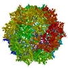
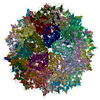
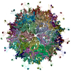
Point symmetry: (Schoenflies symbol: I (icosahedral))
 Movie
Movie Controller


 Open data
 Basic information
 Components
 Keywords
 Function and homology information
 Adeno-associated virus 3B
 X-RAY DIFFRACTION /
  Authors
 Citation
 Structure visualization
 Downloads & links
 Other downloads


 PDBj
PDBj


 Assembly