Loading
PDBj
MenuPDBj@FacebookPDBj@TwitterPDBj@YouTubewwPDB FoundationwwPDB
RCSB PDBPDBeBMRBAdv. SearchSearch help
PDB: 13 results

6VPM
DownloadVisualize
BU of 6vpm by Molmil
TPX2 residues 7-20 fused to Aurora A residues 116-389 with C290 disulfide bonded to compound 8-34, and in complex with AMP-PNP
Descriptor: ADENOSINE MONOPHOSPHATE, MAGNESIUM ION, MALONIC ACID, ...
Authors:Lim, D.C, Yaffe, M.B.
Deposit date:2020-02-03
Release date:2020-08-05
Last modified:2024-04-03
Method:X-RAY DIFFRACTION (1.58 Å)
Cite:Redox priming promotes Aurora A activation during mitosis.
Sci.Signal., 13, 2020
6VPL
DownloadVisualize
BU of 6vpl by Molmil
TPX2 residues 7-20 fused to Aurora A residues 116-389 with C290 disulfide bonded to compound 7-80, and in complex with AMP-PNP
Descriptor: GLYCEROL, MAGNESIUM ION, MALONIC ACID, ...
Authors:Lim, D.C, Yaffe, M.B.
Deposit date:2020-02-03
Release date:2020-08-05
Last modified:2024-04-03
Method:X-RAY DIFFRACTION (1.86 Å)
Cite:Redox priming promotes Aurora A activation during mitosis.
Sci.Signal., 13, 2020
6VPI
DownloadVisualize
BU of 6vpi by Molmil
TPX2 residues 7-20 fused to Aurora A residues 116-389 C247V + D256N + C319V triple mutant disulfide homodimer in complex with AMP-PNP
Descriptor: MAGNESIUM ION, MALONIC ACID, PHOSPHOAMINOPHOSPHONIC ACID-ADENYLATE ESTER, ...
Authors:Lim, D.C, Yaffe, M.B.
Deposit date:2020-02-03
Release date:2020-08-05
Last modified:2023-10-11
Method:X-RAY DIFFRACTION (2 Å)
Cite:Redox priming promotes Aurora A activation during mitosis.
Sci.Signal., 13, 2020
5LXM
DownloadVisualize
BU of 5lxm by Molmil
Crystal structure of Aurora-A bound to a hydrocarbon-stapled proteomimetic of TPX2
Descriptor: 1-ETHOXY-2-(2-ETHOXYETHOXY)ETHANE, 2-(N-MORPHOLINO)-ETHANESULFONIC ACID, ADENOSINE-5'-DIPHOSPHATE, ...
Authors:McIntyre, P.J, Bayliss, R.
Deposit date:2016-09-22
Release date:2016-11-02
Last modified:2024-02-07
Method:X-RAY DIFFRACTION (2.08 Å)
Cite:A TPX2 Proteomimetic Has Enhanced Affinity for Aurora-A Due to Hydrocarbon Stapling of a Helix.
ACS Chem. Biol., 11, 2016
6VPJ
DownloadVisualize
BU of 6vpj by Molmil
TPX2 residues 7-20 fused to Aurora A residues 116-389 C247V + C319V double mutant dephosphorylated, and in complex with AMP-PNP
Descriptor: PHOSPHOAMINOPHOSPHONIC ACID-ADENYLATE ESTER, TPX2 fragment - Aurora A kinase domain fusion, alpha-D-glucopyranose-(1-1)-alpha-D-glucopyranose
Authors:Lim, D.C, Yaffe, M.B.
Deposit date:2020-02-03
Release date:2020-08-05
Last modified:2024-04-03
Method:X-RAY DIFFRACTION (2.1 Å)
Cite:Redox priming promotes Aurora A activation during mitosis.
Sci.Signal., 13, 2020
6VPH
DownloadVisualize
BU of 6vph by Molmil
TPX2 residues 7-20 fused to Aurora A residues 116-389 modified with cacodylate and in complex with AMP-PNP
Descriptor: CHLORIDE ION, DITHIANE DIOL, GLYCEROL, ...
Authors:Lim, D.C, Yaffe, M.B.
Deposit date:2020-02-03
Release date:2020-08-05
Last modified:2023-10-11
Method:X-RAY DIFFRACTION (2.14 Å)
Cite:Redox priming promotes Aurora A activation during mitosis.
Sci.Signal., 13, 2020
3E5A
DownloadVisualize
BU of 3e5a by Molmil
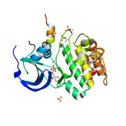
Crystal structure of Aurora A in complex with VX-680 and TPX2
Descriptor: CYCLOPROPANECARBOXYLIC ACID {4-[4-(4-METHYL-PIPERAZIN-1-YL)-6-(5-METHYL-2H-PYRAZOL-3-YLAMINO)-PYRIMIDIN-2-YLSULFANYL]-PHENYL}-AMIDE, SULFATE ION, Serine/threonine-protein kinase 6, ...
Authors:Zhao, B, Smallwood, A, Lai, Z.
Deposit date:2008-08-13
Release date:2008-10-28
Last modified:2023-08-30
Method:X-RAY DIFFRACTION (2.3 Å)
Cite:Modulation of kinase-inhibitor interactions by auxiliary protein binding: crystallography studies on Aurora A interactions with VX-680 and with TPX2.
Protein Sci., 17, 2008
3HA6
DownloadVisualize
BU of 3ha6 by Molmil
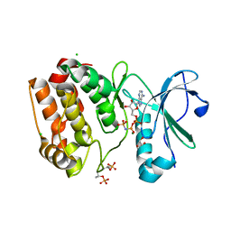
Crystal structure of aurora A in complex with TPX2 and compound 10
Descriptor: N~2~-(3,4-dimethoxyphenyl)-N~4~-[2-(2-fluorophenyl)ethyl]-N~6~-quinolin-6-yl-1,3,5-triazine-2,4,6-triamine, Serine/threonine-protein kinase 6, Targeting protein for Xklp2
Authors:Zhao, B, Clark, M.A.
Deposit date:2009-05-01
Release date:2009-08-04
Last modified:2011-07-13
Method:X-RAY DIFFRACTION (2.36 Å)
Cite:Design, synthesis and selection of DNA-encoded small-molecule libraries.
Nat.Chem.Biol., 5, 2009
1OL5
DownloadVisualize
BU of 1ol5 by Molmil
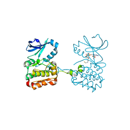
Structure of Aurora-A 122-403, phosphorylated on Thr287, Thr288 and bound to TPX2 1-43
Descriptor: ADENOSINE-5'-DIPHOSPHATE, MAGNESIUM ION, RESTRICTED EXPRESSION PROLIFERATION ASSOCIATED PROTEIN 100, ...
Authors:Bayliss, R, Conti, E.
Deposit date:2003-08-06
Release date:2003-10-30
Last modified:2024-05-01
Method:X-RAY DIFFRACTION (2.5 Å)
Cite:Structural Basis of Aurora-A Activation by Tpx2 at the Mitotic Spindle
Mol.Cell, 12, 2003
6VPG
DownloadVisualize
BU of 6vpg by Molmil
TPX2 residues 7-20 fused to Aurora A residues 116-389 in complex with AMP-PNP
Descriptor: CHLORIDE ION, GLYCEROL, MAGNESIUM ION, ...
Authors:Lim, D.C, Yaffe, M.B.
Deposit date:2020-02-03
Release date:2020-08-05
Last modified:2023-10-11
Method:X-RAY DIFFRACTION (2.64 Å)
Cite:Redox priming promotes Aurora A activation during mitosis.
Sci.Signal., 13, 2020
6XKA
DownloadVisualize
BU of 6xka by Molmil
TPX2 residues 7-20 fused to Aurora A residues 116-389 dephosphorylated, and CoAlated on C290
Descriptor: COENZYME A, TPX2 fragment - Aurora A kinase domain fusion
Authors:Lim, D.C, Yaffe, M.B.
Deposit date:2020-06-26
Release date:2020-08-05
Last modified:2023-10-18
Method:X-RAY DIFFRACTION (2.65 Å)
Cite:Redox priming promotes Aurora A activation during mitosis.
Sci.Signal., 13, 2020
4C3P
DownloadVisualize
BU of 4c3p by Molmil
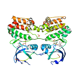
Structure of dephosphorylated Aurora A (122-403) bound to TPX2 and AMPPCP
Descriptor: AURORA KINASE A, PHOSPHOMETHYLPHOSPHONIC ACID ADENYLATE ESTER, SULFATE ION, ...
Authors:Zorba, A, Kutter, S, Kern, D.
Deposit date:2013-08-26
Release date:2014-05-28
Last modified:2023-12-20
Method:X-RAY DIFFRACTION (2.69 Å)
Cite:Molecular Mechanism of Aurora a Kinase Autophosphorylation and its Allosteric Activation by Tpx2.
Elife, 3, 2014
6BJC
DownloadVisualize
BU of 6bjc by Molmil
TPX2_mini decorated GMPCPP-microtubule
Descriptor: GUANOSINE-5'-TRIPHOSPHATE, MAGNESIUM ION, PHOSPHOMETHYLPHOSPHONIC ACID GUANYLATE ESTER, ...
Authors:Zhang, R, Nogales, E.
Deposit date:2017-11-05
Release date:2017-11-22
Last modified:2024-03-13
Method:ELECTRON MICROSCOPY (3.3 Å)
Cite:Structural insight into TPX2-stimulated microtubule assembly.
Elife, 6, 2017

223532

PDB entries from 2024-08-07

PDB statisticsPDBj update infoContact PDBjnumon