4LR6
 
 | | Structure of BRD4 bromodomain 1 with a 3-methyl-4-phenylisoxazol-5-amine fragment | | 分子名称: | 3-methyl-4-phenyl-1,2-oxazol-5-amine, Bromodomain-containing protein 4, FORMIC ACID | | 著者 | Jayaram, H, Poy, F, Gehling, V, Hewitt, M, Vaswani, R, Leblanc, Y, Cote, A, Nasveschuk, C, Taylor, A, Harmange, J.-C, Audia, J, Pardo, E, Joshi, S, Sandy, P, Mertz, J, Sims, R, Bergeron, L, Bryant, B, Ravichandran, S, Yellapuntala, S, Nandana, B.S, Birudukota, S, Albrecht, B, Bellon, S. | | 登録日 | 2013-07-19 | | 公開日 | 2013-08-07 | | 最終更新日 | 2023-09-20 | | 実験手法 | X-RAY DIFFRACTION (1.29 Å) | | 主引用文献 | Discovery, Design, and Optimization of Isoxazole Azepine BET Inhibitors.
ACS Med Chem Lett, 4, 2013
|
|
7VF8
 | | Crystal Structure of HasAp with Co-5-octaethyloxaporphyrinium cation | | 分子名称: | CITRIC ACID, Co-5-octaethyloxaporphyrinium cation, DI(HYDROXYETHYL)ETHER, ... | | 著者 | Takiguchi, A, Sakakibara, E, Sugimoto, H, Shoji, O, Shinokubo, H. | | 登録日 | 2021-09-10 | | 公開日 | 2022-09-14 | | 最終更新日 | 2023-11-29 | | 実験手法 | X-RAY DIFFRACTION (1.35 Å) | | 主引用文献 | Crystal Structure of HasAp with Co-5-octaethyloxaporphyrinium cation
To Be Published
|
|
5E2M
 | | Crystal structure of human carbonic anhydrase isozyme I with 3-(cyclooctylamino)-2,5,6-trifluoro-4-[(2-hydroxyethyl)sulfonyl]benzenesulfonamide | | 分子名称: | 3-(cyclooctylamino)-2,5,6-trifluoro-4-[(2-hydroxyethyl)sulfonyl]benzenesulfonamide, ACETATE ION, Carbonic anhydrase 1, ... | | 著者 | Manakova, E, Smirnov, A, Grazulis, S. | | 登録日 | 2015-10-01 | | 公開日 | 2016-10-05 | | 最終更新日 | 2024-01-10 | | 実験手法 | X-RAY DIFFRACTION (1.41 Å) | | 主引用文献 | Intrinsic Thermodynamics and Structures of 2,4- and 3,4-Substituted Fluorinated Benzenesulfonamides Binding to Carbonic Anhydrases.
ChemMedChem, 12, 2017
|
|
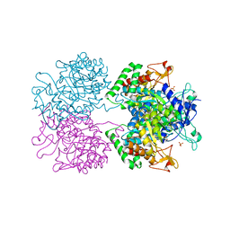
4D4M
 | | human PFKFB3 in complex with a pyrrolopyrimidone compound | | 分子名称: | 6-O-phosphono-beta-D-fructofuranose, 6-PHOSPHOFRUCTO-2-KINASE/FRUCTOSE-2,6-BISPHOSPHATASE 3, 7-(4-bromophenyl)-5-phenyl-3,7-dihydro-4H-pyrrolo[2,3-d]pyrimidin-4-one, ... | | 著者 | Stgallay, S.A, Bennett, N, Critchlow, S, Davies, G, Debreczeni, J.E, Evans, N, Holdgate, G, Jones, N.P, Leach, L, Maman, S, McLoughlin, S, Preston, M, Rigoreau, L, Thomas, A, Walker, G, Walsch, J, Ward, R.A, Wheatley, E, Winter, J. | | 登録日 | 2014-10-29 | | 公開日 | 2016-01-20 | | 最終更新日 | 2024-05-08 | | 実験手法 | X-RAY DIFFRACTION (2.32 Å) | | 主引用文献 | Identifying a Novel Series of Pfkfb3 Inhibitors as a Metabolic Approach to Treating Cancer from Hts, Biophysical and Biochemical Methods
To be Published
|
|
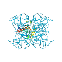
4MB3
 | | Crystal structure of E153Q mutant of cold-adapted chitinase from Moritella marina | | 分子名称: | (4S)-2-METHYL-2,4-PENTANEDIOL, CHLORIDE ION, Chitinase 60, ... | | 著者 | Malecki, P.H, Vorgias, C.E, Rypniewski, W. | | 登録日 | 2013-08-19 | | 公開日 | 2014-03-19 | | 最終更新日 | 2023-11-08 | | 実験手法 | X-RAY DIFFRACTION (1.55 Å) | | 主引用文献 | Crystal structures of substrate-bound chitinase from the psychrophilic bacterium Moritella marina and its structure in solution
Acta Crystallogr.,Sect.D, 70, 2014
|
|
7VF7
 | | Crystal Structure of HasAp with Co-octaethylporphyrin | | 分子名称: | Co-octaethylporphyrin, DI(HYDROXYETHYL)ETHER, GLYCEROL, ... | | 著者 | Takiguchi, A, Sakakibara, E, Sugimoto, H, Shoji, O, Shinokubo, H. | | 登録日 | 2021-09-10 | | 公開日 | 2022-09-14 | | 最終更新日 | 2023-11-29 | | 実験手法 | X-RAY DIFFRACTION (1.35 Å) | | 主引用文献 | Crystal Structure of HasAp with Co-octaethylporphyrin
To Be Published
|
|
1BEA
 | | BIFUNCTIONAL HAGEMAN FACTOR/AMYLASE INHIBITOR FROM MAIZE | | 分子名称: | BIFUNCTIONAL AMYLASE/SERINE PROTEASE INHIBITOR | | 著者 | Behnke, C.A, Yee, V.C, Le Trong, I, Pedersen, L.C, Stenkamp, R.E, Kim, S.S, Reeck, G.R, Teller, D.C. | | 登録日 | 1998-05-13 | | 公開日 | 1998-08-12 | | 最終更新日 | 2023-08-02 | | 実験手法 | X-RAY DIFFRACTION (1.95 Å) | | 主引用文献 | Structural determinants of the bifunctional corn Hageman factor inhibitor: x-ray crystal structure at 1.95 A resolution.
Biochemistry, 37, 1998
|
|
3NQU
 | |
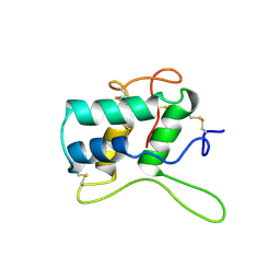
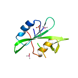
1AX3
 | | SOLUTION NMR STRUCTURE OF B. SUBTILIS IIAGLC, 16 STRUCTURES | | 分子名称: | GLUCOSE PERMEASE IIA DOMAIN | | 著者 | Chen, Y, Case, D.A, Reizer, J, Saier Junior, M.H, Wright, P.E. | | 登録日 | 1997-10-25 | | 公開日 | 1998-06-17 | | 最終更新日 | 2024-05-22 | | 実験手法 | SOLUTION NMR | | 主引用文献 | High-resolution solution structure of Bacillus subtilis IIAglc.
Proteins, 31, 1998
|
|
1BCT
 | |
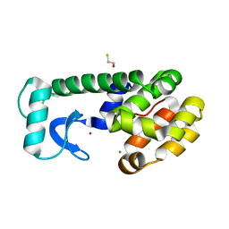
5XZ2
 | | Crystal structure of adenylate kinase | | 分子名称: | Adenylate kinase isoenzyme 1, BIS(ADENOSINE)-5'-PENTAPHOSPHATE, SULFATE ION | | 著者 | Moon, S, Bae, E, Kim, J. | | 登録日 | 2017-07-11 | | 公開日 | 2018-07-18 | | 最終更新日 | 2023-11-22 | | 実験手法 | X-RAY DIFFRACTION (1.75 Å) | | 主引用文献 | Structural analyses of adenylate kinases from Antarctic and tropical fishes for understanding cold adaptation of enzymes
Sci Rep, 7, 2017
|
|
376D
 | | A ZIPPER-LIKE DNA DUPLEX D(GCGAAAGCT) | | 分子名称: | COBALT HEXAMMINE(III), DNA (5'-D(*GP*(CBR)P*GP*AP*AP*AP*GP*CP*T)-3') | | 著者 | Cruse, W.B.T, Shepard, W, Prange, T, delalFortelle, E, Fourme, R. | | 登録日 | 1998-01-22 | | 公開日 | 1999-10-26 | | 最終更新日 | 2024-04-03 | | 実験手法 | X-RAY DIFFRACTION (2.1 Å) | | 主引用文献 | A zipper-like duplex in DNA: the crystal structure of d(GCGAAAGCT) at 2.1 A resolution.
Structure, 6, 1998
|
|
5E7Z
 | | 3-deoxy-D-arabino-heptulosonate 7-phosphate synthase from Mycobacterium tuberculosis in complex with D/L-tryptophan and D-phenylalanine | | 分子名称: | 3-deoxy-D-arabinoheptulosonate-7-phosphate synthase, D-PHENYLALANINE, D-TRYPTOPHAN, ... | | 著者 | Reichau, S, Jiao, W, Parker, E.J. | | 登録日 | 2015-10-13 | | 公開日 | 2016-06-01 | | 最終更新日 | 2023-11-15 | | 実験手法 | X-RAY DIFFRACTION (2.4 Å) | | 主引用文献 | Probing the Sophisticated Synergistic Allosteric Regulation of Aromatic Amino Acid Biosynthesis in Mycobacterium tuberculosis Using -Amino Acids.
Plos One, 11, 2016
|
|
5E8D
 | | Crystal structure of human epiregulin in complex with the Fab fragment of murine monoclonal antibody 9E5 | | 分子名称: | CHLORIDE ION, GLYCEROL, Proepiregulin, ... | | 著者 | Kado, Y, Mizohata, E, Nagatoishi, S, Iijima, M, Shinoda, K, Miyafusa, T, Nakayama, T, Yoshizumi, T, Sugiyama, A, Kawamura, T, Lee, Y.H, Matsumura, H, Doi, H, Fujitani, H, Kodama, T, Shibasaki, Y, Tsumoto, K, Inoue, T. | | 登録日 | 2015-10-14 | | 公開日 | 2015-12-09 | | 最終更新日 | 2020-02-19 | | 実験手法 | X-RAY DIFFRACTION (2.5 Å) | | 主引用文献 | Epiregulin Recognition Mechanisms by Anti-epiregulin Antibody 9E5: STRUCTURAL, FUNCTIONAL, AND MOLECULAR DYNAMICS SIMULATION ANALYSES
J.Biol.Chem., 291, 2016
|
|
5E40
 | | 3-Deoxy-D-arabino-heptulosonate 7-phosphate synthase from Mycobacterium tuberculosis with D-tyrosine bound in the phenylalanine binding site | | 分子名称: | 3-deoxy-D-arabinoheptulosonate-7-phosphate synthase, D-TYROSINE, GLYCEROL, ... | | 著者 | Reichau, S, Jiao, W, Parker, E.J. | | 登録日 | 2015-10-05 | | 公開日 | 2016-06-01 | | 最終更新日 | 2023-09-27 | | 実験手法 | X-RAY DIFFRACTION (2.05 Å) | | 主引用文献 | Probing the Sophisticated Synergistic Allosteric Regulation of Aromatic Amino Acid Biosynthesis in Mycobacterium tuberculosis Using -Amino Acids.
Plos One, 11, 2016
|
|
4D4J
 | | human PFKFB3 in complex with a pyrrolopyrimidone compound | | 分子名称: | 5-(4-bromophenyl)-7-phenyl-3,7-dihydro-4H-pyrrolo[2,3-d]pyrimidin-4-one, 6-O-phosphono-beta-D-fructofuranose, 6-PHOSPHOFRUCTO-2-KINASE/FRUCTOSE-2,6-BISPHOSPHATASE 3, ... | | 著者 | Stgallay, S.A, Bennett, N, Critchlow, S, Davies, G, Debreczeni, J.E, Evans, N, Holdgate, G, Jones, N.P, Leach, L, Maman, S, McLoughlin, S, Preston, M, Rigoreau, L, Thomas, A, Walker, G, Walsch, J, Ward, R.A, Wheatley, E, Winter, J. | | 登録日 | 2014-10-29 | | 公開日 | 2016-01-20 | | 最終更新日 | 2024-05-08 | | 実験手法 | X-RAY DIFFRACTION (3 Å) | | 主引用文献 | Identifying a Novel Series of Pfkfb3 Inhibitors as a Metabolic Approach to Treating Cancer from Hts, Biophysical and Biochemical Methods
To be Published
|
|
1P13
 | | Crystal Structure of the Src SH2 Domain Complexed with Peptide (SDpYANFK) | | 分子名称: | CACODYLATE ION, Peptide, Proto-oncogene tyrosine-protein kinase Src | | 著者 | Sonnenburg, E.D, Bilwes, A, Hunter, T, Noel, J.P. | | 登録日 | 2003-04-11 | | 公開日 | 2003-08-19 | | 最終更新日 | 2011-07-13 | | 実験手法 | X-RAY DIFFRACTION (1.63 Å) | | 主引用文献 | The structure of the membrane distal phosphatase domain of RPTPalpha reveals interdomain flexibility and an SH2 domain interaction region.
Biochemistry, 42, 2003
|
|
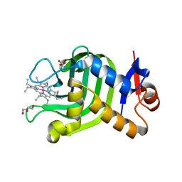
1P36
 | | T4 LYOSZYME CORE REPACKING MUTANT I100V/TA | | 分子名称: | BETA-MERCAPTOETHANOL, CHLORIDE ION, LYSOZYME, ... | | 著者 | Mooers, B.H, Datta, D, Baase, W.A, Zollars, E.S, Mayo, S.L, Matthews, B.W. | | 登録日 | 2003-04-16 | | 公開日 | 2003-10-07 | | 最終更新日 | 2023-08-16 | | 実験手法 | X-RAY DIFFRACTION (1.45 Å) | | 主引用文献 | Repacking the Core of T4 lysozyme by automated design
J.Mol.Biol., 332, 2003
|
|
3EPO
 | | Crystal structure of Caulobacter crescentus ThiC complexed with HMP-P | | 分子名称: | (4-AMINO-2-METHYLPYRIMIDIN-5-YL)METHYL DIHYDROGEN PHOSPHATE, Thiamine biosynthesis protein thiC | | 著者 | Li, S, Chatterjee, A, Zhang, Y, Grove, T.L, Lee, M, Krebs, C, Booker, S.J, Begley, T.P, Ealick, S.E. | | 登録日 | 2008-09-29 | | 公開日 | 2008-10-28 | | 最終更新日 | 2024-02-21 | | 実験手法 | X-RAY DIFFRACTION (2.1 Å) | | 主引用文献 | Reconstitution of ThiC in thiamine pyrimidine biosynthesis expands the radical SAM superfamily
Nat.Chem.Biol., 4, 2008
|
|
1INO
 | |
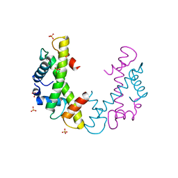
1EYN
 | | Structure of mura liganded with the extrinsic fluorescence probe ANS | | 分子名称: | 8-ANILINO-1-NAPHTHALENE SULFONATE, GLYCEROL, UDP-N-ACETYLGLUCOSAMINE 1-CARBOXYVINYLTRANSFERASE | | 著者 | Schonbrunn, E, Eschenburg, S, Luger, K, Kabsch, W, Amrhein, N. | | 登録日 | 2000-05-07 | | 公開日 | 2000-06-09 | | 最終更新日 | 2023-08-09 | | 実験手法 | X-RAY DIFFRACTION (1.7 Å) | | 主引用文献 | Structural basis for the interaction of the fluorescence probe 8-anilino-1-naphthalene sulfonate (ANS) with the antibiotic target MurA.
Proc.Natl.Acad.Sci.USA, 97, 2000
|
|
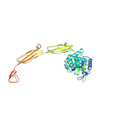
4LOR
 | | C1s CUB1-EGF-CUB2 in complex with a collagen-like peptide from C1q | | 分子名称: | 2-acetamido-2-deoxy-beta-D-glucopyranose, CALCIUM ION, Complement C1s subcomponent heavy chain, ... | | 著者 | Wallis, R, Venkatraman Girija, U, Moody, P.C.E, Marshall, J.E. | | 登録日 | 2013-07-13 | | 公開日 | 2013-08-07 | | 最終更新日 | 2020-07-29 | | 実験手法 | X-RAY DIFFRACTION (2.5 Å) | | 主引用文献 | Structural basis of the C1q/C1s interaction and its central role in assembly of the C1 complex of complement activation.
Proc.Natl.Acad.Sci.USA, 110, 2013
|
|
3S88
 | | Crystal structure of Sudan Ebolavirus Glycoprotein (strain Gulu) bound to 16F6 | | 分子名称: | 16F6 - Heavy chain, 16F6 - Light chain, 2-acetamido-2-deoxy-beta-D-glucopyranose-(1-4)-2-acetamido-2-deoxy-beta-D-glucopyranose, ... | | 著者 | Saphire, E.O, Dias, J.M, Bale, S. | | 登録日 | 2011-05-27 | | 公開日 | 2011-12-07 | | 最終更新日 | 2023-09-13 | | 実験手法 | X-RAY DIFFRACTION (3.351 Å) | | 主引用文献 | A shared structural solution for neutralizing ebolaviruses.
Nat.Struct.Mol.Biol., 18, 2011
|
|
2HN2
 | |
5DVY
 | | 2.95 Angstrom Crystal Structure of the Dimeric Form of Penicillin Binding Protein 2 Prime from Enterococcus faecium | | 分子名称: | 2-AMINO-2-HYDROXYMETHYL-PROPANE-1,3-DIOL, Penicillin binding protein 2 prime, SULFATE ION | | 著者 | Minasov, G, Wawrzak, Z, Shuvalova, L, Dubrovska, I, Flores, K, Filippova, E, Grimshaw, S, Kwon, K, Anderson, W.F, Center for Structural Genomics of Infectious Diseases (CSGID) | | 登録日 | 2015-09-21 | | 公開日 | 2015-10-07 | | 実験手法 | X-RAY DIFFRACTION (2.95 Å) | | 主引用文献 | 2.95 Angstrom Crystal Structure of the Dimeric Form of Penicillin Binding Protein 2 Prime from Enterococcus faecium.
To Be Published
|
|


