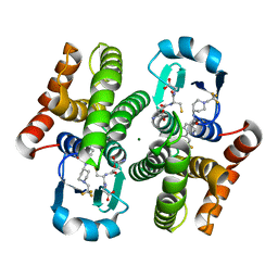
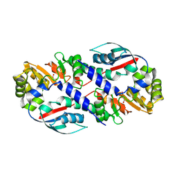
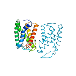
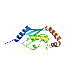
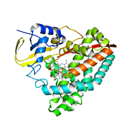
3KXG
 
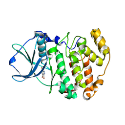 | | Crystal structure of Z. mays CK2 kinase alpha subunit in complex with the inhibitor 3,4,5,6,7-pentabromo-1H-indazole (K64) | | 分子名称: | 3,4,5,6,7-pentabromo-1H-indazole, Casein kinase II subunit alpha | | 著者 | Papinutto, E, Franchin, C, Battistutta, R. | | 登録日 | 2009-12-03 | | 公開日 | 2010-11-17 | | 最終更新日 | 2017-11-01 | | 実験手法 | X-RAY DIFFRACTION (1.7 Å) | | 主引用文献 | ATP site-directed inhibitors of protein kinase CK2: an update.
Curr Top Med Chem, 11, 2011
|
|
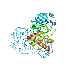
5GRT
 | | HUMAN GLUTATHIONE REDUCTASE A34E, R37W MUTANT, GLUTATHIONYLSPERMIDINE COMPLEX | | 分子名称: | FLAVIN-ADENINE DINUCLEOTIDE, GLUTATHIONE REDUCTASE, GLUTATHIONYLSPERMIDINE DISULFIDE | | 著者 | Stoll, V.S, Simpson, S.J, Krauth-Siegel, R.L, Walsh, C.T, Pai, E.F. | | 登録日 | 1997-02-12 | | 公開日 | 1997-08-12 | | 最終更新日 | 2023-08-09 | | 実験手法 | X-RAY DIFFRACTION (2.4 Å) | | 主引用文献 | Glutathione reductase turned into trypanothione reductase: structural analysis of an engineered change in substrate specificity.
Biochemistry, 36, 1997
|
|
3KX9
 | |
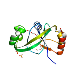
5GS3
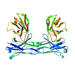 | | Crystal structure of diabody | | 分子名称: | diabody protein | | 著者 | Kim, J.H, Song, D.H, Youn, S.J, Kim, J.W, Cho, G, Lee, H, Lee, J.O. | | 登録日 | 2016-08-13 | | 公開日 | 2016-10-12 | | 最終更新日 | 2023-11-08 | | 実験手法 | X-RAY DIFFRACTION (1.698 Å) | | 主引用文献 | Crystal structure of mono- and bi-specific diabodies and reduction of their structural flexibility by introduction of disulfide bridges at the Fv interface.
Sci Rep, 6, 2016
|
|
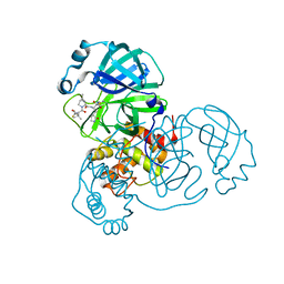
3KXO
 | | An orally active inhibitor bound at the active site of HPGDS | | 分子名称: | 6-(3-fluorophenyl)-N-[1-(2,2,2-trifluoroethyl)piperidin-4-yl]pyridine-3-carboxamide, GLUTATHIONE, Glutathione-requiring prostaglandin D synthase, ... | | 著者 | Kiefer, J.R, Day, J.E, Thorarensen, A. | | 登録日 | 2009-12-03 | | 公開日 | 2010-09-01 | | 最終更新日 | 2024-02-21 | | 実験手法 | X-RAY DIFFRACTION (2.1 Å) | | 主引用文献 | Discovery of an oral potent selective inhibitor of hematopoietic prostaglandin D synthase
TO BE PUBLISHED
|
|
5GSC
 | |
5GSR
 | | Mouse MHC class I H-2Kd with a MERS-CoV-derived peptide I5A | | 分子名称: | 9-mer peptide from Spike protein, Beta-2-microglobulin, H-2 class I histocompatibility antigen, ... | | 著者 | Liu, K, Chai, Y, Qi, J, Tan, W, Liu, W.J, Gao, G.F. | | 登録日 | 2016-08-17 | | 公開日 | 2017-04-26 | | 実験手法 | X-RAY DIFFRACTION (2.198 Å) | | 主引用文献 | Protective T Cell Responses Featured by Concordant Recognition of Middle East Respiratory Syndrome Coronavirus-Derived CD8+ T Cell Epitopes and Host MHC.
J. Immunol., 198, 2017
|
|
5GSZ
 | | Crystal Structure of the KIF19A Motor Domain Complexed with Mg-ADP | | 分子名称: | ADENOSINE-5'-DIPHOSPHATE, Kinesin-like protein KIF19, MAGNESIUM ION | | 著者 | Wang, D, Nitta, R, Hirokawa, N. | | 登録日 | 2016-08-18 | | 公開日 | 2016-09-28 | | 最終更新日 | 2020-02-26 | | 実験手法 | X-RAY DIFFRACTION (2.72 Å) | | 主引用文献 | Motility and microtubule depolymerization mechanisms of the Kinesin-8 motor, KIF19A
Elife, 5, 2016
|
|
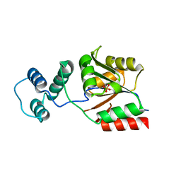
3L0G
 | |
5GT7
 | | Crystal Structure of Arg-bound CASTOR1 | | 分子名称: | ARGININE, GATS-like protein 3, MALONATE ION | | 著者 | Guo, L, Deng, D. | | 登録日 | 2016-08-18 | | 公開日 | 2017-08-23 | | 最終更新日 | 2024-03-20 | | 実験手法 | X-RAY DIFFRACTION (2.048 Å) | | 主引用文献 | Crystal structures of arginine sensor CASTOR1 in arginine-bound and ligand free states
Biochem. Biophys. Res. Commun., 508, 2019
|
|
5GTZ
 | |
5GU8
 | | Structure of biotin carboxyl carrier protein from pyrococcus horikoshi OT3 (delta N79) wild type | | 分子名称: | 149aa long hypothetical methylmalonyl-CoA decarboxylase gamma chain, SODIUM ION | | 著者 | Yamada, K, Kunishima, N, Matsuura, Y, Nakai, K, Naitow, H, Fukasawa, Y, Tomii, K. | | 登録日 | 2016-08-26 | | 公開日 | 2017-08-30 | | 最終更新日 | 2023-11-08 | | 実験手法 | X-RAY DIFFRACTION (1.8 Å) | | 主引用文献 | Designing better diffracting crystals of biotin carboxyl carrier protein from Pyrococcus horikoshii by a mutation based on the crystal-packing propensity of amino acids.
Acta Crystallogr D Struct Biol, 73, 2017
|
|
5GUF
 | |
5H4P
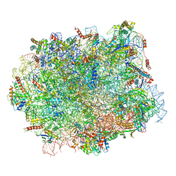 | | Structural snapshot of cytoplasmic pre-60S ribosomal particles bound with Nmd3, Lsg1, Tif6 and Reh1 | | 分子名称: | 25S ribosomal RNA, 5.8S ribosomal RNA, 5S ribosomal RNA, ... | | 著者 | Ma, C, Wu, S, Li, N, Chen, Y, Yan, K, Li, Z, Zheng, L, Lei, J, Woolford, J.L, Gao, N. | | 登録日 | 2016-11-01 | | 公開日 | 2017-01-25 | | 最終更新日 | 2024-03-20 | | 実験手法 | ELECTRON MICROSCOPY (3.07 Å) | | 主引用文献 | Structural snapshot of cytoplasmic pre-60S ribosomal particles bound by Nmd3, Lsg1, Tif6 and Reh1
Nat. Struct. Mol. Biol., 24, 2017
|
|
3L1N
 | | Crystal structure of Mp1p ligand binding domain 2 complexd with palmitic acid | | 分子名称: | Cell wall antigen, PALMITIC ACID | | 著者 | Liao, S, Tung, E.T, Zheng, W, Chong, K, Xu, Y, Bartlam, M, Rao, Z, Yuen, K.Y. | | 登録日 | 2009-12-14 | | 公開日 | 2010-01-05 | | 最終更新日 | 2021-11-10 | | 実験手法 | X-RAY DIFFRACTION (1.3 Å) | | 主引用文献 | Crystal structure of the Mp1p ligand binding domain 2 reveals its function as a fatty acid-binding protein.
J.Biol.Chem., 285, 2010
|
|
7SF3
 | | SARS-CoV-2 Main Protease (Mpro) in Complex with ML1006m | | 分子名称: | (1R,2S,5S)-N-{(2S,3R)-3-hydroxy-4-(methylamino)-4-oxo-1-[(3S)-2-oxopyrrolidin-3-yl]butan-2-yl}-6,6-dimethyl-3-[3-methyl-N-(trifluoroacetyl)-L-valyl]-3-azabicyclo[3.1.0]hexane-2-carboxamide, 3C-like proteinase, CHLORIDE ION | | 著者 | Westberg, M, Fernandez, D, Lin, M.Z. | | 登録日 | 2021-10-02 | | 公開日 | 2022-10-05 | | 最終更新日 | 2024-04-17 | | 実験手法 | X-RAY DIFFRACTION (1.75 Å) | | 主引用文献 | An orally bioavailable SARS-CoV-2 main protease inhibitor exhibits improved affinity and reduced sensitivity to mutations.
Sci Transl Med, 16, 2024
|
|
5GOM
 | | Truncated mitofusin-1, transition-like state | | 分子名称: | 2,3-DIHYDROXY-1,4-DITHIOBUTANE, GUANOSINE-5'-DIPHOSPHATE, Mitofusin-1 | | 著者 | Cao, Y.L, Gao, S. | | 登録日 | 2016-07-27 | | 公開日 | 2017-02-01 | | 最終更新日 | 2024-03-20 | | 実験手法 | X-RAY DIFFRACTION (2.802 Å) | | 主引用文献 | MFN1 structures reveal nucleotide-triggered dimerization critical for mitochondrial fusion
Nature, 542, 2017
|
|
3L1Y
 | |
7SFH
 | | SARS-CoV-2 Main Protease (Mpro) in Complex with ML102 | | 分子名称: | (1R,2S,5S)-N-{(2S,3R)-4-amino-3-hydroxy-4-oxo-1-[(3S)-2-oxopyrrolidin-3-yl]butan-2-yl}-6,6-dimethyl-3-(3-phenylpropanoyl)-3-azabicyclo[3.1.0]hexane-2-carboxamide, 3C-like proteinase, CALCIUM ION | | 著者 | Westberg, M, Fernandez, D, Lin, M.Z. | | 登録日 | 2021-10-03 | | 公開日 | 2022-10-05 | | 最終更新日 | 2023-10-18 | | 実験手法 | X-RAY DIFFRACTION (1.4 Å) | | 主引用文献 | Rational design of a new class of protease inhibitors for the potential treatment of coronavirus diseases
To Be Published
|
|
5GUN
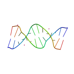 | | Crystal structure of d(GTGGAATGGAAC) | | 分子名称: | COBALT (II) ION, DNA (5'-D(*GP*TP*GP*GP*AP*AP*TP*GP*GP*AP*AP*C)-3') | | 著者 | Satange, R.B, Kao, Y.F, Hou, M.H. | | 登録日 | 2016-08-29 | | 公開日 | 2017-08-30 | | 最終更新日 | 2024-03-20 | | 実験手法 | X-RAY DIFFRACTION (2.588 Å) | | 主引用文献 | Parity-dependent hairpin configurations of repetitive DNA sequence promote slippage associated with DNA expansion
Proc. Natl. Acad. Sci. U.S.A., 114, 2017
|
|
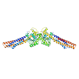
5GOX
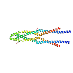 | | Eukaryotic Rad50 Functions as A Rod-shaped Dimer | | 分子名称: | DNA repair protein RAD50, GLYCEROL, ZINC ION | | 著者 | Park, Y.B, Hohl, M, Padjasek, M, Jeong, E, Jin, K.S, Krezel, A, Petrini, J.H.J, Cho, Y. | | 登録日 | 2016-07-30 | | 公開日 | 2017-02-01 | | 最終更新日 | 2017-03-15 | | 実験手法 | X-RAY DIFFRACTION (2.405 Å) | | 主引用文献 | Eukaryotic Rad50 functions as a rod-shaped dimer
Nat. Struct. Mol. Biol., 24, 2017
|
|
6OOW
 | |
3L27
 | | Crystal structure of Zaire Ebola VP35 interferon inhibitory domain R312A mutant | | 分子名称: | CHLORIDE ION, GLYCEROL, PHOSPHATE ION, ... | | 著者 | Leung, D.W, Prins, K.C, Borek, D.M, Farahbakhsh, M, Tufariello, J.M, Ramanan, P, Nix, J.C, Helgeson, L.A, Otwinowski, Z, Honzatko, R.B, Basler, C.F, Amarasinghe, G.K. | | 登録日 | 2009-12-14 | | 公開日 | 2010-01-26 | | 最終更新日 | 2023-09-06 | | 実験手法 | X-RAY DIFFRACTION (1.95 Å) | | 主引用文献 | Structural basis for dsRNA recognition and interferon antagonism by Ebola VP35.
Nat.Struct.Mol.Biol., 17, 2010
|
|
5GUS
 | | Crystal structure of ASCH domain from Zymomonas mobilis | | 分子名称: | 3,6,9,12,15,18,21-HEPTAOXATRICOSANE-1,23-DIOL, CHLORIDE ION, Helix-turn-helix domain-containing protein, ... | | 著者 | Ha, S.C, Park, S.Y, Kim, J.S. | | 登録日 | 2016-08-31 | | 公開日 | 2017-08-30 | | 最終更新日 | 2024-03-20 | | 実験手法 | X-RAY DIFFRACTION (1.951 Å) | | 主引用文献 | Crystal structure of an ASCH protein from Zymomonas mobilis and its ribonuclease activity specific for single-stranded RNA.
Sci Rep, 7, 2017
|
|
3KZX
 | |


