6G67
 
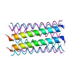 | |
7D3I
 
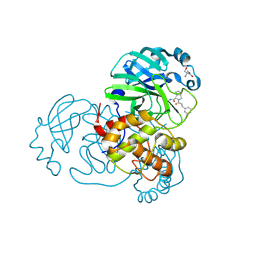 | | Crystal structure of SARS-CoV-2 main protease in complex with MI-23 | | 分子名称: | (3~{S},3~{a}~{S},6~{a}~{R})-2-[3-[3,5-bis(fluoranyl)phenyl]propanoyl]-~{N}-[(2~{S})-1-oxidanylidene-3-[(3~{S})-2-oxidanylidenepyrrolidin-3-yl]propan-2-yl]-3,3~{a},4,5,6,6~{a}-hexahydro-1~{H}-cyclopenta[c]pyrrole-3-carboxamide, 2-AMINO-2-HYDROXYMETHYL-PROPANE-1,3-DIOL, 3C-like proteinase | | 著者 | Zeng, R, Li, Y.S, Qiao, J.X, Wang, Y.F, Yang, S.Y, Lei, J. | | 登録日 | 2020-09-19 | | 公開日 | 2020-10-07 | | 最終更新日 | 2023-11-29 | | 実験手法 | X-RAY DIFFRACTION (2.004 Å) | | 主引用文献 | SARS-CoV-2 M pro inhibitors with antiviral activity in a transgenic mouse model.
Science, 371, 2021
|
|
6G6A
 
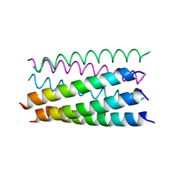 | |
5TWZ
 
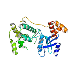 | |
8OZS
 
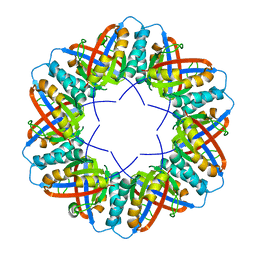 | | Populus tremula stable protein 1 with N-terminal binding peptide extension with hemin | | 分子名称: | Stable protein 1 | | 著者 | Sklyar, J, Zeibaq, Y, Bachar, O, Yehezkeli, O, Adir, N. | | 登録日 | 2023-05-09 | | 公開日 | 2024-03-27 | | 実験手法 | X-RAY DIFFRACTION (2.4 Å) | | 主引用文献 | A Bioengineered Stable Protein 1-Hemin Complex with Enhanced Peroxidase-Like Catalytic Properties
Small Science, 2023
|
|
7DC8
 
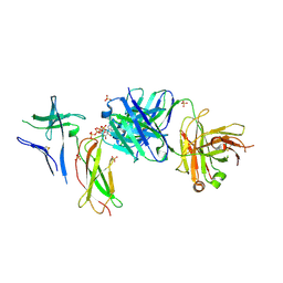 | | Crystal structure of Switch Ab Fab and hIL6R in complex with ATP | | 分子名称: | ADENOSINE-5'-TRIPHOSPHATE, Interleukin-6 receptor subunit alpha, SULFATE ION, ... | | 著者 | Kadono, S, Fukami, T.A, Kawauchi, H, Torizawa, T, Mimoto, F. | | 登録日 | 2020-10-23 | | 公開日 | 2021-01-13 | | 最終更新日 | 2024-10-30 | | 実験手法 | X-RAY DIFFRACTION (2.757 Å) | | 主引用文献 | Exploitation of Elevated Extracellular ATP to Specifically Direct Antibody to Tumor Microenvironment.
Cell Rep, 33, 2020
|
|
6FRV
 
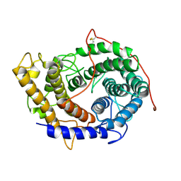 | | Structure of the catalytic domain of Aspergillus niger Glucoamylase | | 分子名称: | 2-acetamido-2-deoxy-beta-D-glucopyranose, 2-acetamido-2-deoxy-beta-D-glucopyranose-(1-4)-2-acetamido-2-deoxy-beta-D-glucopyranose, Glucoamylase, ... | | 著者 | Roth, C, Moroz, O.V, Ariza, A, Friis, E.P, Davies, G.J, Wilson, K.S. | | 登録日 | 2018-02-16 | | 公開日 | 2018-05-09 | | 最終更新日 | 2024-10-23 | | 実験手法 | X-RAY DIFFRACTION (2.3 Å) | | 主引用文献 | Structural insight into industrially relevant glucoamylases: flexible positions of starch-binding domains.
Acta Crystallogr D Struct Biol, 74, 2018
|
|
5DSG
 
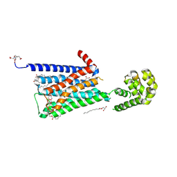 | | Structure of the M4 muscarinic acetylcholine receptor (M4-mT4L) bound to tiotropium | | 分子名称: | (1R,2R,4S,5S,7S)-7-{[hydroxy(dithiophen-2-yl)acetyl]oxy}-9,9-dimethyl-3-oxa-9-azoniatricyclo[3.3.1.0~2,4~]nonane, (2R)-2,3-dihydroxypropyl (9Z)-octadec-9-enoate, 1-(2-METHOXY-ETHOXY)-2-{2-[2-(2-METHOXY-ETHOXY]-ETHOXY}-ETHANE, ... | | 著者 | Thal, D.M, Kobilka, B.K, Sexton, P.M, Christopoulos, A. | | 登録日 | 2015-09-17 | | 公開日 | 2016-03-16 | | 最終更新日 | 2024-03-13 | | 実験手法 | X-RAY DIFFRACTION (2.6 Å) | | 主引用文献 | Crystal structures of the M1 and M4 muscarinic acetylcholine receptors.
Nature, 531, 2016
|
|
6G6D
 
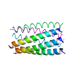 | |
6VFW
 
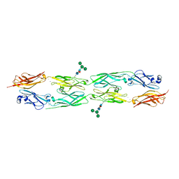 | | Crystal structure of human delta protocadherin 10 EC1-EC4 | | 分子名称: | CALCIUM ION, Protocadherin-10, alpha-D-mannopyranose, ... | | 著者 | Harrison, O.J, Brasch, J, Shapiro, L. | | 登録日 | 2020-01-06 | | 公開日 | 2020-03-11 | | 最終更新日 | 2024-10-23 | | 実験手法 | X-RAY DIFFRACTION (3.6 Å) | | 主引用文献 | Family-wide Structural and Biophysical Analysis of Binding Interactions among Non-clustered delta-Protocadherins.
Cell Rep, 30, 2020
|
|
8OZO
 
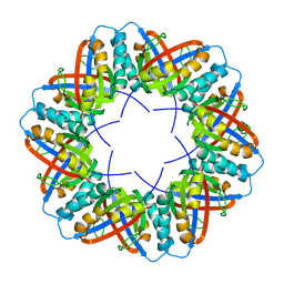 | | Populus tremula stable protein 1 with N-terminal binding peptide extension | | 分子名称: | Stable protein 1 | | 著者 | Sklyar, J, Zeibaq, Y, Bachar, O, Yehezkeli, O, Adir, N. | | 登録日 | 2023-05-09 | | 公開日 | 2024-03-27 | | 実験手法 | X-RAY DIFFRACTION (2.4 Å) | | 主引用文献 | A Bioengineered Stable Protein 1-Hemin Complex with Enhanced Peroxidase-Like Catalytic Properties
Small Science, 2024
|
|
6G6G
 
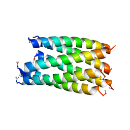 | |
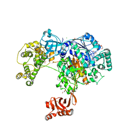
5L6I
 
 | |
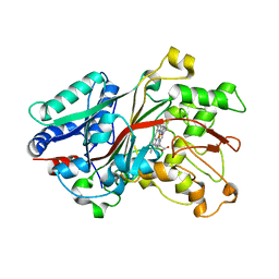
6FSL
 
 | |
5TXY
 
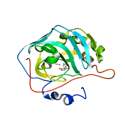 | | Identification of a New Zinc Binding Chemotype of by Fragment Screening on human carbonic anhydrase | | 分子名称: | (5R)-5-phenyl-1,3-oxazolidine-2,4-dione, Carbonic anhydrase 2, FORMIC ACID, ... | | 著者 | Ren, B, Peat, T.S, Poulsen, S.-A. | | 登録日 | 2016-11-17 | | 公開日 | 2017-08-30 | | 最終更新日 | 2024-03-06 | | 実験手法 | X-RAY DIFFRACTION (1.206 Å) | | 主引用文献 | Identification of a New Zinc Binding Chemotype by Fragment Screening.
J. Med. Chem., 60, 2017
|
|
5HWP
 
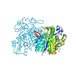 | | MvaS with acetylated Cys115 in complex with coenzyme A | | 分子名称: | COENZYME A, GLYCEROL, Hydroxymethylglutaryl-CoA synthase, ... | | 著者 | Bock, T, Kasten, J, Blankenfeldt, W. | | 登録日 | 2016-01-29 | | 公開日 | 2016-05-11 | | 最終更新日 | 2016-07-13 | | 実験手法 | X-RAY DIFFRACTION (2.2 Å) | | 主引用文献 | Crystal Structure of the HMG-CoA Synthase MvaS from the Gram-Negative Bacterium Myxococcus xanthus.
Chembiochem, 17, 2016
|
|
6XP7
 
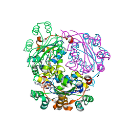 | |
8ODO
 
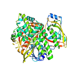 | | Structure of human guanylylated RTCB in complex with Archease | | 分子名称: | GUANOSINE-5'-MONOPHOSPHATE, MANGANESE (II) ION, PHOSPHATE ION, ... | | 著者 | Kopp, J, Gerber, J.L, Peschek, J. | | 登録日 | 2023-03-09 | | 公開日 | 2024-03-27 | | 最終更新日 | 2024-11-06 | | 実験手法 | X-RAY DIFFRACTION (2.2 Å) | | 主引用文献 | Structural and mechanistic insights into activation of the human RNA ligase RTCB by Archease.
Nat Commun, 15, 2024
|
|
5TYA
 
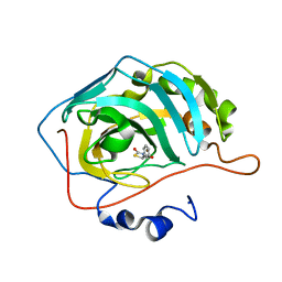 | | Identification of a New Zinc Binding Chemotype by Fragment Screening | | 分子名称: | (5R)-5-phenyl-1,3-thiazolidine-2,4-dione, Carbonic anhydrase 2, ZINC ION | | 著者 | Peat, T.S, Poulsen, S.A, Ren, B, Dolezal, O, Woods, L.A, Mujumdar, P, Chrysanthopoulos, P.K. | | 登録日 | 2016-11-18 | | 公開日 | 2017-08-30 | | 最終更新日 | 2023-10-04 | | 実験手法 | X-RAY DIFFRACTION (1.5 Å) | | 主引用文献 | Identification of a New Zinc Binding Chemotype by Fragment Screening.
J. Med. Chem., 60, 2017
|
|
6XPC
 
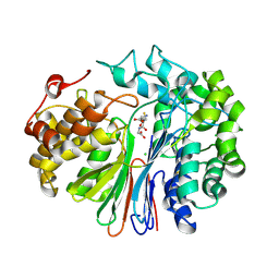 | |
6MNZ
 
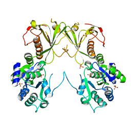 | | Crystal structure of RibBX, a two domain 3,4-dihydroxy-2-butanone 4-phosphate synthase from A. baumannii. | | 分子名称: | 3,4-dihydroxy-2-butanone 4-phosphate synthase, CHLORIDE ION, SULFATE ION | | 著者 | Wang, J, Gonzalez-Gutierrez, G, Giedroc, D.P. | | 登録日 | 2018-10-03 | | 公開日 | 2019-04-17 | | 最終更新日 | 2024-03-13 | | 実験手法 | X-RAY DIFFRACTION (2.66 Å) | | 主引用文献 | Multi-metal Restriction by Calprotectin Impacts De Novo Flavin Biosynthesis in Acinetobacter baumannii.
Cell Chem Biol, 26, 2019
|
|
7DIF
 
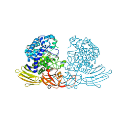 | | GH127 beta-L-arabinofuranosidase HypBA1 covalently complexed with beta-L-arabinofuranose-configured cyclophellitol at 1.75-angstrom resolution | | 分子名称: | (1S,2S,3R,4R)-3-(hydroxymethyl)cyclopentane-1,2,4-triol, Non-reducing end beta-L-arabinofuranosidase, POTASSIUM ION, ... | | 著者 | Amaki, S, McGregor, N.G.S, Arakawa, T, Yamada, C, Borlandelli, V, Overkleeft, H.S, Davies, G.J, Fushinobu, S. | | 登録日 | 2020-11-19 | | 公開日 | 2021-01-27 | | 最終更新日 | 2024-10-23 | | 実験手法 | X-RAY DIFFRACTION (1.75 Å) | | 主引用文献 | Cysteine Nucleophiles in Glycosidase Catalysis: Application of a Covalent beta-l-Arabinofuranosidase Inhibitor.
Angew.Chem.Int.Ed.Engl., 60, 2021
|
|
6G6M
 
 | | Crystal structure of the computationally designed Tako8 protein in P42212 | | 分子名称: | SULFATE ION, Tako8 | | 著者 | Noguchi, H, Addy, C, Simoncini, D, Van Meervelt, L, Schiex, T, Zhang, K.Y.J, Tame, J.R.H, Voet, A.R.D. | | 登録日 | 2018-04-01 | | 公開日 | 2018-11-28 | | 最終更新日 | 2024-05-08 | | 実験手法 | X-RAY DIFFRACTION (1.7 Å) | | 主引用文献 | Computational design of symmetrical eight-bladed beta-propeller proteins.
IUCrJ, 6, 2019
|
|
5L6W
 
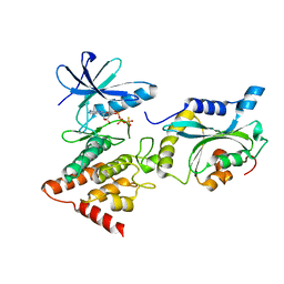 | | Structure Of the LIMK1-ATPgammaS-CFL1 Complex | | 分子名称: | Cofilin-1, LIM domain kinase 1, PHOSPHOTHIOPHOSPHORIC ACID-ADENYLATE ESTER | | 著者 | Salah, E, Mathea, S, Oerum, S, Newman, J.A, Tallant, C, Adamson, R, Canning, P, Beltrami, A, von Delft, F, Arrowsmith, C.H, Edwards, A.M, Bountra, C, Knapp, S, Bullock, A.N. | | 登録日 | 2016-05-31 | | 公開日 | 2016-06-08 | | 最終更新日 | 2024-01-10 | | 実験手法 | X-RAY DIFFRACTION (2.53 Å) | | 主引用文献 | Structure Of the LIMK1-ATPgammaS-CFL1 Complex
To Be Published
|
|
6VGW
 
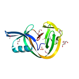 | | Crystal structure of VidaL intein (selenomethionine variant) | | 分子名称: | GLYCEROL, SULFATE ION, VidaL | | 著者 | Burton, A.J, Haugbro, M, Parisi, E, Muir, T.W. | | 登録日 | 2020-01-09 | | 公開日 | 2020-05-27 | | 最終更新日 | 2024-11-06 | | 実験手法 | X-RAY DIFFRACTION (1.51 Å) | | 主引用文献 | Live-cell protein engineering with an ultra-short split intein.
Proc.Natl.Acad.Sci.USA, 117, 2020
|
|
