1NU4
 
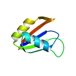
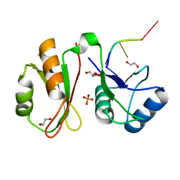
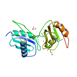
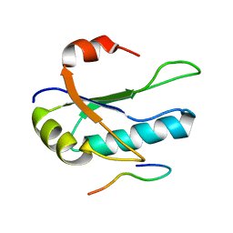
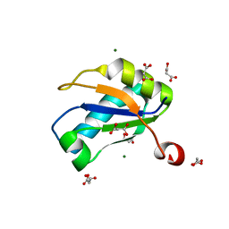 | | U1A RNA binding domain at 1.8 angstrom resolution reveals a pre-organized C-terminal helix | | 分子名称: | MAGNESIUM ION, MALONIC ACID, U1A RNA binding domain | | 著者 | Rupert, P.B, Xiao, H, Ferre-D'Amare, A.R. | | 登録日 | 2003-01-30 | | 公開日 | 2003-02-14 | | 最終更新日 | 2023-08-16 | | 実験手法 | X-RAY DIFFRACTION (1.8 Å) | | 主引用文献 | U1A RNA-binding domain at 1.8 A resolution.
Acta Crystallogr.,Sect.D, 59, 2003
|
|
6XH0
 | |
6XH3
 | |
6XLV
 | |
1NO8
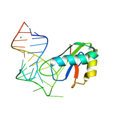
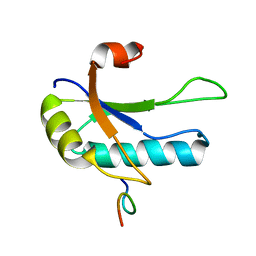
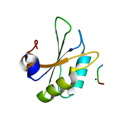
 | | SOLUTION STRUCTURE OF THE NUCLEAR FACTOR ALY RBD DOMAIN | | 分子名称: | ALY | | 著者 | Perez-Alvarado, G.C, Martinez-Yamout, M, Allen, M.M, Grosschedl, R, Dyson, H.J, Wright, P.E. | | 登録日 | 2003-01-15 | | 公開日 | 2003-08-12 | | 最終更新日 | 2024-05-22 | | 実験手法 | SOLUTION NMR | | 主引用文献 | Structure of the Nuclear Factor ALY: Insights into Post-Transcriptional
Regulatory and mRNA Nuclear Export Processes
Biochemistry, 42, 2003
|
|
1OPI
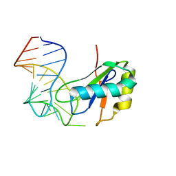
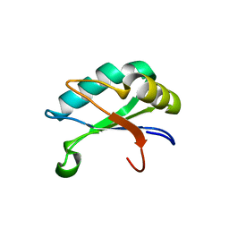
 | | SOLUTION STRUCTURE OF THE THIRD RNA RECOGNITION MOTIF (RRM) OF U2AF65 IN COMPLEX WITH AN N-TERMINAL SF1 PEPTIDE | | 分子名称: | SPLICING FACTOR SF1, SPLICING FACTOR U2AF 65 KDA SUBUNIT | | 著者 | Selenko, P, Gregorovic, G, Sprangers, R, Stier, G, Rhani, Z, Kramer, A, Sattler, M. | | 登録日 | 2003-03-05 | | 公開日 | 2004-03-16 | | 最終更新日 | 2024-05-22 | | 実験手法 | SOLUTION NMR | | 主引用文献 | Structural basis for the molecular recognition between human splicing factors U2AF65 and SF1/mBBP
Mol.Cell, 11, 2003
|
|
1OIA
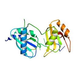
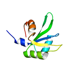
 | | U1A rnp domain 1-95 | | 分子名称: | U1 SMALL NUCLEAR RIBONUCLEOPROTEIN A | | 著者 | Nagai, K, Evans, P.R. | | 登録日 | 2003-06-12 | | 公開日 | 2003-06-26 | | 最終更新日 | 2024-05-08 | | 実験手法 | X-RAY DIFFRACTION (2.4 Å) | | 主引用文献 | Crystal Structure of the RNA-Binding Domain of the U1 Small Nuclear Ribonucleoprotein A
Nature, 348, 1990
|
|
1P1T
 | |
6GX6
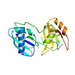
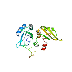
 | | Crystal structure of IMP3 RRM12 in complex with RNA (ACAC) | | 分子名称: | 1,2-ETHANEDIOL, Insulin-like growth factor 2 mRNA-binding protein 3, PHOSPHATE ION, ... | | 著者 | Jia, M, Gut, H, Chao, A.J. | | 登録日 | 2018-06-26 | | 公開日 | 2018-09-05 | | 最終更新日 | 2024-01-17 | | 実験手法 | X-RAY DIFFRACTION (2 Å) | | 主引用文献 | Structural basis of IMP3 RRM12 recognition of RNA.
RNA, 24, 2018
|
|
3G9C
 | | Crystal structure of the product Bacillus anthracis glmS ribozyme | | 分子名称: | 2-amino-2-deoxy-6-O-phosphono-alpha-D-glucopyranose, GLMS RIBOZYME, MAGNESIUM ION, ... | | 著者 | Strobel, S.A, Cochrane, J.C, Lipchock, S.V, Smith, K.D. | | 登録日 | 2009-02-13 | | 公開日 | 2009-11-03 | | 最終更新日 | 2024-02-21 | | 実験手法 | X-RAY DIFFRACTION (2.9 Å) | | 主引用文献 | Structural and chemical basis for glucosamine 6-phosphate binding and activation of the glmS ribozyme
Biochemistry, 48, 2009
|
|
3G96
 | | Crystal structure of the Bacillus anthracis glmS ribozyme bound to MaN6P | | 分子名称: | 2-amino-2-deoxy-6-O-phosphono-alpha-D-mannopyranose, GLMS RIBOZYME, MAGNESIUM ION, ... | | 著者 | Strobel, S.A, Cochrane, J.C, Lipchock, S.V, Smith, K.D. | | 登録日 | 2009-02-12 | | 公開日 | 2009-11-03 | | 最終更新日 | 2024-02-21 | | 実験手法 | X-RAY DIFFRACTION (3.01 Å) | | 主引用文献 | Structural and chemical basis for glucosamine 6-phosphate binding and activation of the glmS ribozyme
Biochemistry, 48, 2009
|
|
6XH1
 | |
6XLW
 | | Crystal structure of U2AF65 bound to AdML splice site sequence | | 分子名称: | DI(HYDROXYETHYL)ETHER, DNA/RNA (5'-R(P*UP*UP*(UD)P*UP*U)-D(P*(BRU))-R(P*CP*C)-3'), GLYCEROL, ... | | 著者 | Maji, D, Jenkins, J.L, Kielkopf, C.L. | | 登録日 | 2020-06-29 | | 公開日 | 2020-10-07 | | 最終更新日 | 2024-05-22 | | 実験手法 | X-RAY DIFFRACTION (1.5 Å) | | 主引用文献 | Representative cancer-associated U2AF2 mutations alter RNA interactions and splicing.
J.Biol.Chem., 295, 2020
|
|
3G8T
 | | Crystal structure of the G33A mutant Bacillus anthracis glmS ribozyme bound to GlcN6P | | 分子名称: | 2-amino-2-deoxy-6-O-phosphono-alpha-D-glucopyranose, MAGNESIUM ION, RNA (5'-R(*AP*(A2M)P*GP*CP*GP*CP*CP*AP*GP*AP*AP*CP*U)-3'), ... | | 著者 | Strobel, S.A, Cochrane, J.C, Lipchock, S.V, Smith, K.D. | | 登録日 | 2009-02-12 | | 公開日 | 2009-11-03 | | 最終更新日 | 2024-02-21 | | 実験手法 | X-RAY DIFFRACTION (3 Å) | | 主引用文献 | Structural and chemical basis for glucosamine 6-phosphate binding and activation of the glmS ribozyme
Biochemistry, 48, 2009
|
|
6XH2
 | |
6XLX
 | |
1O0P
 | | Solution Structure of the third RNA Recognition Motif (RRM) of U2AF65 in complex with an N-terminal SF1 peptide | | 分子名称: | Splicing Factor SF1, Splicing factor U2AF 65 kDa subunit | | 著者 | Selenko, P, Gregorovic, G, Sprangers, R, Stier, G, Rhani, Z, Kramer, A, Sattler, M. | | 登録日 | 2003-02-24 | | 公開日 | 2003-05-06 | | 最終更新日 | 2024-05-22 | | 実験手法 | SOLUTION NMR | | 主引用文献 | Structural Basis for the molecular recognition between human splicing factors U2AF65 and SF1/mBBP
Mol.Cell, 11, 2003
|
|
6HIP
 | | Structure of SPF45 UHM bound to HIV-1 Rev ULM | | 分子名称: | HIV-1 Rev (41-49), SODIUM ION, Splicing factor 45, ... | | 著者 | Pabis, M, Corsini, L, Sattler, M. | | 登録日 | 2018-08-30 | | 公開日 | 2019-03-27 | | 最終更新日 | 2024-01-17 | | 実験手法 | X-RAY DIFFRACTION (1.2 Å) | | 主引用文献 | Modulation of HIV-1 gene expression by binding of a ULM motif in the Rev protein to UHM-containing splicing factors.
Nucleic Acids Res., 47, 2019
|
|
3EGZ
 | | Crystal structure of an in vitro evolved tetracycline aptamer and artificial riboswitch | | 分子名称: | 7-CHLOROTETRACYCLINE, MAGNESIUM ION, Tetracycline aptamer and artificial riboswitch, ... | | 著者 | Xiao, H, Edwards, T.E, Ferre-D'Amare, A.R. | | 登録日 | 2008-09-11 | | 公開日 | 2008-10-28 | | 最終更新日 | 2021-10-20 | | 実験手法 | X-RAY DIFFRACTION (2.2 Å) | | 主引用文献 | Structural basis for specific, high-affinity tetracycline binding by an in vitro evolved aptamer and artificial riboswitch
Chem.Biol., 15, 2008
|
|
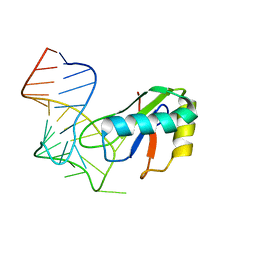
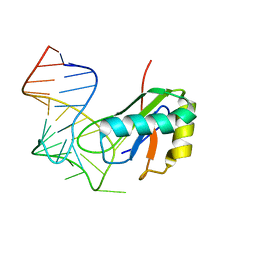
1RKJ
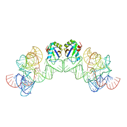
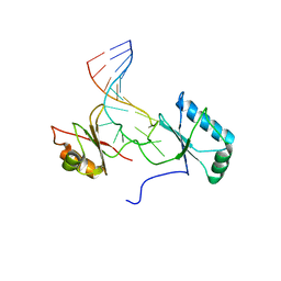 | | Solution structure of the complex formed by the two N-terminal RNA-binding domains of nucleolin and a pre-rRNA target | | 分子名称: | 5'-R(*GP*GP*AP*UP*GP*CP*CP*UP*CP*CP*CP*GP*AP*GP*UP*GP*CP*AP*UP*CP*C)-3', Nucleolin | | 著者 | Johansson, C, Finger, L.D, Trantirek, L, Mueller, T.D, Kim, S, Laird-Offringa, I.A, Feigon, J. | | 登録日 | 2003-11-21 | | 公開日 | 2004-04-27 | | 最終更新日 | 2024-05-22 | | 実験手法 | SOLUTION NMR | | 主引用文献 | Solution structure of the complex formed by the two N-terminal RNA-binding domains of nucleolin and a pre-rRNA target.
J.Mol.Biol., 337, 2004
|
|
3G8S
 | | Crystal structure of the pre-cleaved Bacillus anthracis glmS ribozyme | | 分子名称: | GLMS RIBOZYME, MAGNESIUM ION, RNA (5'-R(*AP*(A2M)P*GP*CP*GP*CP*CP*AP*GP*AP*AP*CP*U)-3'), ... | | 著者 | Strobel, S.A, Cochrane, J.C, Lipchock, S.V, Smith, K.D. | | 登録日 | 2009-02-12 | | 公開日 | 2009-11-03 | | 最終更新日 | 2024-02-21 | | 実験手法 | X-RAY DIFFRACTION (3.1 Å) | | 主引用文献 | Structural and chemical basis for glucosamine 6-phosphate binding and activation of the glmS ribozyme
Biochemistry, 48, 2009
|
|
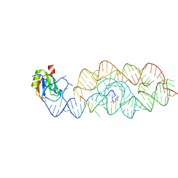
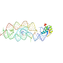
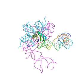
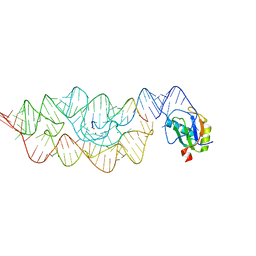
3HHN
 | | Crystal structure of class I ligase ribozyme self-ligation product, in complex with U1A RBD | | 分子名称: | Class I ligase ribozyme, self-ligation product, MAGNESIUM ION, ... | | 著者 | Shechner, D.M, Grant, R.A, Bagby, S.C, Bartel, D.P. | | 登録日 | 2009-05-15 | | 公開日 | 2009-11-24 | | 最終更新日 | 2024-02-21 | | 実験手法 | X-RAY DIFFRACTION (2.987 Å) | | 主引用文献 | Crystal structure of the catalytic core of an RNA-polymerase ribozyme.
Science, 326, 2009
|
|
3HI9
 | |
1PO6
 | |
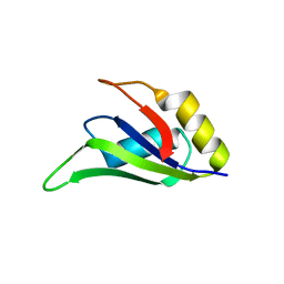
6KOR
 | | Crystal structure of the RRM domain of SYNCRIP | | 分子名称: | Heterogeneous nuclear ribonucleoprotein Q | | 著者 | Chen, Y, Chan, J, Chen, W, Jobichen, C. | | 登録日 | 2019-08-12 | | 公開日 | 2020-01-22 | | 最終更新日 | 2023-11-22 | | 実験手法 | X-RAY DIFFRACTION (2.602 Å) | | 主引用文献 | SYNCRIP, a new player in pri-let-7a processing.
Rna, 26, 2020
|
|


