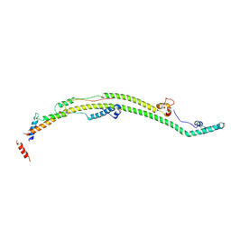
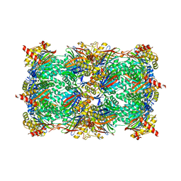
8HI5
 
 | |
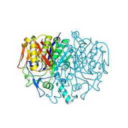
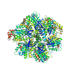
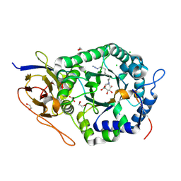
4WC9
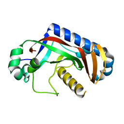 | | Catalytic domain of mouse 2',3'-cyclic nucleotide 3'- phosphodiesterase, with mutation F235L | | 分子名称: | 2',3'-cyclic-nucleotide 3'-phosphodiesterase | | 著者 | Myllykoski, M, Raasakka, A, Kursula, P. | | 登録日 | 2014-09-04 | | 公開日 | 2015-09-23 | | 最終更新日 | 2024-01-10 | | 実験手法 | X-RAY DIFFRACTION (2 Å) | | 主引用文献 | Determinants of ligand binding and catalytic activity in the myelin enzyme 2',3'-cyclic nucleotide 3'-phosphodiesterase.
Sci Rep, 5, 2015
|
|
8H9Q
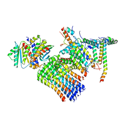 | | Human ATP synthase state 3b subregion 3 | | 分子名称: | ATP synthase F(0) complex subunit B1, mitochondrial, ATP synthase F(0) complex subunit C1, ... | | 著者 | Lai, Y, Zhang, Y, Liu, F, Gao, Y, Gong, H, Rao, Z. | | 登録日 | 2022-10-25 | | 公開日 | 2023-05-31 | | 最終更新日 | 2024-07-03 | | 実験手法 | ELECTRON MICROSCOPY (3.47 Å) | | 主引用文献 | Structure of the human ATP synthase.
Mol.Cell, 83, 2023
|
|
1RK5
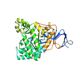 | | The D-aminoacylase mutant D366A in complex with 100mM CuCl2 | | 分子名称: | ACETATE ION, COPPER (II) ION, D-aminoacylase, ... | | 著者 | Lai, W.L, Chou, L.Y, Ting, C.Y, Tsai, Y.C, Liaw, S.H. | | 登録日 | 2003-11-20 | | 公開日 | 2004-04-20 | | 最終更新日 | 2023-10-25 | | 実験手法 | X-RAY DIFFRACTION (1.8 Å) | | 主引用文献 | The functional role of the binuclear metal center in D-aminoacylase: one-metal activation and second-metal attenuation.
J.Biol.Chem., 279, 2004
|
|
1RRV
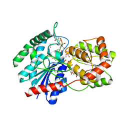 | | X-ray crystal structure of TDP-vancosaminyltransferase GtfD as a complex with TDP and the natural substrate, desvancosaminyl vancomycin. | | 分子名称: | DESVANCOSAMINYL VANCOMYCIN, GLYCEROL, GLYCOSYLTRANSFERASE GTFD, ... | | 著者 | Mulichak, A.M, Lu, W, Losey, H.C, Walsh, C.T, Garavito, R.M. | | 登録日 | 2003-12-09 | | 公開日 | 2004-05-18 | | 最終更新日 | 2023-11-15 | | 実験手法 | X-RAY DIFFRACTION (2 Å) | | 主引用文献 | Crystal Structure of Vancosaminyltransferase Gtfd from the Vancomycin Biosynthetic Pathway: Interactions with Acceptor and Nucleotide Ligands
Biochemistry, 43, 2004
|
|
1B3N
 | | BETA-KETOACYL CARRIER PROTEIN SYNTHASE AS A DRUG TARGET, IMPLICATIONS FROM THE CRYSTAL STRUCTURE OF A COMPLEX WITH THE INHIBITOR CERULENIN. | | 分子名称: | (2S, 3R)-3-HYDROXY-4-OXO-7,10-TRANS,TRANS-DODECADIENAMIDE, PROTEIN (KETOACYL ACYL CARRIER PROTEIN SYNTHASE 2) | | 著者 | Moche, M, Schneider, G, Edwards, P, Dehesh, K, Lindqvist, Y. | | 登録日 | 1998-12-14 | | 公開日 | 1999-04-06 | | 最終更新日 | 2023-12-27 | | 実験手法 | X-RAY DIFFRACTION (2.65 Å) | | 主引用文献 | Structure of the complex between the antibiotic cerulenin and its target, beta-ketoacyl-acyl carrier protein synthase.
J.Biol.Chem., 274, 1999
|
|
1RU5
 | |
4G2U
 | | Crystal Structure Analysis of Ostertagia ostertagi ASP-1 | | 分子名称: | 2-acetamido-2-deoxy-beta-D-glucopyranose, Ancylostoma-secreted protein-like protein, SULFATE ION | | 著者 | Weeks, S.D, Borloo, J, Geldhof, P, Vercruysse, J, Strelkov, S.V. | | 登録日 | 2012-07-13 | | 公開日 | 2013-03-27 | | 最終更新日 | 2023-11-08 | | 実験手法 | X-RAY DIFFRACTION (1.85 Å) | | 主引用文献 | Structure of Ostertagia ostertagi ASP-1: insights into disulfide-mediated cyclization and dimerization
Acta Crystallogr.,Sect.D, 69, 2013
|
|
5L3U
 | | Thermolysin in complex with JC149 (MPD cryo protectant) | | 分子名称: | CALCIUM ION, DIMETHYL SULFOXIDE, N~2~-[(R)-({[(benzyloxy)carbonyl]amino}methyl)(hydroxy)phosphoryl]-N-[(2R)-2,3,3-trimethylbutyl]-L-leucinamide, ... | | 著者 | Krimmer, S.G, Cramer, J, Heine, A, Klebe, G. | | 登録日 | 2016-05-24 | | 公開日 | 2016-12-21 | | 最終更新日 | 2024-01-10 | | 実験手法 | X-RAY DIFFRACTION (1.23 Å) | | 主引用文献 | Rational Design of Thermodynamic and Kinetic Binding Profiles by Optimizing Surface Water Networks Coating Protein-Bound Ligands.
J. Med. Chem., 59, 2016
|
|
8H9R
 | | Human ATP synthase state 3b subregion 2 | | 分子名称: | ATP synthase F(0) complex subunit B1, mitochondrial, ATP synthase subunit d, ... | | 著者 | Lai, Y, Zhang, Y, Liu, F, Gao, Y, Gong, H, Rao, Z. | | 登録日 | 2022-10-25 | | 公開日 | 2023-05-31 | | 最終更新日 | 2024-07-03 | | 実験手法 | ELECTRON MICROSCOPY (3.97 Å) | | 主引用文献 | Structure of the human ATP synthase.
Mol.Cell, 83, 2023
|
|
8H9P
 | | Human ATP synthase F1 domain, state 3b | | 分子名称: | ADENOSINE-5'-DIPHOSPHATE, ADENOSINE-5'-TRIPHOSPHATE, ATP synthase subunit O, ... | | 著者 | Lai, Y, Zhang, Y, Liu, F, Gao, Y, Gong, H, Rao, Z. | | 登録日 | 2022-10-25 | | 公開日 | 2023-05-31 | | 最終更新日 | 2024-07-03 | | 実験手法 | ELECTRON MICROSCOPY (3.02 Å) | | 主引用文献 | Structure of the human ATP synthase.
Mol.Cell, 83, 2023
|
|
1R17
 | | Crystal Structure Analysis of S.epidermidis adhesin SdrG binding to Fibrinogen (adhesin-ligand complex) | | 分子名称: | CALCIUM ION, fibrinogen-binding protein SdrG, fibrinopeptide B | | 著者 | Ponnuraj, K, Bowden, M.G, Davis, S, Gurusiddappa, S, Moore, D, Choe, D, Xu, Y, Hook, M, Narayana, S.V.L. | | 登録日 | 2003-09-23 | | 公開日 | 2003-10-28 | | 最終更新日 | 2024-02-14 | | 実験手法 | X-RAY DIFFRACTION (1.86 Å) | | 主引用文献 | A "dock, lock and latch" Structural Model for a Staphylococcal Adhesin Binding to Fibrinogen
Cell(Cambridge,Mass.), 115, 2003
|
|
5L5J
 | |
6IF1
 | | Crystal structure of Ube2K and K48-linked di-ubiquitin complex | | 分子名称: | Ubiquitin, Ubiquitin-conjugating enzyme E2 K | | 著者 | Lee, J.-G, Youn, H.-S, Lee, Y, An, J.Y, Park, K.R, Kang, J.Y, Lim, J.J, Eom, S.H. | | 登録日 | 2018-09-18 | | 公開日 | 2018-11-21 | | 最終更新日 | 2023-11-22 | | 実験手法 | X-RAY DIFFRACTION (2.466 Å) | | 主引用文献 | Crystal structure of the Ube2K/E2-25K and K48-linked di-ubiquitin complex provides structural insight into the mechanism of K48-specific ubiquitin chain synthesis.
Biochem. Biophys. Res. Commun., 506, 2018
|
|
5L62
 | | Yeast 20S proteasome with human beta5c (1-138) and human beta6 (97-111; 118-133) in complex with epoxyketone inhibitor 16 | | 分子名称: | (2~{S})-3-(1~{H}-indol-3-yl)-~{N}-[(2~{S},3~{S},4~{R})-4-methyl-3,5-bis(oxidanyl)-1-phenyl-pentan-2-yl]-2-[[(2~{R})-2-(2-morpholin-4-ylethanoylamino)propanoyl]amino]propanamide, 2-(N-MORPHOLINO)-ETHANESULFONIC ACID, CHLORIDE ION, ... | | 著者 | Groll, M, Huber, E.M. | | 登録日 | 2016-05-28 | | 公開日 | 2016-11-09 | | 最終更新日 | 2024-01-10 | | 実験手法 | X-RAY DIFFRACTION (2.8 Å) | | 主引用文献 | A humanized yeast proteasome identifies unique binding modes of inhibitors for the immunosubunit beta 5i.
EMBO J., 35, 2016
|
|
8H9U
 | | Human ATP synthase state 3a (combined) | | 分子名称: | ADENOSINE-5'-DIPHOSPHATE, ADENOSINE-5'-TRIPHOSPHATE, ATP synthase F(0) complex subunit B1, ... | | 著者 | Lai, Y, Zhang, Y, Liu, F, Gao, Y, Gong, H, Rao, Z. | | 登録日 | 2022-10-25 | | 公開日 | 2023-05-31 | | 最終更新日 | 2024-07-03 | | 実験手法 | ELECTRON MICROSCOPY (2.61 Å) | | 主引用文献 | Structure of the human ATP synthase.
Mol.Cell, 83, 2023
|
|
1B7Y
 | | PHENYLALANYL TRNA SYNTHETASE COMPLEXED WITH PHENYLALANINYL-ADENYLATE | | 分子名称: | ADENOSINE-5'-[PHENYLALANINOL-PHOSPHATE], MAGNESIUM ION, PROTEIN (PHENYLALANYL-TRNA SYNTHETASE) | | 著者 | Reshetnikova, L, Moor, N, Lavrik, O, Vassylyev, D.G. | | 登録日 | 1999-01-26 | | 公開日 | 2000-01-26 | | 最終更新日 | 2023-08-09 | | 実験手法 | X-RAY DIFFRACTION (2.5 Å) | | 主引用文献 | Crystal structures of phenylalanyl-tRNA synthetase complexed with phenylalanine and a phenylalanyl-adenylate analogue.
J.Mol.Biol., 287, 1999
|
|
8H9V
 | | Human ATP synthase state 3b (combined) | | 分子名称: | ADENOSINE-5'-DIPHOSPHATE, ADENOSINE-5'-TRIPHOSPHATE, ATP synthase F(0) complex subunit B1, ... | | 著者 | Lai, Y, Zhang, Y, Liu, F, Gao, Y, Gong, H, Rao, Z. | | 登録日 | 2022-10-25 | | 公開日 | 2023-05-31 | | 最終更新日 | 2024-07-03 | | 実験手法 | ELECTRON MICROSCOPY (3.02 Å) | | 主引用文献 | Structure of the human ATP synthase.
Mol.Cell, 83, 2023
|
|
1SAK
 | |
8HI4
 | |
5LB5
 | | Crystal structure of human RECQL5 helicase in complex with ADP/Mg (tricilinc form). | | 分子名称: | ADENOSINE-5'-DIPHOSPHATE, ATP-dependent DNA helicase Q5, DIMETHYL SULFOXIDE, ... | | 著者 | Newman, J.A, Aitkenhead, H, Savitsky, P, Krojer, T, von Delft, F, Arrowsmith, C.H, Edwards, A.M, Bountra, C, Gileadi, O, Structural Genomics Consortium (SGC) | | 登録日 | 2016-06-15 | | 公開日 | 2016-07-06 | | 最終更新日 | 2024-01-10 | | 実験手法 | X-RAY DIFFRACTION (2 Å) | | 主引用文献 | Insights into the RecQ helicase mechanism revealed by the structure of the helicase domain of human RECQL5.
Nucleic Acids Res., 45, 2017
|
|
5L9Z
 | | Crystal structure of human heparanase nucleophile mutant (E343Q), in complex with unreacted glucuronic acid configured aziridine probe JJB355 | | 分子名称: | (1~{R},2~{S},3~{R},4~{S},5~{S},6~{R})-7-[8-[(azanylidene-{4}-azanylidene)amino]octyl]-3,4,5-tris(oxidanyl)-7-azabicyclo[4.1.0]heptane-2-carboxylic acid, 1,2-ETHANEDIOL, 2-acetamido-2-deoxy-beta-D-glucopyranose, ... | | 著者 | Wu, L, Jin, Y, Davies, G.J. | | 登録日 | 2016-06-13 | | 公開日 | 2017-05-31 | | 最終更新日 | 2024-01-10 | | 実験手法 | X-RAY DIFFRACTION (1.57 Å) | | 主引用文献 | Activity-based probes for functional interrogation of retaining beta-glucuronidases.
Nat. Chem. Biol., 13, 2017
|
|
1SAE
 | |
4V83
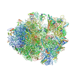 | | Crystal structure of a complex containing domain 3 from the PSIV IGR IRES RNA bound to the 70S ribosome. | | 分子名称: | 23S ribosomal RNA, 30S ribosomal protein S10, 30S ribosomal protein S11, ... | | 著者 | Zhu, J, Korostelev, A, Costantino, D, Noller, H.F, Kieft, J.S. | | 登録日 | 2010-12-13 | | 公開日 | 2014-07-09 | | 最終更新日 | 2014-12-10 | | 実験手法 | X-RAY DIFFRACTION (3.5 Å) | | 主引用文献 | Crystal structures of complexes containing domains from two viral internal ribosome entry site (IRES) RNAs bound to the 70S ribosome.
Proc.Natl.Acad.Sci.USA, 108, 2011
|
|
8H9N
 | | Human ATP synthase state 3a subregion 2 | | 分子名称: | ATP synthase F(0) complex subunit B1, mitochondrial, ATP synthase subunit d, ... | | 著者 | Lai, Y, Zhang, Y, Liu, F, Gao, Y, Gong, H, Rao, Z. | | 登録日 | 2022-10-25 | | 公開日 | 2023-05-31 | | 最終更新日 | 2024-07-03 | | 実験手法 | ELECTRON MICROSCOPY (3.56 Å) | | 主引用文献 | Structure of the human ATP synthase.
Mol.Cell, 83, 2023
|
|


