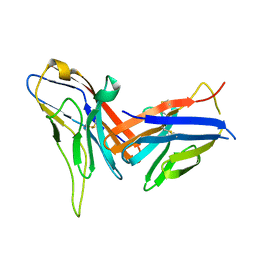
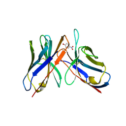
5MIX
 
 | |
7ST5
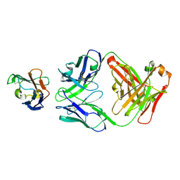
 | | Structure of Fab CC-95251 in complex with SIRP-alpha | | 分子名称: | 1,2-ETHANEDIOL, Fab CC-95251 anti-SIRP-alpha heavy chain, Fab CC-95251 anti-SIRP-alpha light chain, ... | | 著者 | Fenalti, G. | | 登録日 | 2021-11-12 | | 公開日 | 2023-05-17 | | 最終更新日 | 2024-10-16 | | 実験手法 | X-RAY DIFFRACTION (2.2 Å) | | 主引用文献 | Discovery and Preclinical Activity of BMS-986351, an Antibody to SIRP alpha That Enhances Macrophage-mediated Tumor Phagocytosis When Combined with Opsonizing Antibodies.
Cancer Res Commun, 4, 2024
|
|
4AIZ
 | | Crystallographic structure of 3mJL2 from the germinal line lambda 3 | | 分子名称: | 1,5:6,10-dianhydro-3,4,7,8-tetradeoxy-2,9-bis-C-(hydroxymethyl)-L-manno-decitol, CITRIC ACID, SULFATE ION, ... | | 著者 | Villalba, M.I, Luna, O.D, Rudino-Pinera, E, Sanchez, R, Sanchez-Lopez, R, Rojas-Trejo, S, Olamendi-Portugal, T, Fernandez-Velasco, D.A, Becerril, B. | | 登録日 | 2012-02-15 | | 公開日 | 2013-03-06 | | 最終更新日 | 2024-11-13 | | 実験手法 | X-RAY DIFFRACTION (1.75 Å) | | 主引用文献 | Site-Directed Mutagenesis Reveals Regions Implicated in the Stability and Fiber Formation of Human Lambda3R Light Chains.
J.Biol.Chem., 290, 2015
|
|
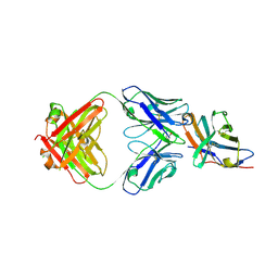
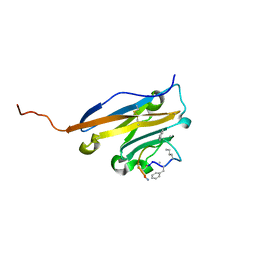
43C9
 | |
5OWP
 | | Crystal structure of glycopeptide "GVTSAfPDT*RPAP" in complex with scFv-SM3 | | 分子名称: | 1,2-ETHANEDIOL, 2-acetamido-2-deoxy-alpha-D-galactopyranose, 5,6-DIHYDRO-BENZO[H]CINNOLIN-3-YLAMINE, ... | | 著者 | Bermejo, I.A, Albuquerque, I.S, Somovilla, V.J, Martinez-Saez, N, Castro-Lopez, J, Garcia-Martin, F, Hinou, H, Nishimura, S, Jimenez-Barbero, J, Asensio, J.L, Avenoza, A, Busto, J.H, Hurtado-Guerrero, R, Peregrina, J.M, Bernardes, G.J, Corzana, F. | | 登録日 | 2017-09-02 | | 公開日 | 2017-12-13 | | 最終更新日 | 2024-10-23 | | 実験手法 | X-RAY DIFFRACTION (1.85 Å) | | 主引用文献 | The Use of Fluoroproline in MUC1 Antigen Enables Efficient Detection of Antibodies in Patients with Prostate Cancer.
J. Am. Chem. Soc., 139, 2017
|
|
5NIU
 | | Structure of human Programmed cell death 1 ligand 1 (PD-L1) with low molecular mass inhibitor | | 分子名称: | (2~{R})-2-[[2-[(3-cyanophenyl)methoxy]-4-[[3-(2,3-dihydro-1,4-benzodioxin-6-yl)-2-methyl-phenyl]methoxy]-5-methyl-phenyl]methylamino]-3-oxidanyl-propanoic acid, 1,2-ETHANEDIOL, Programmed cell death 1 ligand 1 | | 著者 | Zak, K.M, Grudnik, P, Skalniak, L, Dubin, G, Holak, T.A. | | 登録日 | 2017-03-27 | | 公開日 | 2017-12-06 | | 最終更新日 | 2024-11-13 | | 実験手法 | X-RAY DIFFRACTION (2.01 Å) | | 主引用文献 | Small-molecule inhibitors of PD-1/PD-L1 immune checkpoint alleviate the PD-L1-induced exhaustion of T-cells.
Oncotarget, 8, 2017
|
|
8VTE
 | |
8VTD
 | |
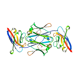
5N7B
 | | Understanding the singular conformational landscape of the Tn antigens: Sulfur-for- oxygen substitution in the glycosidic linkage provides new insights into molecular recognition by an antibody | | 分子名称: | 1,2-ETHANEDIOL, 2-acetamido-2-deoxy-alpha-D-galactopyranose, APD(CG6)RP(NH2) peptide, ... | | 著者 | Companon, I, Martinez-Saez, N, Castro-Lopez, J, Jimenez-Barbero, J, Bernardes, G.J.L, Busto, J.H, Avenoza, A, Jimenez-Oses, G, Hurtado-Guerrero, R, Peregrina, J.M, Corzana, F. | | 登録日 | 2017-02-20 | | 公開日 | 2018-06-27 | | 最終更新日 | 2024-01-17 | | 実験手法 | X-RAY DIFFRACTION (1.7 Å) | | 主引用文献 | Structure-Based Design of Potent Tumor-Associated Antigens: Modulation of Peptide Presentation by Single-Atom O/S or O/Se Substitutions at the Glycosidic Linkage.
J.Am.Chem.Soc., 141, 2019
|
|
5N2F
 | | Structure of PD-L1/small-molecule inhibitor complex | | 分子名称: | 4-[[4-[[3-(2,3-dihydro-1,4-benzodioxin-6-yl)-2-methyl-phenyl]methoxy]-2,5-bis(fluoranyl)phenyl]methylamino]-3-oxidanylidene-butanoic acid, Programmed cell death 1 ligand 1 | | 著者 | Guzik, K, Zak, K.M, Grudnik, P, Dubin, G, Holak, T.A. | | 登録日 | 2017-02-07 | | 公開日 | 2017-06-28 | | 最終更新日 | 2024-11-13 | | 実験手法 | X-RAY DIFFRACTION (1.7 Å) | | 主引用文献 | Small-Molecule Inhibitors of the Programmed Cell Death-1/Programmed Death-Ligand 1 (PD-1/PD-L1) Interaction via Transiently Induced Protein States and Dimerization of PD-L1.
J. Med. Chem., 60, 2017
|
|
7SU1
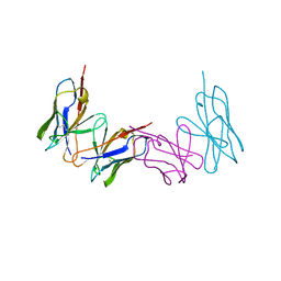
 | | Crystal structure of an acidic pH-selective Ipilimumab variant Ipi.106 in complex with CTLA-4 | | 分子名称: | 2-acetamido-2-deoxy-beta-D-glucopyranose, Cytotoxic T-lymphocyte protein 4, Fab heavy chain, ... | | 著者 | Lee, P.S, Chau, B, Strop, P. | | 登録日 | 2021-11-15 | | 公開日 | 2022-03-02 | | 最終更新日 | 2024-10-23 | | 実験手法 | X-RAY DIFFRACTION (2.53 Å) | | 主引用文献 | Improved therapeutic index of an acidic pH-selective antibody.
Mabs, 14, 2022
|
|
8X6B
 | | Crystal structure of immune receptor PVRIG in complex with ligand Nectin-2 | | 分子名称: | 2-acetamido-2-deoxy-beta-D-glucopyranose, Nectin-2, Transmembrane protein PVRIG | | 著者 | Hu, S.T, Han, P, Wang, H, Qi, J.X. | | 登録日 | 2023-11-21 | | 公開日 | 2024-04-24 | | 最終更新日 | 2024-11-13 | | 実験手法 | X-RAY DIFFRACTION (2 Å) | | 主引用文献 | Structural basis for the immune recognition and selectivity of the immune receptor PVRIG for ligand Nectin-2.
Structure, 32, 2024
|
|
7SU0
 | | Crystal structure of an acidic pH-selective Ipilimumab variant Ipi.105 in complex with CTLA-4 | | 分子名称: | 2-acetamido-2-deoxy-beta-D-glucopyranose, CITRATE ANION, Cytotoxic T-lymphocyte protein 4, ... | | 著者 | Lee, P.S, Chau, B, Strop, P. | | 登録日 | 2021-11-15 | | 公開日 | 2022-03-02 | | 最終更新日 | 2024-10-16 | | 実験手法 | X-RAY DIFFRACTION (2.41 Å) | | 主引用文献 | Improved therapeutic index of an acidic pH-selective antibody.
Mabs, 14, 2022
|
|
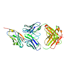
3ZHD
 | |
3ZHL
 | |
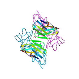
43CA
 | |
5O45
 | | Structure of human PD-L1 in complex with inhibitor | | 分子名称: | PHE-MEA-9KK-SAR-ASP-VAL-MEA-TYR-SAR-TRP-TYR-LEU-CCS-GLY-NH2, Programmed cell death 1 ligand 1 | | 著者 | Magiera, K, Grudnik, P, Dubin, G, Holak, T.A. | | 登録日 | 2017-05-26 | | 公開日 | 2017-09-20 | | 最終更新日 | 2024-01-17 | | 実験手法 | X-RAY DIFFRACTION (0.99 Å) | | 主引用文献 | Bioactive Macrocyclic Inhibitors of the PD-1/PD-L1 Immune Checkpoint.
Angew. Chem. Int. Ed. Engl., 56, 2017
|
|
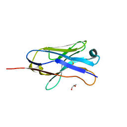
7YTE
 | |
1WZ1
 | | Crystal structure of the Fv fragment complexed with dansyl-lysine | | 分子名称: | Ig heavy chain, Ig light chain, N~6~-{[5-(DIMETHYLAMINO)-1-NAPHTHYL]SULFONYL}-L-LYSINE | | 著者 | Nakasako, M, Oka, T, Mashumo, M, Takahashi, H, Shimada, I, Yamaguchi, Y, Kato, K, Arata, Y. | | 登録日 | 2005-02-21 | | 公開日 | 2006-01-31 | | 最終更新日 | 2024-10-23 | | 実験手法 | X-RAY DIFFRACTION (1.85 Å) | | 主引用文献 | Conformational dynamics of complementarity-determining region H3 of an anti-dansyl Fv fragment in the presence of its hapten
J.Mol.Biol., 351, 2005
|
|
7YDS
 | | The structure of the bispecific antibody targeted PD-L1 and 4-1BB | | 分子名称: | Anti-PDL1-VH-CH1, Anti-PDL1-VL-CL, Programmed cell death 1 ligand 1 | | 著者 | Gao, Y, Zhu, M, Liu, W.T, Cheng, L.S, Zhu, Z.L, Niu, L.W. | | 登録日 | 2022-07-04 | | 公開日 | 2023-07-19 | | 最終更新日 | 2024-10-23 | | 実験手法 | X-RAY DIFFRACTION (2.3 Å) | | 主引用文献 | A bispecific antibody targeted PD-L1 and 4-1BB induces a potent antitumor immune activity in colorectal cancer without systemic toxicity
To Be Published
|
|
7ZOZ
 | | Crystal structure of Siglec-15 in complex with Fab | | 分子名称: | Anti-Siglec 15 Fab HC, Anti-Siglec 15 Fab LC, Sialic acid-binding Ig-like lectin 15 | | 著者 | Lenza, M.P, Oyenarte, I, Jimenez-Barbero, J, Ereno-Orbea, J. | | 登録日 | 2022-04-26 | | 公開日 | 2023-06-28 | | 最終更新日 | 2024-11-20 | | 実験手法 | X-RAY DIFFRACTION (2.104 Å) | | 主引用文献 | Structural insights into Siglec-15 reveal glycosylation dependency for its interaction with T cells through integrin CD11b.
Nat Commun, 14, 2023
|
|
1WTL
 | |
1VES
 | |
1VER
 | |
8AS0
 | | PD-1 extracellular domain in complex with Fab fragment from D12 antibody | | 分子名称: | 2-acetamido-2-deoxy-beta-D-glucopyranose, 2-acetamido-2-deoxy-beta-D-glucopyranose-(1-4)-2-acetamido-2-deoxy-beta-D-glucopyranose, 2-acetamido-2-deoxy-beta-D-glucopyranose-(1-4)-[alpha-L-fucopyranose-(1-6)]2-acetamido-2-deoxy-beta-D-glucopyranose, ... | | 著者 | Ongaro, T, Scietti, L, Pluss, L, Peissert, F, Villa, A, Puca, E, De Luca, R, Neri, D, Forneris, F. | | 登録日 | 2022-08-17 | | 公開日 | 2022-11-23 | | 最終更新日 | 2024-10-23 | | 実験手法 | X-RAY DIFFRACTION (3.5 Å) | | 主引用文献 | Selection of a PD-1 blocking antibody from a novel fully human phage display library.
Protein Sci., 31, 2022
|
|


