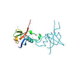
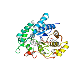
7K3I
 
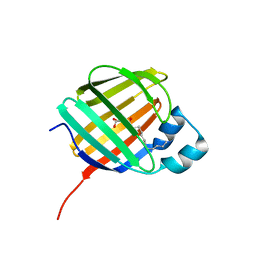 | | Cellular retinol-binding protein 2 (CRBP2) in complex with 2-lauroylglycerol | | 分子名称: | 1,3-dihydroxypropan-2-yl dodecanoate, Retinol-binding protein 2 | | 著者 | Adams, C, Silvaroli, J.A, Banarjee, S, Golczak, M. | | 登録日 | 2020-09-11 | | 公開日 | 2021-03-10 | | 最終更新日 | 2023-10-18 | | 実験手法 | X-RAY DIFFRACTION (1.2 Å) | | 主引用文献 | Molecular basis for the interaction of cellular retinol binding protein 2 (CRBP2) with nonretinoid ligands.
J.Lipid Res., 62, 2021
|
|
7JZ5
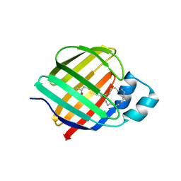 | | Cellular retinol-binding protein 2 (CRBP2) in complex with 1-arachodonoyl-1-thio-glycerol | | 分子名称: | Retinol-binding protein 2, S-[(2R)-2,3-dihydroxypropyl] (5Z,8Z,11Z,14Z)-icosa-5,8,11,14-tetraenethioate | | 著者 | Silvaroli, J.A, Banarjee, S, Golczak, M. | | 登録日 | 2020-09-01 | | 公開日 | 2021-03-10 | | 最終更新日 | 2023-10-18 | | 実験手法 | X-RAY DIFFRACTION (1.567 Å) | | 主引用文献 | Molecular basis for the interaction of cellular retinol binding protein 2 (CRBP2) with nonretinoid ligands.
J.Lipid Res., 62, 2021
|
|
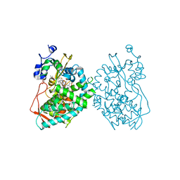
7JQD
 | | Crystal Structure of PAC1r in complex with peptide antagonist | | 分子名称: | Peptide-43, Pituitary adenylate cyclase-activating polypeptide type I receptor | | 著者 | Piper, D.E, Hu, E, Fang-Tsao, H. | | 登録日 | 2020-08-10 | | 公開日 | 2021-03-24 | | 最終更新日 | 2023-10-18 | | 実験手法 | X-RAY DIFFRACTION (2.7 Å) | | 主引用文献 | Discovery of Selective Pituitary Adenylate Cyclase 1 Receptor (PAC1R) Antagonist Peptides Potent in a Maxadilan/PACAP38-Induced Increase in Blood Flow Pharmacodynamic Model.
J.Med.Chem., 64, 2021
|
|
2J51
 | | Crystal structure of Human STE20-like kinase bound to 5-Amino-3-((4-(aminosulfonyl)phenyl)amino) -N-(2,6-difluorophenyl)-1H-1,2,4-triazole- 1-carbothioamide | | 分子名称: | 1,2-ETHANEDIOL, 5-AMINO-3-{[4-(AMINOSULFONYL)PHENYL]AMINO}-N-(2,6-DIFLUOROPHENYL)-1H-1,2,4-TRIAZOLE-1-CARBOTHIOAMIDE, STE20-LIKE SERINE/THREONINE-PROTEIN KINASE, ... | | 著者 | Pike, A.C.W, Rellos, P, Fedorov, O, Keates, T, Salah, E, Savitsky, P, Papagrigoriou, E, Turnbull, A.P, Debreczeni, J.E, von Delft, F, Arrowsmith, C, Edwards, A, Weigelt, J, Sundstrom, M, Knapp, S. | | 登録日 | 2006-09-08 | | 公開日 | 2006-10-03 | | 最終更新日 | 2023-12-13 | | 実験手法 | X-RAY DIFFRACTION (2.1 Å) | | 主引用文献 | Activation Segment Dimerization: A Mechanism for Kinase Autophosphorylation of Non-Consensus Sites.
Embo J., 27, 2008
|
|
4M2T
 | | Corrected Structure of Mouse P-glycoprotein bound to QZ59-SSS | | 分子名称: | (4S,11S,18S)-4,11,18-tri(propan-2-yl)-6,13,20-triselena-3,10,17,22,23,24-hexaazatetracyclo[17.2.1.1~5,8~.1~12,15~]tetracosa-1(21),5(24),7,12(23),14,19(22)-hexaene-2,9,16-trione, Multidrug resistance protein 1A | | 著者 | Li, J, Jaimes, K.F, Aller, S.G. | | 登録日 | 2013-08-05 | | 公開日 | 2013-11-13 | | 最終更新日 | 2024-02-28 | | 実験手法 | X-RAY DIFFRACTION (4.35 Å) | | 主引用文献 | Refined structures of mouse P-glycoprotein.
Protein Sci., 23, 2014
|
|
6S67
 | | Structure of the Fluorescent Protein AausFP1 from Aequorea cf. australis at pH 7.0 | | 分子名称: | Aequorea cf. australis fluorescent protein 1 (AausFP1), GLYCEROL | | 著者 | Depernet, H, Gotthard, G, Lambert, G.G, Shaner, N.C, Royant, A. | | 登録日 | 2019-07-02 | | 公開日 | 2020-07-22 | | 最終更新日 | 2024-01-24 | | 実験手法 | X-RAY DIFFRACTION (2.47 Å) | | 主引用文献 | Aequorea's secrets revealed: New fluorescent proteins with unique properties for bioimaging and biosensing.
Plos Biol., 18, 2020
|
|
5EHD
 | | Crystal structure of human nucleophosmin-core in complex with cytochrome c | | 分子名称: | CHLORIDE ION, Nucleophosmin | | 著者 | Bernardo-Garcia, N, Hermoso, J.A, Gonzalez-Arzola, K, Diaz-Moreno, I, De la Rosa, M.A. | | 登録日 | 2015-10-28 | | 公開日 | 2016-11-09 | | 最終更新日 | 2024-01-10 | | 実験手法 | X-RAY DIFFRACTION (2.55 Å) | | 主引用文献 | Crystal structure of human nucleophosmin-core in complex with cytochrome c
To Be Published
|
|
4EQU
 | | Human STK-10 (LOK) kinase domain in DFG-out conformation with inhibitor DSA-7 | | 分子名称: | CALCIUM ION, N-{3-[(3-{4-[(4-methoxyphenyl)amino]-1,3,5-triazin-2-yl}pyridin-2-yl)amino]-4-methylphenyl}-3-(trifluoromethyl)benzamide, PENTAETHYLENE GLYCOL, ... | | 著者 | Merritt, E.A, Larson, E.T. | | 登録日 | 2012-04-19 | | 公開日 | 2012-11-07 | | 最終更新日 | 2012-12-19 | | 実験手法 | X-RAY DIFFRACTION (2 Å) | | 主引用文献 | Affinity-Based Probes Based on Type II Kinase Inhibitors.
J.Am.Chem.Soc., 134, 2012
|
|
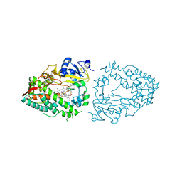
5F0J
 | | Structure of retromer VPS26-VPS35 subunits bound to SNX3 | | 分子名称: | 1,2-ETHANEDIOL, GLYCEROL, SULFATE ION, ... | | 著者 | Lucas, M, Gershlick, D, Vidaurrazaga, A, Rojas, A.L, Bonifacino, J.S, Hierro, A. | | 登録日 | 2015-11-27 | | 公開日 | 2016-12-07 | | 最終更新日 | 2024-01-10 | | 実験手法 | X-RAY DIFFRACTION (2.7 Å) | | 主引用文献 | Structural Mechanism for Cargo Recognition by the Retromer Complex.
Cell, 167, 2016
|
|
4EW1
 | | High resolution structure of human glycinamide ribonucleotide transformylase in apo form. | | 分子名称: | PHOSPHATE ION, SULFATE ION, Trifunctional purine biosynthetic protein adenosine-3 | | 著者 | Connelly, S, DeMartino, K, Boger, D.L, Wilson, I.A. | | 登録日 | 2012-04-26 | | 公開日 | 2013-07-31 | | 最終更新日 | 2023-09-13 | | 実験手法 | X-RAY DIFFRACTION (1.522 Å) | | 主引用文献 | Biological and Structural Evaluation of 10R- and 10S-Methylthio-DDACTHF Reveals a New Role for Sulfur in Inhibition of Glycinamide Ribonucleotide Transformylase.
Biochemistry, 52, 2013
|
|
5EW8
 | | FIBROBLAST GROWTH FACTOR RECEPTOR 1 IN COMPLEX WITH JNJ-4275693 | | 分子名称: | Fibroblast growth factor receptor 1, SULFATE ION, ~{N}'-(3,5-dimethoxyphenyl)-~{N}'-[3-(1-methylpyrazol-4-yl)quinoxalin-6-yl]-~{N}-propan-2-yl-ethane-1,2-diamine | | 著者 | Ogg, D, Breed, J. | | 登録日 | 2015-11-20 | | 公開日 | 2016-03-30 | | 最終更新日 | 2024-05-08 | | 実験手法 | X-RAY DIFFRACTION (1.63 Å) | | 主引用文献 | Landscape of activating cancer mutations in FGFR kinases and their differential responses to inhibitors in clinical use.
Oncotarget, 7, 2016
|
|
4E74
 | | Crystal structure of the RHO GTPASE BINDING DOMAIN of Plexin A4A | | 分子名称: | Plexin-A4, UNKNOWN ATOM OR ION | | 著者 | Guan, X, Wang, H, Tempel, W, Dong, A, Tong, Y, Arrowsmith, C.H, Edwards, A.M, Bountra, C, Weigelt, J, Park, H, Structural Genomics Consortium (SGC) | | 登録日 | 2012-03-16 | | 公開日 | 2012-03-28 | | 最終更新日 | 2023-09-13 | | 実験手法 | X-RAY DIFFRACTION (1.58 Å) | | 主引用文献 | Crystal structure of the RHO GTPASE BINDING DOMAIN of Plexin A4A
to be published
|
|
5F0L
 | | Structure of retromer VPS26-VPS35 subunits bound to SNX3 and DMT1 | | 分子名称: | 1,2-ETHANEDIOL, GLYCEROL, Natural resistance-associated macrophage protein 2, ... | | 著者 | Lucas, M, Gershlick, D, Vidaurrazaga, A, Rojas, A.L, Bonifacino, J.S, Hierro, A. | | 登録日 | 2015-11-27 | | 公開日 | 2016-12-07 | | 最終更新日 | 2024-01-10 | | 実験手法 | X-RAY DIFFRACTION (3.2 Å) | | 主引用文献 | Structural Mechanism for Cargo Recognition by the Retromer Complex.
Cell, 167, 2016
|
|
4EW3
 | | The structure of human glycinamide ribonucleotide transformylase in complex with 10R-methylthio-DDATHF. | | 分子名称: | N-({4-[(1R)-4-(2,4-diamino-6-oxo-1,6-dihydropyrimidin-5-yl)-1-(methylsulfanyl)butyl]phenyl}carbonyl)-L-glutamic acid, PHOSPHATE ION, SULFATE ION, ... | | 著者 | Connelly, S, DeMartino, K, Boger, D.L, Wilson, I.A. | | 登録日 | 2012-04-26 | | 公開日 | 2013-07-31 | | 最終更新日 | 2023-09-13 | | 実験手法 | X-RAY DIFFRACTION (1.702 Å) | | 主引用文献 | Biological and Structural Evaluation of 10R- and 10S-Methylthio-DDACTHF Reveals a New Role for Sulfur in Inhibition of Glycinamide Ribonucleotide Transformylase.
Biochemistry, 52, 2013
|
|
4I5X
 | | Crystal Structure Of AKR1B10 Complexed With NADP+ And Flufenamic acid | | 分子名称: | 2-[[3-(TRIFLUOROMETHYL)PHENYL]AMINO] BENZOIC ACID, Aldo-keto reductase family 1 member B10, NADP NICOTINAMIDE-ADENINE-DINUCLEOTIDE PHOSPHATE | | 著者 | Zhang, L, Zheng, X, Chen, S, Zhai, J, Zhang, H, Zhao, Y. | | 登録日 | 2012-11-29 | | 公開日 | 2013-10-23 | | 最終更新日 | 2024-04-03 | | 実験手法 | X-RAY DIFFRACTION (2.1 Å) | | 主引用文献 | Inhibitor selectivity between aldo-keto reductase superfamily members AKR1B10 and AKR1B1: Role of Trp112 (Trp111)
Febs Lett., 587, 2013
|
|
5F6L
 | | The crystal structure of MLL1 (N3861I/Q3867L) in complex with RbBP5 and Ash2L | | 分子名称: | Histone-lysine N-methyltransferase 2A, Retinoblastoma-binding protein 5, S-ADENOSYL-L-HOMOCYSTEINE, ... | | 著者 | Li, Y, Lei, M, Chen, Y. | | 登録日 | 2015-12-06 | | 公開日 | 2016-02-24 | | 最終更新日 | 2023-11-08 | | 実験手法 | X-RAY DIFFRACTION (1.9 Å) | | 主引用文献 | Structural basis for activity regulation of MLL family methyltransferases.
Nature, 530, 2016
|
|
7KHW
 | |
3T6G
 | |
2IYD
 | |
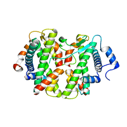
4L1X
 | | Crystal Structuer of Human 3-alpha Hydroxysteroid Dehydrogenase Type 3 V54L Mutant in Complex with NADP+ and Progesterone | | 分子名称: | Aldo-keto reductase family 1 member C2, NADP NICOTINAMIDE-ADENINE-DINUCLEOTIDE PHOSPHATE, PROGESTERONE, ... | | 著者 | Zhang, B, Hu, X.-J, Lin, S.-X. | | 登録日 | 2013-06-03 | | 公開日 | 2014-04-16 | | 最終更新日 | 2023-11-08 | | 実験手法 | X-RAY DIFFRACTION (2 Å) | | 主引用文献 | Human 3-alpha hydroxysteroid dehydrogenase type 3 (3 alpha-HSD3): The V54L mutation restricting the steroid alternative binding and enhancing the 20 alpha-HSD activity
J.Steroid Biochem.Mol.Biol., 141, 2014
|
|
7KVI
 | | Human CYP3A4 bound to an inhibitor | | 分子名称: | Cytochrome P450 3A4, GLYCEROL, PROTOPORPHYRIN IX CONTAINING FE, ... | | 著者 | Sevrioukova, I. | | 登録日 | 2020-11-28 | | 公開日 | 2021-01-20 | | 最終更新日 | 2023-10-18 | | 実験手法 | X-RAY DIFFRACTION (2.55 Å) | | 主引用文献 | Rational Design of CYP3A4 Inhibitors: A One-Atom Linker Elongation in Ritonavir-Like Compounds Leads to a Marked Improvement in the Binding Strength.
Int J Mol Sci, 22, 2021
|
|
7KVJ
 | | Human CYP3A4 bound to an inhibitor | | 分子名称: | Cytochrome P450 3A4, PROTOPORPHYRIN IX CONTAINING FE, tert-butyl [(2S)-1-{[(2S)-1-oxo-3-phenyl-1-{[3-(pyridin-3-yl)propyl]amino}propan-2-yl]sulfanyl}-3-phenylpropan-2-yl]carbamate | | 著者 | Sevrioukova, I. | | 登録日 | 2020-11-28 | | 公開日 | 2021-01-20 | | 最終更新日 | 2023-10-18 | | 実験手法 | X-RAY DIFFRACTION (2.65 Å) | | 主引用文献 | Rational Design of CYP3A4 Inhibitors: A One-Atom Linker Elongation in Ritonavir-Like Compounds Leads to a Marked Improvement in the Binding Strength.
Int J Mol Sci, 22, 2021
|
|
4E45
 | | Crystal structure of the hMHF1/hMHF2 Histone-Fold Tetramer in Complex with Fanconi Anemia Associated Helicase hFANCM | | 分子名称: | Centromere protein S, Centromere protein X, Fanconi anemia group M protein, ... | | 著者 | Fox III, D, Zhao, Y, Yang, W, Weidong, W. | | 登録日 | 2012-03-12 | | 公開日 | 2013-03-20 | | 最終更新日 | 2023-09-13 | | 実験手法 | X-RAY DIFFRACTION (2 Å) | | 主引用文献 | Crystal Structures Reveal that FANCM remodels the MHF Tetramer in favor of binding Branched DNA
To be Published
|
|
7KVQ
 | | Human CYP3A4 bound to an inhibitor | | 分子名称: | 1,2-ETHANEDIOL, Cytochrome P450 3A4, GLYCEROL, ... | | 著者 | Sevrioukova, I. | | 登録日 | 2020-11-28 | | 公開日 | 2021-01-20 | | 最終更新日 | 2023-10-18 | | 実験手法 | X-RAY DIFFRACTION (2.75 Å) | | 主引用文献 | Rational Design of CYP3A4 Inhibitors: A One-Atom Linker Elongation in Ritonavir-Like Compounds Leads to a Marked Improvement in the Binding Strength.
Int J Mol Sci, 22, 2021
|
|
1X4X
 | | Solution structure of the 6th fibronectin type III domain from human fibronectin type III domain containing protein 3 | | 分子名称: | Fibronectin type-III domain containing protein 3a | | 著者 | Tomizawa, T, Kigawa, T, Koshiba, S, Inoue, M, Yokoyama, S, RIKEN Structural Genomics/Proteomics Initiative (RSGI) | | 登録日 | 2005-05-15 | | 公開日 | 2005-11-15 | | 最終更新日 | 2024-05-29 | | 実験手法 | SOLUTION NMR | | 主引用文献 | Solution structure of the 6th fibronectin type III domain from human fibronectin type III domain containing protein 3
To be Published
|
|


