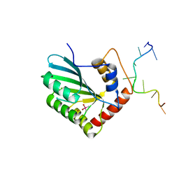
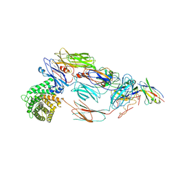
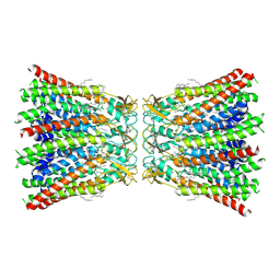
4UQQ
 
 | | Electron density map of GluK2 desensitized state in complex with 2S,4R-4-methylglutamate | | 分子名称: | GLUTAMATE RECEPTOR IONOTROPIC, KAINATE 2, GLUTAMIC ACID | | 著者 | Meyerson, J.R, Kumar, J, Chittori, S, Rao, P, Pierson, J, Bartesaghi, A, Mayer, M.L, Subramaniam, S. | | 登録日 | 2014-06-24 | | 公開日 | 2014-08-13 | | 最終更新日 | 2017-08-02 | | 実験手法 | ELECTRON MICROSCOPY (7.6 Å) | | 主引用文献 | Structural Mechanism of Glutamate Receptor Activation and Desensitization
Nature, 514, 2014
|
|
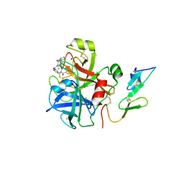
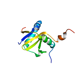
5UNO
 | | Crystal Structure of Hip1 (Rv2224c) | | 分子名称: | Carboxylesterase A | | 著者 | Naffin-Olivos, J.L, Daab, A, White, A, Goldfarb, N, Milne, A.C, Liu, D, Dunn, B.M, Rengarajan, J, Petsko, G.A, Ringe, D. | | 登録日 | 2017-01-31 | | 公開日 | 2017-04-12 | | 最終更新日 | 2019-12-11 | | 実験手法 | X-RAY DIFFRACTION (2.603 Å) | | 主引用文献 | Structure Determination of Mycobacterium tuberculosis Serine Protease Hip1 (Rv2224c).
Biochemistry, 56, 2017
|
|
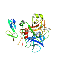
5ZVA
 | | APOBEC3F Chimeric Catalytic Domain in Complex with DNA(dC9) | | 分子名称: | APEBEC3F/ssDNA-C9, CACODYLATE ION, DNA (5'-D(*AP*TP*TP*TP*TP*CP*AP*AP*CP*T)-3'), ... | | 著者 | Cheng, C, Zhang, T.L, Wang, C.X, Lan, W.X, Ding, J.P, Cao, C.Y. | | 登録日 | 2018-05-09 | | 公開日 | 2018-11-21 | | 最終更新日 | 2018-11-28 | | 実験手法 | X-RAY DIFFRACTION (2.3 Å) | | 主引用文献 | Crystal Structure of Cytidine Deaminase Human APOBEC3F Chimeric Catalytic Domain in Complex with DNA
Chin.J.Chem., 36, 2018
|
|
8H05
 | |
8H03
 | |
8H04
 | |
6XYO
 | | Multiple system atrophy Type I alpha-synuclein filament | | 分子名称: | Alpha-synuclein | | 著者 | Schweighauser, M, Shi, Y, Tarutani, A, Kametani, F, Murzin, A.G, Ghetti, B, Matsubara, T, Tomita, T, Ando, T, Hasegawa, K, Murayama, S, Yoshida, M, Hasegawa, M, Scheres, S.H.W, Goedert, M. | | 登録日 | 2020-01-30 | | 公開日 | 2020-02-12 | | 最終更新日 | 2024-05-22 | | 実験手法 | ELECTRON MICROSCOPY (2.6 Å) | | 主引用文献 | Structures of alpha-synuclein filaments from multiple system atrophy.
Nature, 585, 2020
|
|
6XYP
 | | Multiple system atrophy Type II-1 alpha-synuclein filament | | 分子名称: | Alpha-synuclein | | 著者 | Schweighauser, M, Shi, Y, Tarutani, A, Kametani, F, Murzin, A.G, Ghetti, B, Matsubara, T, Tomita, T, Ando, T, Hasegawa, K, Murayama, S, Yoshida, M, Hasegawa, M, Scheres, S.H.W, Goedert, M. | | 登録日 | 2020-01-30 | | 公開日 | 2020-02-12 | | 最終更新日 | 2024-05-22 | | 実験手法 | ELECTRON MICROSCOPY (3.29 Å) | | 主引用文献 | Structures of alpha-synuclein filaments from multiple system atrophy.
Nature, 585, 2020
|
|
6XYQ
 | | Multiple system atrophy Type II-2 alpha-synuclein filament | | 分子名称: | Alpha-synuclein | | 著者 | Schweighauser, M, Shi, Y, Tarutani, A, Kametani, F, Murzin, A.G, Ghetti, B, Matsubara, T, Tomita, T, Ando, T, Hasegawa, K, Murayama, S, Yoshida, M, Hasegawa, M, Scheres, S.H.W, Goedert, M. | | 登録日 | 2020-01-30 | | 公開日 | 2020-02-12 | | 最終更新日 | 2024-05-22 | | 実験手法 | ELECTRON MICROSCOPY (3.09 Å) | | 主引用文献 | Structures of alpha-synuclein filaments from multiple system atrophy.
Nature, 585, 2020
|
|
3NSW
 | |
2VVU
 | | Aminopyrrolidine Factor Xa inhibitor | | 分子名称: | 5-chloro-N-[(3R)-1-(2-{[2-fluoro-4-(2-oxopyridin-1(2H)-yl)phenyl]amino}-2-oxoethyl)pyrrolidin-3-yl]thiophene-2-carboxamide, ACTIVATED FACTOR XA HEAVY CHAIN, CALCIUM ION, ... | | 著者 | Groebke-Zbinden, K, Banner, D.W, Benz, J.M, Blasco, F, Decoret, G, Himber, J, Kuhn, B, Panday, N, Ricklin, F, Risch, P, Schlatter, D, Stahl, M, Unger, R, Haap, W. | | 登録日 | 2008-06-11 | | 公開日 | 2009-07-07 | | 最終更新日 | 2023-12-13 | | 実験手法 | X-RAY DIFFRACTION (2.3 Å) | | 主引用文献 | Design of Novel Aminopyrrolidine Factor Xa Inhibitors from a Screening Hit.
Eur.J.Med.Chem., 44, 2009
|
|
2W3I
 | | Crystal Structure of FXa in complex with 4,4-disubstituted pyrrolidine-1,2-dicarboxamide inhibitor 2 | | 分子名称: | (2R,4S)-N^1^-(4-chlorophenyl)-4-(2,4-difluorophenyl)-4-hydroxy-N^2^-(2-oxo-2H-1,3'-bipyridin-6'-yl)pyrrolidine-1,2-dicarboxamide, CALCIUM ION, COAGULATION FACTOR X, ... | | 著者 | Zhang, E, Mochalkin, I, Casimiro-Garcia, A, Van Huis, C.A. | | 登録日 | 2008-11-12 | | 公開日 | 2009-04-07 | | 最終更新日 | 2023-12-13 | | 実験手法 | X-RAY DIFFRACTION (1.9 Å) | | 主引用文献 | Exploration of 4,4-Disubstituted Pyrrolidine-1,2-Dicarboxamides as Potent, Orally Active Factor Xa Inhibitors with Extended Duration of Action.
Bioorg.Med.Chem., 17, 2009
|
|
6CU7
 | | Alpha Synuclein fibril formed by full length protein - Rod Polymorph | | 分子名称: | Alpha-synuclein | | 著者 | Li, B, Hatami, A, Ge, P, Murray, K.A, Sheth, P, Zhang, M, Nair, G, Sawaya, M.R, Zhu, C, Broad, M, Shin, W.S, Ye, S, John, V, Eisenberg, D.S, Zhou, Z.H, Jiang, L. | | 登録日 | 2018-03-23 | | 公開日 | 2018-09-12 | | 最終更新日 | 2024-03-13 | | 実験手法 | ELECTRON MICROSCOPY (3.5 Å) | | 主引用文献 | Cryo-EM of full-length alpha-synuclein reveals fibril polymorphs with a common structural kernel.
Nat Commun, 9, 2018
|
|
7B2P
 | | Cryo-EM structure of complement C4b in complex with nanobody B5 | | 分子名称: | 2-acetamido-2-deoxy-beta-D-glucopyranose, 2-acetamido-2-deoxy-beta-D-glucopyranose-(1-4)-2-acetamido-2-deoxy-beta-D-glucopyranose, Complement C4 alpha chain, ... | | 著者 | Oosterheert, W, De la O Becerra, K.I, Gros, P. | | 登録日 | 2020-11-27 | | 公開日 | 2022-03-02 | | 最終更新日 | 2022-05-04 | | 実験手法 | ELECTRON MICROSCOPY (3.43 Å) | | 主引用文献 | Multifaceted Activities of Seven Nanobodies against Complement C4b.
J Immunol., 208, 2022
|
|
7B2M
 | | Cryo-EM structure of complement C4b in complex with nanobody E3 | | 分子名称: | 2-acetamido-2-deoxy-beta-D-glucopyranose, Anti-C4b nanobody E3, Complement C4 alpha chain, ... | | 著者 | Oosterheert, W, De la O Becerra, K.I, van den Bos, R.M, Gros, P. | | 登録日 | 2020-11-27 | | 公開日 | 2022-03-02 | | 最終更新日 | 2022-05-04 | | 実験手法 | ELECTRON MICROSCOPY (3.39 Å) | | 主引用文献 | Multifaceted Activities of Seven Nanobodies against Complement C4b.
J Immunol., 208, 2022
|
|
7B2Q
 | | Cryo-EM structure of complement C4b in complex with nanobody B12 | | 分子名称: | 2-acetamido-2-deoxy-beta-D-glucopyranose, 2-acetamido-2-deoxy-beta-D-glucopyranose-(1-4)-2-acetamido-2-deoxy-beta-D-glucopyranose, Anti-C4b nanobody B12, ... | | 著者 | Oosterheert, W, De la O Becerra, K.I, Gros, P. | | 登録日 | 2020-11-27 | | 公開日 | 2022-03-02 | | 最終更新日 | 2022-05-04 | | 実験手法 | ELECTRON MICROSCOPY (3.76 Å) | | 主引用文献 | Multifaceted Activities of Seven Nanobodies against Complement C4b.
J Immunol., 208, 2022
|
|
7OZH
 | |
7OZG
 | |
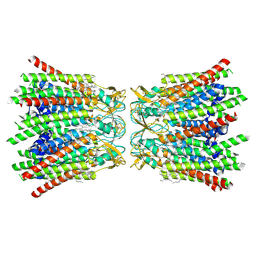
2KCF
 | |
2KC8
 | | Structure of E. coli toxin RelE (R81A/R83A) mutant in complex with antitoxin RelBc (K47-L79) peptide | | 分子名称: | Antitoxin RelB, Toxin relE | | 著者 | Li, G, Zhang, Y, Inouye, M, Ikura, M. | | 登録日 | 2008-12-17 | | 公開日 | 2009-03-17 | | 最終更新日 | 2024-05-22 | | 実験手法 | SOLUTION NMR | | 主引用文献 | Inhibitory mechanism of Escherichia coli RelE-RelB toxin-antitoxin module involves a helix displacement near an mRNA interferase active site.
J.Biol.Chem., 284, 2009
|
|
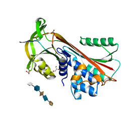
2I81
 | | Crystal Structure of Plasmodium vivax 2-Cys Peroxiredoxin, Reduced | | 分子名称: | 2-Cys Peroxiredoxin | | 著者 | Artz, J.D, Qiu, W, Dong, A, Lew, J, Ren, H, Zhao, Y, Kozieradski, I, Edwards, A.M, Arrowsmith, C.H, Weigelt, J, Sundstrom, M, Bochkarev, A, Hui, R, Structural Genomics Consortium (SGC) | | 登録日 | 2006-08-31 | | 公開日 | 2006-09-19 | | 最終更新日 | 2023-08-30 | | 実験手法 | X-RAY DIFFRACTION (2.45 Å) | | 主引用文献 | Crystal Structure of Plasmodium vivax 2-Cys Peroxiredoxin, Reduced
To be published
|
|
3DY0
 | | Crystal Structure of Cleaved PCI Bound to Heparin | | 分子名称: | 2-O-sulfo-alpha-L-idopyranuronic acid-(1-4)-2-deoxy-6-O-sulfo-2-(sulfoamino)-alpha-D-glucopyranose-(1-4)-2-O-sulfo-alpha-L-idopyranuronic acid-(1-4)-2-deoxy-6-O-sulfo-2-(sulfoamino)-alpha-D-glucopyranose-(1-4)-2-O-sulfo-alpha-L-idopyranuronic acid, C-terminus Plasma serine protease inhibitor, GLYCEROL, ... | | 著者 | Li, W, Huntington, J.A. | | 登録日 | 2008-07-25 | | 公開日 | 2008-10-28 | | 最終更新日 | 2023-08-30 | | 実験手法 | X-RAY DIFFRACTION (1.55 Å) | | 主引用文献 | The heparin binding site of protein C inhibitor is protease-dependent.
J.Biol.Chem., 283, 2008
|
|
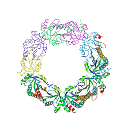
7QEQ
 | | human Connexin 26 dodecamer at 90mmHg PCO2, pH7.4 | | 分子名称: | DODECYL-BETA-D-MALTOSIDE, Gap junction beta-2 protein, PHOSPHATIDYLETHANOLAMINE | | 著者 | Brotherton, D.H, Cameron, A.D, Savva, C.G, Ragan, T.J. | | 登録日 | 2021-12-03 | | 公開日 | 2022-03-30 | | 最終更新日 | 2022-05-18 | | 実験手法 | ELECTRON MICROSCOPY (1.9 Å) | | 主引用文献 | Conformational changes and CO 2 -induced channel gating in connexin26.
Structure, 30, 2022
|
|
7QER
 | | human Connexin 26 dodecamer at 55mm Hg PCO2, pH7.4 | | 分子名称: | DODECYL-BETA-D-MALTOSIDE, Gap junction beta-2 protein, PHOSPHATIDYLETHANOLAMINE | | 著者 | Brotherton, D.H, Cameron, A.D, Savva, C.G, Ragan, T.J. | | 登録日 | 2021-12-03 | | 公開日 | 2022-03-30 | | 最終更新日 | 2022-05-18 | | 実験手法 | ELECTRON MICROSCOPY (2.2 Å) | | 主引用文献 | Conformational changes and CO 2 -induced channel gating in connexin26.
Structure, 30, 2022
|
|
7DKJ
 | |


