7AL3
 
 | |
7D77
 | | Cryo-EM structure of the cortisol-bound adhesion receptor GPR97-Go complex | | 分子名称: | (11alpha,14beta)-11,17,21-trihydroxypregn-4-ene-3,20-dione, Adhesion G protein-coupled receptor G3; GPR97, CHOLESTEROL, ... | | 著者 | Ping, Y, Mao, C, Xiao, P, Zhao, R, Jiang, Y, Yang, Z, An, W, Shen, D, Yang, F, Zhang, H, Qu, C, Shen, Q, Tian, C, Li, Z, Li, S, Wang, G, Tao, X, Wen, X, Zhong, Y, Yang, J, Yi, F, Yu, X, Xu, E, Zhang, Y, Sun, J. | | 登録日 | 2020-10-03 | | 公開日 | 2021-02-03 | | 最終更新日 | 2021-02-10 | | 実験手法 | ELECTRON MICROSCOPY (2.9 Å) | | 主引用文献 | Structures of the glucocorticoid-bound adhesion receptor GPR97-G o complex.
Nature, 589, 2021
|
|
7D76
 | | Cryo-EM structure of the beclomethasone-bound adhesion receptor GPR97-Go complex | | 分子名称: | (8~{S},9~{R},10~{S},11~{S},13~{S},14~{S},16~{S},17~{R})-9-chloranyl-10,13,16-trimethyl-11,17-bis(oxidanyl)-17-(2-oxidanylethanoyl)-6,7,8,11,12,14,15,16-octahydrocyclopenta[a]phenanthren-3-one, Adhesion G protein-coupled receptor G3; GPR97, CHOLESTEROL, ... | | 著者 | Ping, Y, Mao, C, Xiao, P, Zhao, R, Jiang, Y, Yang, Z, An, W, Shen, D, Yang, F, Zhang, H, Qu, C, Shen, Q, Tian, C, Li, Z, Li, S, Wang, G, Tao, X, Wen, X, Zhong, Y, Yang, J, Yi, F, Yu, X, Xu, E, Zhang, Y, Sun, J. | | 登録日 | 2020-10-03 | | 公開日 | 2021-02-03 | | 最終更新日 | 2021-02-10 | | 実験手法 | ELECTRON MICROSCOPY (3.1 Å) | | 主引用文献 | Structures of the glucocorticoid-bound adhesion receptor GPR97-G o complex.
Nature, 589, 2021
|
|
7AKV
 | | The cryo-EM structure of the Vag8-C1 inhibitor complex | | 分子名称: | Plasma protease C1 inhibitor, Vag8 | | 著者 | Johnson, S, Lea, S.M, Deme, J.C, Furlong, E, Dhillon, A. | | 登録日 | 2020-10-02 | | 公開日 | 2021-06-16 | | 実験手法 | ELECTRON MICROSCOPY (3.6 Å) | | 主引用文献 | Molecular Basis for Bordetella pertussis Interference with Complement, Coagulation, Fibrinolytic, and Contact Activation Systems: the Cryo-EM Structure of the Vag8-C1 Inhibitor Complex.
Mbio, 12, 2021
|
|
7AKO
 | | Crystal structure of CHK1 kinase domain in complex with a CLASPIN phosphopeptide | | 分子名称: | 1,2-ETHANEDIOL, Claspin, STAUROSPORINE, ... | | 著者 | Day, M, Oliver, A.W, Pearl, L.H. | | 登録日 | 2020-10-01 | | 公開日 | 2021-04-14 | | 最終更新日 | 2024-01-31 | | 実験手法 | X-RAY DIFFRACTION (1.8 Å) | | 主引用文献 | Structural basis for recruitment of the CHK1 DNA damage kinase by the CLASPIN scaffold protein.
Structure, 29, 2021
|
|
7AKM
 | | Crystal structure of CHK1 kinase domain in complex with ATPyS | | 分子名称: | 1,2-ETHANEDIOL, CITRIC ACID, MAGNESIUM ION, ... | | 著者 | Day, M, Oliver, A.W, Pearl, L.H. | | 登録日 | 2020-10-01 | | 公開日 | 2021-04-14 | | 最終更新日 | 2024-01-31 | | 実験手法 | X-RAY DIFFRACTION (1.93 Å) | | 主引用文献 | Structural basis for recruitment of the CHK1 DNA damage kinase by the CLASPIN scaffold protein.
Structure, 29, 2021
|
|
7AKH
 | | Structure of DYRK2 in complex with compound 58 | | 分子名称: | 4-[3-[(2~{S})-2-(6-bromanylpyridin-2-yl)oxypropyl]-2-methyl-imidazo[4,5-b]pyridin-5-yl]pyridine-2,6-diamine, CHLORIDE ION, Dual specificity tyrosine-phosphorylation-regulated kinase 2 | | 著者 | Dokurno, P, Surgenor, A.E, Kotschy, A. | | 登録日 | 2020-10-01 | | 公開日 | 2021-05-26 | | 最終更新日 | 2024-01-31 | | 実験手法 | X-RAY DIFFRACTION (2.85 Å) | | 主引用文献 | Structure-Guided Discovery of Potent and Selective DYRK1A Inhibitors.
J.Med.Chem., 64, 2021
|
|
7AKL
 | | Structure of DYRK1A in complex with compound 50 | | 分子名称: | 4-(2,3-dimethyl-1-benzofuran-5-yl)pyridine-2,6-diamine, CHLORIDE ION, Dual specificity tyrosine-phosphorylation-regulated kinase 1A | | 著者 | Dokurno, P, Surgenor, A.E, Kotschy, A. | | 登録日 | 2020-10-01 | | 公開日 | 2021-05-26 | | 最終更新日 | 2024-01-31 | | 実験手法 | X-RAY DIFFRACTION (2 Å) | | 主引用文献 | Structure-Guided Discovery of Potent and Selective DYRK1A Inhibitors.
J.Med.Chem., 64, 2021
|
|
7AKK
 | | Structure of a complement factor-receptor complex | | 分子名称: | 2-acetamido-2-deoxy-beta-D-glucopyranose, Complement C3 beta chain, Complement C3b alpha' chain, ... | | 著者 | Fernandez, F.J, Santos-Lopez, J, Martinez-Barricarte, R, Querol-Garcia, J, Navas-Yuste, S, Savko, M, Shepard, W.E, Rodriguez de Cordoba, S, Vega, M.C. | | 登録日 | 2020-10-01 | | 公開日 | 2022-04-13 | | 最終更新日 | 2024-01-31 | | 実験手法 | X-RAY DIFFRACTION (3.395 Å) | | 主引用文献 | The crystal structure of iC3b-CR3 alpha I reveals a modular recognition of the main opsonin iC3b by the CR3 integrin receptor
Nat Commun, 13, 2022
|
|
7D6L
 | |
7AKG
 | |
7AKE
 | | Structure of DYRK1A in complex with compound 58 | | 分子名称: | 4-[3-[(2~{S})-2-(6-bromanylpyridin-2-yl)oxypropyl]-2-methyl-imidazo[4,5-b]pyridin-5-yl]pyridine-2,6-diamine, CHLORIDE ION, Dual specificity tyrosine-phosphorylation-regulated kinase 1A | | 著者 | Dokurno, P, Surgenor, A.E, Kotschy, A. | | 登録日 | 2020-09-30 | | 公開日 | 2021-05-26 | | 最終更新日 | 2024-01-31 | | 実験手法 | X-RAY DIFFRACTION (2.3 Å) | | 主引用文献 | Structure-Guided Discovery of Potent and Selective DYRK1A Inhibitors.
J.Med.Chem., 64, 2021
|
|
7AKB
 | | Structure of DYRK1A in complex with compound 56 | | 分子名称: | 4-[3-[2-(6-bromanylpyridin-2-yl)oxyethyl]-2-methyl-imidazo[4,5-b]pyridin-5-yl]pyridine-2,6-diamine, Dual specificity tyrosine-phosphorylation-regulated kinase 1A | | 著者 | Dokurno, P, Surgenor, A.E, Kotschy, A. | | 登録日 | 2020-09-30 | | 公開日 | 2021-05-26 | | 最終更新日 | 2024-01-31 | | 実験手法 | X-RAY DIFFRACTION (2.8 Å) | | 主引用文献 | Structure-Guided Discovery of Potent and Selective DYRK1A Inhibitors.
J.Med.Chem., 64, 2021
|
|
7AKF
 | | Structure of DYRK2 in complex with compound 50 | | 分子名称: | 4-(2,3-dimethyl-1-benzofuran-5-yl)pyridine-2,6-diamine, CHLORIDE ION, Dual specificity tyrosine-phosphorylation-regulated kinase 2 | | 著者 | Dokurno, P, Surgenor, A.E, Kotschy, A. | | 登録日 | 2020-09-30 | | 公開日 | 2021-05-26 | | 最終更新日 | 2024-01-31 | | 実験手法 | X-RAY DIFFRACTION (2.6 Å) | | 主引用文献 | Structure-Guided Discovery of Potent and Selective DYRK1A Inhibitors.
J.Med.Chem., 64, 2021
|
|
7AKA
 | | Structure of DYRK1A in complex with compound 54 | | 分子名称: | 4-[2-methyl-3-[(2~{R})-2-pyridin-2-yloxypropyl]imidazo[4,5-b]pyridin-5-yl]pyridine-2,6-diamine, CHLORIDE ION, DIMETHYL SULFOXIDE, ... | | 著者 | Dokurno, P, Surgenor, A.E, Kotschy, A. | | 登録日 | 2020-09-30 | | 公開日 | 2021-05-26 | | 最終更新日 | 2024-01-31 | | 実験手法 | X-RAY DIFFRACTION (1.9 Å) | | 主引用文献 | Structure-Guided Discovery of Potent and Selective DYRK1A Inhibitors.
J.Med.Chem., 64, 2021
|
|
7KAC
 | | Crystal structure of HPK1 (MAP4K1) kinase in complex with 5-{[4-{[(1S)-2-HYDROXY-1-PHENYLETHYL]AMINO}-5-(1,3,4-OXADIAZOL-2-YL)PYRIMIDIN-2-YL]AMINO}-3,3-DIMETHYL-2-BENZOFURAN-1(3H)-ONE | | 分子名称: | 5-{[4-{[(1S)-2-hydroxy-1-phenylethyl]amino}-5-(1,3,4-oxadiazol-2-yl)pyrimidin-2-yl]amino}-3,3-dimethyl-2-benzofuran-1(3H)-one, Isoform 2 of Mitogen-activated protein kinase kinase kinase kinase 1 | | 著者 | Sheriff, S. | | 登録日 | 2020-09-30 | | 公開日 | 2021-01-13 | | 最終更新日 | 2023-10-18 | | 実験手法 | X-RAY DIFFRACTION (1.85 Å) | | 主引用文献 | Using yeast surface display to engineer a soluble and crystallizable construct of hematopoietic progenitor kinase 1 (HPK1).
Acta Crystallogr.,Sect.F, 77, 2021
|
|
7AJJ
 | | bovine ATP synthase dimer state3:state3 | | 分子名称: | 1,2-DIPALMITOYL-PHOSPHATIDYL-GLYCEROLE, ATP synthase F(0) complex subunit B1, mitochondrial, ... | | 著者 | Spikes, T.E, Montgomery, M.G, Walker, J.E. | | 登録日 | 2020-09-29 | | 公開日 | 2021-02-03 | | 最終更新日 | 2021-02-24 | | 実験手法 | ELECTRON MICROSCOPY (13.1 Å) | | 主引用文献 | Interface mobility between monomers in dimeric bovine ATP synthase participates in the ultrastructure of inner mitochondrial membranes.
Proc.Natl.Acad.Sci.USA, 118, 2021
|
|
7AJH
 | | bovine ATP synthase dimer state3:state1 | | 分子名称: | 1,2-DIPALMITOYL-PHOSPHATIDYL-GLYCEROLE, ATP synthase F(0) complex subunit B1, mitochondrial, ... | | 著者 | Spikes, T.E, Montgomery, M.G, Walker, J.E. | | 登録日 | 2020-09-29 | | 公開日 | 2021-02-03 | | 最終更新日 | 2021-02-24 | | 実験手法 | ELECTRON MICROSCOPY (9.7 Å) | | 主引用文献 | Interface mobility between monomers in dimeric bovine ATP synthase participates in the ultrastructure of inner mitochondrial membranes.
Proc.Natl.Acad.Sci.USA, 118, 2021
|
|
7AJD
 | | bovine ATP synthase dimer state1:state3 | | 分子名称: | 1,2-DIPALMITOYL-PHOSPHATIDYL-GLYCEROLE, ATP synthase F(0) complex subunit B1, mitochondrial, ... | | 著者 | Spikes, T.E, Montgomery, M.G, Walker, J.E. | | 登録日 | 2020-09-29 | | 公開日 | 2021-02-03 | | 最終更新日 | 2021-02-24 | | 実験手法 | ELECTRON MICROSCOPY (9 Å) | | 主引用文献 | Interface mobility between monomers in dimeric bovine ATP synthase participates in the ultrastructure of inner mitochondrial membranes.
Proc.Natl.Acad.Sci.USA, 118, 2021
|
|
7K9Y
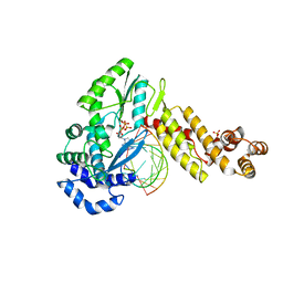 | | GsI-IIC RT Template-Switching Complex (twinned) | | 分子名称: | 2'-DEOXYADENOSINE 5'-TRIPHOSPHATE, DNA (5'-D(*CP*TP*CP*CP*AP*GP*GP*CP*AP*AP*C)-3'), MAGNESIUM ION, ... | | 著者 | Stamos, J.L, Lentzsch, A.M. | | 登録日 | 2020-09-29 | | 公開日 | 2021-08-18 | | 最終更新日 | 2023-10-18 | | 実験手法 | X-RAY DIFFRACTION (3.2 Å) | | 主引用文献 | Structural basis for template switching by a group II intron-encoded non-LTR-retroelement reverse transcriptase.
J.Biol.Chem., 297, 2021
|
|
7AJC
 | | bovine ATP synthase dimer state1:state2 | | 分子名称: | 1,2-DIPALMITOYL-PHOSPHATIDYL-GLYCEROLE, ATP synthase F(0) complex subunit B1, mitochondrial, ... | | 著者 | Spikes, T.E, Montgomery, M.G, Walker, J.E. | | 登録日 | 2020-09-29 | | 公開日 | 2021-02-03 | | 最終更新日 | 2021-02-24 | | 実験手法 | ELECTRON MICROSCOPY (11.9 Å) | | 主引用文献 | Interface mobility between monomers in dimeric bovine ATP synthase participates in the ultrastructure of inner mitochondrial membranes.
Proc.Natl.Acad.Sci.USA, 118, 2021
|
|
7AJK
 | | Crystal structure of CRYI-B Rac1 complex | | 分子名称: | CYFIP-related Rac1 interactor B, MAGNESIUM ION, PHOSPHOAMINOPHOSPHONIC ACID-GUANYLATE ESTER, ... | | 著者 | Yelland, T, Anh, L, Insall, R, Machesky, L, Ismail, S. | | 登録日 | 2020-09-29 | | 公開日 | 2020-11-18 | | 最終更新日 | 2024-01-31 | | 実験手法 | X-RAY DIFFRACTION (3.1 Å) | | 主引用文献 | Structural Basis of CYRI-B Direct Competition with Scar/WAVE Complex for Rac1.
Structure, 29, 2021
|
|
7AJB
 | | bovine ATP synthase dimer state1:state1 | | 分子名称: | 1,2-DIPALMITOYL-PHOSPHATIDYL-GLYCEROLE, ATP synthase F(0) complex subunit B1, mitochondrial, ... | | 著者 | Spikes, T.E, Montgomery, M.G, Walker, J.E. | | 登録日 | 2020-09-29 | | 公開日 | 2021-02-03 | | 最終更新日 | 2021-02-24 | | 実験手法 | ELECTRON MICROSCOPY (9.2 Å) | | 主引用文献 | Interface mobility between monomers in dimeric bovine ATP synthase participates in the ultrastructure of inner mitochondrial membranes.
Proc.Natl.Acad.Sci.USA, 118, 2021
|
|
7AJU
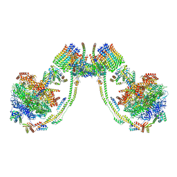
 | | Cryo-EM structure of the 90S-exosome super-complex (state Post-A1-exosome) | | 分子名称: | 13 kDa ribonucleoprotein-associated protein, 18S rRNA, 40S ribosomal protein S1-A, ... | | 著者 | Cheng, J, Lau, B, Flemming, D, Venuta, G.L, Berninghausen, O, Beckmann, R, Hurt, E. | | 登録日 | 2020-09-29 | | 公開日 | 2020-12-30 | | 最終更新日 | 2024-05-01 | | 実験手法 | ELECTRON MICROSCOPY (3.8 Å) | | 主引用文献 | Structure of the Maturing 90S Pre-ribosome in Association with the RNA Exosome.
Mol.Cell, 81, 2021
|
|
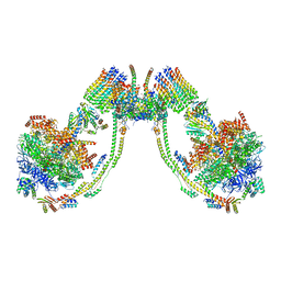
7AJT
 | | Cryo-EM structure of the 90S-exosome super-complex (state Pre-A1-exosome) | | 分子名称: | 13 kDa ribonucleoprotein-associated protein, 18S rRNA, 40S ribosomal protein S1-A, ... | | 著者 | Cheng, J, Lau, B, Flemming, D, Venuta, G.L, Berninghausen, O, Beckmann, R, Hurt, E. | | 登録日 | 2020-09-29 | | 公開日 | 2020-12-30 | | 最終更新日 | 2021-02-03 | | 実験手法 | ELECTRON MICROSCOPY (4.6 Å) | | 主引用文献 | Structure of the Maturing 90S Pre-ribosome in Association with the RNA Exosome.
Mol.Cell, 81, 2021
|
|


