3FER
 
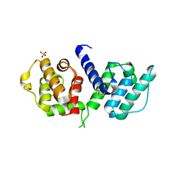
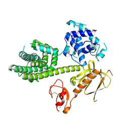
 | | Crystal structure of n-terminal actin-binding domain from human filamin b (tandem ch-domains). northeast structural genomics consortium target hr5571a. | | 分子名称: | ACETIC ACID, Filamin-B | | 著者 | Kuzin, A.P, Abashidze, M, Seetharaman, R, Shastry, R, Sahdev, S, Ciccosanti, C, Xiao, R, Everett, J.K, Huang, Y, Acton, T, Rost, B, Montelione, G.T, Tong, L, Hunt, J.F, Northeast Structural Genomics Consortium (NESG) | | 登録日 | 2008-11-30 | | 公開日 | 2009-01-06 | | 最終更新日 | 2023-11-22 | | 実験手法 | X-RAY DIFFRACTION (2.4 Å) | | 主引用文献 | Crystal structure of n-terminal actin-binding domain from human filamin b (tandem ch-domains). northeast structural genomics consortium target hr5571a.
To be Published
|
|
3F7P
 | |
3CO1
 | |
2WA6
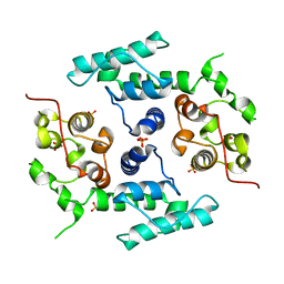
 | | Structure of the W148R mutant of human filamin b actin binding domain at 1.95 Angstrom resolution | | 分子名称: | CARBONATE ION, FILAMIN-B | | 著者 | Sawyer, G.M, Clark, A.R, Robertson, S.P, Sutherland-Smith, A.J. | | 登録日 | 2009-02-03 | | 公開日 | 2009-06-23 | | 最終更新日 | 2023-12-13 | | 実験手法 | X-RAY DIFFRACTION (1.95 Å) | | 主引用文献 | Disease-Associated Substitutions in the Filamin B Actin Binding Domain Confer Enhanced Actin Binding Affinity in the Absence of Major Structural Disturbance: Insights from the Crystal Structures of Filamin B Actin Binding Domains.
J.Mol.Biol., 390, 2009
|
|
2WA5
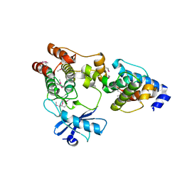
 | | Crystal structure of human filamin B actin binding domain at 1.9 Angstroms resolution | | 分子名称: | CARBONATE ION, FILAMIN-B, SULFATE ION | | 著者 | Sawyer, G.M, Clark, A.R, Robertson, S.P, Sutherland-Smith, A.J. | | 登録日 | 2009-02-03 | | 公開日 | 2009-06-23 | | 最終更新日 | 2023-12-13 | | 実験手法 | X-RAY DIFFRACTION (1.9 Å) | | 主引用文献 | Disease-Associated Substitutions in the Filamin B Actin Binding Domain Confer Enhanced Actin Binding Affinity in the Absence of Major Structural Disturbance: Insights from the Crystal Structures of Filamin B Actin Binding Domains.
J.Mol.Biol., 390, 2009
|
|
2WA7
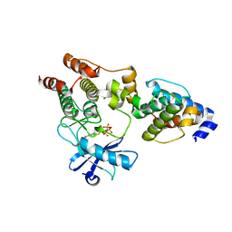
 | | Structure of the M202V mutant of human filamin b actin binding domain at 1.85 Angstrom resolution | | 分子名称: | CACODYLATE ION, CARBONATE ION, FILAMIN-B | | 著者 | Sawyer, G.M, Clark, A.R, Robertson, S.P, Sutherland-Smith, A.J. | | 登録日 | 2009-02-03 | | 公開日 | 2009-06-23 | | 最終更新日 | 2023-12-13 | | 実験手法 | X-RAY DIFFRACTION (1.85 Å) | | 主引用文献 | Disease-Associated Substitutions in the Filamin B Actin Binding Domain Confer Enhanced Actin Binding Affinity in the Absence of Major Structural Disturbance: Insights from the Crystal Structures of Filamin B Actin Binding Domains.
J.Mol.Biol., 390, 2009
|
|
3HOR
 | | Structure of the actin-binding domain of human filamin A (reduced) | | 分子名称: | Filamin-A, PHOSPHATE ION | | 著者 | Clark, A.R, Sawyer, G.M, Robertson, S.P, Sutherland-Smith, A.J. | | 登録日 | 2009-06-03 | | 公開日 | 2009-10-13 | | 最終更新日 | 2023-11-01 | | 実験手法 | X-RAY DIFFRACTION (2.7 Å) | | 主引用文献 | Skeletal dysplasias due to filamin A mutations result from a gain-of-function mechanism distinct from allelic neurological disorders
Hum.Mol.Genet., 18, 2009
|
|
3HOC
 | | Structure of the actin-binding domain of human filamin A mutant E254K | | 分子名称: | Filamin-A, PHOSPHATE ION | | 著者 | Clark, A.R, Sawyer, G.M, Robertson, S.P, Sutherland-Smith, A.J. | | 登録日 | 2009-06-02 | | 公開日 | 2009-10-13 | | 最終更新日 | 2023-11-01 | | 実験手法 | X-RAY DIFFRACTION (2.3 Å) | | 主引用文献 | Skeletal dysplasias due to filamin A mutations result from a gain-of-function mechanism distinct from allelic neurological disorders
Hum.Mol.Genet., 18, 2009
|
|
3HOP
 | | Structure of the actin-binding domain of human filamin A | | 分子名称: | Filamin-A, PHOSPHATE ION | | 著者 | Clark, A.R, Sawyer, G.M, Robertson, S.P, Sutherland-Smith, A.J. | | 登録日 | 2009-06-03 | | 公開日 | 2009-10-13 | | 最終更新日 | 2023-11-01 | | 実験手法 | X-RAY DIFFRACTION (2.3 Å) | | 主引用文献 | Skeletal dysplasias due to filamin A mutations result from a gain-of-function mechanism distinct from allelic neurological disorders
Hum.Mol.Genet., 18, 2009
|
|
2WFN
 | |
3KMU
 | |
3KMW
 | |
3KY9
 | | Autoinhibited Vav1 | | 分子名称: | Proto-oncogene vav, ZINC ION | | 著者 | Tomchick, D.R, Rosen, M.K, Machius, M, Yu, B. | | 登録日 | 2009-12-04 | | 公開日 | 2010-02-23 | | 最終更新日 | 2011-07-13 | | 実験手法 | X-RAY DIFFRACTION (2.731 Å) | | 主引用文献 | Structural and Energetic Mechanisms of Cooperative Autoinhibition and Activation of Vav1
Cell(Cambridge,Mass.), 140, 2010
|
|
3LUE
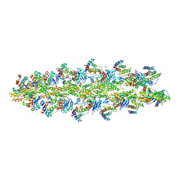 | | Model of alpha-actinin CH1 bound to F-actin | | 分子名称: | Actin, cytoplasmic 1, Alpha-actinin-3 | | 著者 | Galkin, V.E, Orlova, A, Salmazo, A, Djinovic-Carugo, K, Egelman, E.H. | | 登録日 | 2010-02-17 | | 公開日 | 2010-04-28 | | 最終更新日 | 2024-02-21 | | 実験手法 | ELECTRON MICROSCOPY (15 Å) | | 主引用文献 | Opening of tandem calponin homology domains regulates their affinity for F-actin.
Nat.Struct.Mol.Biol., 17, 2010
|
|
3I6X
 | |
2RR8
 | |
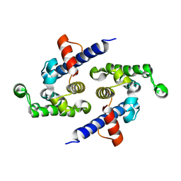
2L3G
 | | Solution NMR Structure of CH domain of Rho guanine nucleotide exchange factor 7 from Homo sapiens, Northeast Structural Genomics Consortium Target HR4495E | | 分子名称: | Rho guanine nucleotide exchange factor 7 | | 著者 | Liu, G, Xiao, R, Janjua, H, Acton, T.B, Ciccosanti, A, Shastry, R, Everett, J, Montelione, G.T, Northeast Structural Genomics Consortium (NESG) | | 登録日 | 2010-09-13 | | 公開日 | 2010-12-15 | | 最終更新日 | 2024-05-01 | | 実験手法 | SOLUTION NMR | | 主引用文献 | Northeast Structural Genomics Consortium Target HR4495E
To be Published
|
|
3REP
 | |
4EDL
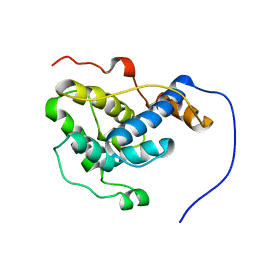
 | | Crystal structure of beta-parvin CH2 domain | | 分子名称: | 1,2-ETHANEDIOL, Beta-parvin | | 著者 | Stiegler, A.L, Draheim, K.M, Li, X, Chayen, N.E, Calderwood, D.A, Boggon, T.J. | | 登録日 | 2012-03-27 | | 公開日 | 2012-08-08 | | 最終更新日 | 2024-02-28 | | 実験手法 | X-RAY DIFFRACTION (2.1 Å) | | 主引用文献 | Structural basis for paxillin binding and focal adhesion targeting of beta-parvin.
J.Biol.Chem., 287, 2012
|
|
4EDN
 | | Crystal structure of beta-parvin CH2 domain in complex with paxillin LD1 motif | | 分子名称: | Beta-parvin, Paxillin, SULFATE ION | | 著者 | Stiegler, A.L, Draheim, K.M, Li, X, Chayen, N.E, Calderwood, D.A, Boggon, T.J. | | 登録日 | 2012-03-27 | | 公開日 | 2012-08-08 | | 最終更新日 | 2013-06-19 | | 実験手法 | X-RAY DIFFRACTION (2.9 Å) | | 主引用文献 | Structural basis for paxillin binding and focal adhesion targeting of beta-parvin.
J.Biol.Chem., 287, 2012
|
|
4EDM
 | | Crystal structure of beta-parvin CH2 domain | | 分子名称: | 1,2-ETHANEDIOL, Beta-parvin | | 著者 | Stiegler, A.L, Draheim, K.M, Li, X, Chayen, N.E, Calderwood, D.A, Boggon, T.J. | | 登録日 | 2012-03-27 | | 公開日 | 2012-08-08 | | 最終更新日 | 2024-02-28 | | 実験手法 | X-RAY DIFFRACTION (2 Å) | | 主引用文献 | Structural basis for paxillin binding and focal adhesion targeting of beta-parvin.
J.Biol.Chem., 287, 2012
|
|
4B7L
 | |
4Q58
 | |
4Q59
 | |
4Q57
 | | Crystal structure of the plectin 1a actin-binding domain/N-terminal domain of calmodulin complex | | 分子名称: | 1,2-ETHANEDIOL, CALCIUM ION, CHLORIDE ION, ... | | 著者 | Song, J.-G, Kostan, J, Grishkovskaya, I, Djinovic-Carugo, K. | | 登録日 | 2014-04-16 | | 公開日 | 2014-07-23 | | 最終更新日 | 2024-02-28 | | 実験手法 | X-RAY DIFFRACTION (1.8 Å) | | 主引用文献 | Crystal structure of the plectin 1a actin-binding domain/N-terminal domain of calmodulin complex
To be Published
|
|


