8T2I
 
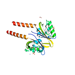
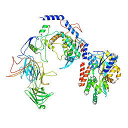 | | Negative stain EM assembly of MYC, JAZ, and NINJA complex | | 分子名称: | AFP homolog 2, Maltose/maltodextrin-binding periplasmic protein, Protein TIFY 10A, ... | | 著者 | Zhou, X.E, Zhang, Y, Zhou, Y, He, Q, Cao, X, Kariapper, L, Suino-Powell, K, Zhu, Y, Zhang, F, Karsten, M. | | 登録日 | 2023-06-06 | | 公開日 | 2023-06-28 | | 最終更新日 | 2023-11-22 | | 実験手法 | ELECTRON MICROSCOPY (10.4 Å) | | 主引用文献 | Assembly of JAZ-JAZ and JAZ-NINJA complexes in jasmonate signaling.
Plant Commun., 4, 2023
|
|
5L8Z
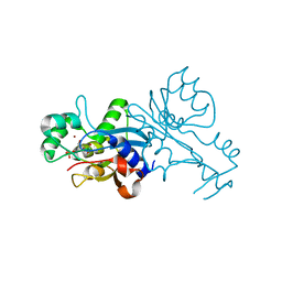
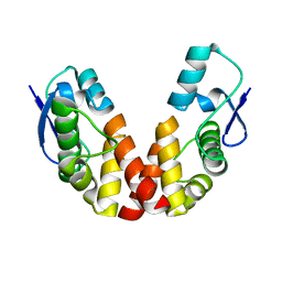
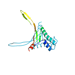 | | Structure of thermostable DNA-binding HU protein from micoplasma Spiroplasma melliferum | | 分子名称: | DNA-binding protein, SODIUM ION | | 著者 | Boyko, K.M, Gorbacheva, M.A, Rakitina, T.V, Korzhenevskiy, D.A, Kamashev, D.E, Vanyushkina, A.A, Lipkin, A.V, Popov, V.O. | | 登録日 | 2016-06-09 | | 公開日 | 2016-06-22 | | 最終更新日 | 2024-01-10 | | 実験手法 | X-RAY DIFFRACTION (1.4 Å) | | 主引用文献 | Structural basis of the high thermal stability of the histone-like HU protein from the mollicute Spiroplasma melliferum KC3.
Sci Rep, 6, 2016
|
|
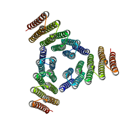
6SB2
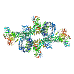 | | cryo-EM structure of mTORC1 bound to active RagA/C GTPases | | 分子名称: | GUANOSINE-5'-DIPHOSPHATE, GUANOSINE-5'-TRIPHOSPHATE, Ras-related GTP-binding protein A, ... | | 著者 | Anandapadamanaban, M, Berndt, A, Masson, G.R, Perisic, O, Williams, R.L. | | 登録日 | 2019-07-18 | | 公開日 | 2019-10-16 | | 最終更新日 | 2024-05-22 | | 実験手法 | ELECTRON MICROSCOPY (6.2 Å) | | 主引用文献 | Architecture of human Rag GTPase heterodimers and their complex with mTORC1.
Science, 366, 2019
|
|
6SB0
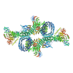 | | cryo-EM structure of mTORC1 bound to PRAS40-fused active RagA/C GTPases | | 分子名称: | GUANOSINE-5'-DIPHOSPHATE, GUANOSINE-5'-TRIPHOSPHATE, Proline-rich AKT1 substrate 1, ... | | 著者 | Anandapadamanaban, M, Berndt, A, Masson, G.R, Perisic, O, Williams, R.L. | | 登録日 | 2019-07-18 | | 公開日 | 2019-10-16 | | 最終更新日 | 2024-05-22 | | 実験手法 | ELECTRON MICROSCOPY (5.5 Å) | | 主引用文献 | Architecture of human Rag GTPase heterodimers and their complex with mTORC1.
Science, 366, 2019
|
|
8JD8
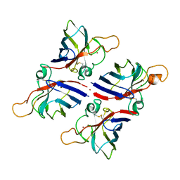 | | Crystal structure of Citrus limon Cu-Zn superoxide dismutase | | 分子名称: | COPPER (II) ION, Superoxide dismutase [Cu-Zn], ZINC ION | | 著者 | Utami, R.A, Yoshida, H, Retnoningrum, D.S, Ismaya, W.T. | | 登録日 | 2023-05-12 | | 公開日 | 2023-12-27 | | 実験手法 | X-RAY DIFFRACTION (1.86 Å) | | 主引用文献 | Direct relationship between dimeric form and activity in the acidic copper-zinc superoxide dismutase from lemon.
Acta Crystallogr.,Sect.F, 79, 2023
|
|
5K7V
 | | Computational Design of Self-Assembling Cyclic Protein Homooligomers | | 分子名称: | Designed protein HR00C3 | | 著者 | Sankaran, B, Zwart, P.H, Fallas, J.A, Pereira, J.H, Ueda, G, Baker, D. | | 登録日 | 2016-05-26 | | 公開日 | 2017-04-12 | | 最終更新日 | 2024-02-28 | | 実験手法 | X-RAY DIFFRACTION (3.165 Å) | | 主引用文献 | Computational design of self-assembling cyclic protein homo-oligomers.
Nat Chem, 9, 2017
|
|
5KBA
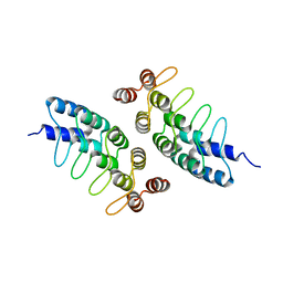 | | Computational Design of Self-Assembling Cyclic Protein Homooligomers | | 分子名称: | Designed protein ANK1C2 | | 著者 | Sankaran, B, Zwart, P.H, Fallas, J.A, Pereira, J.H, Ueda, G, Baker, D. | | 登録日 | 2016-06-02 | | 公開日 | 2017-04-12 | | 最終更新日 | 2024-03-06 | | 実験手法 | X-RAY DIFFRACTION (2.601 Å) | | 主引用文献 | Computational design of self-assembling cyclic protein homo-oligomers.
Nat Chem, 9, 2017
|
|
2PR6
 | |
2PR5
 | |
1VQ2
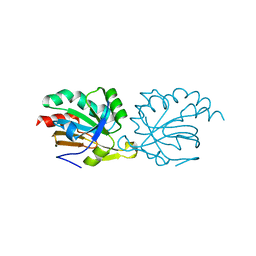
 | | CRYSTAL STRUCTURE OF T4-BACTERIOPHAGE DEOXYCYTIDYLATE DEAMINASE, MUTANT R115E | | 分子名称: | 3,4-DIHYDRO-2'-DEOXYURIDINE-5'-MONOPHOSPHATE, DEOXYCYTIDYLATE DEAMINASE, ZINC ION | | 著者 | Almog, R, Maley, F, Maley, G.F, Maccoll, R, Van Roey, P. | | 登録日 | 2004-12-15 | | 公開日 | 2004-12-21 | | 最終更新日 | 2023-12-27 | | 実験手法 | X-RAY DIFFRACTION (2.2 Å) | | 主引用文献 | Three-Dimensional Structure of the R115E Mutant of T4-Bacteriophage 2'-Deoxycytidylate Deaminase
Biochemistry, 43, 2004
|
|
1VRH
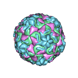 | | HRV14/SDZ 880-061 COMPLEX | | 分子名称: | 2-[4-(2H-1,4-BENZOTHIAZINE-3-YL)-PIPERAZINE-1-LY]-1,3-THIAZOLE-4-CARBOXYLIC ACID ETHYLESTER, RHINOVIRUS 14 | | 著者 | Oren, D.A, Zhang, A, Arnold, E. | | 登録日 | 1996-02-26 | | 公開日 | 1997-02-12 | | 最終更新日 | 2024-05-22 | | 実験手法 | X-RAY DIFFRACTION (3 Å) | | 主引用文献 | Synthesis and activity of piperazine-containing antirhinoviral agents and crystal structure of SDZ 880-061 bound to human rhinovirus 14.
J.Mol.Biol., 259, 1996
|
|
1W9G
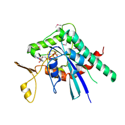
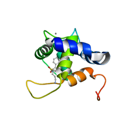
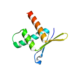 | | Structure of ERH (Enhencer of Rudimentary Gene) | | 分子名称: | ENHANCER OF RUDIMENTARY HOMOLOG | | 著者 | Wan, C, Tempel, W, Liu, Z, Wang, B.-C, Rose, R.B. | | 登録日 | 2004-10-13 | | 公開日 | 2005-04-06 | | 最終更新日 | 2024-05-08 | | 実験手法 | X-RAY DIFFRACTION (2 Å) | | 主引用文献 | Structure of the Conserved Transcriptional Repressor Enhancer of Rudimentary Homolog
Biochemistry, 44, 2005
|
|
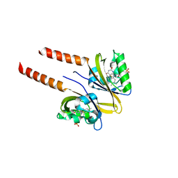
5VGS
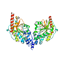
 | | Crystal structure of lachrymatory factor synthase from Allium cepa in complex with crotyl alcohol | | 分子名称: | (2E)-but-2-en-1-ol, (2Z)-but-2-en-1-ol, Lachrymatory-factor synthase | | 著者 | Silvaroli, J.A, Pleshinger, M.J, Banerjee, S, Kiser, P.D, Golczak, M. | | 登録日 | 2017-04-11 | | 公開日 | 2017-07-26 | | 最終更新日 | 2023-10-04 | | 実験手法 | X-RAY DIFFRACTION (1.9 Å) | | 主引用文献 | Enzyme That Makes You Cry-Crystal Structure of Lachrymatory Factor Synthase from Allium cepa.
ACS Chem. Biol., 12, 2017
|
|
1W94
 | |
1VWT
 | | T STATE HUMAN HEMOGLOBIN [ALPHA V96W], ALPHA AQUOMET, BETA DEOXY | | 分子名称: | HEMOGLOBIN, PROTOPORPHYRIN IX CONTAINING FE, SULFATE ION | | 著者 | Puius, Y.A, Zou, M, Ho, N.T, Ho, C, Almo, S.C. | | 登録日 | 1997-03-20 | | 公開日 | 1998-03-25 | | 最終更新日 | 2024-05-22 | | 実験手法 | X-RAY DIFFRACTION (1.9 Å) | | 主引用文献 | Novel water-mediated hydrogen bonds as the structural basis for the low oxygen affinity of the blood substitute candidate rHb(alpha 96Val-->Trp).
Biochemistry, 37, 1998
|
|
2YYF
 | | Purification and structural characterization of a D-amino acid containing conopeptide, marmophine, from Conus marmoreus | | 分子名称: | M-conotoxin mr12 | | 著者 | Huang, F, Du, W, Han, Y, Wang, C. | | 登録日 | 2007-04-29 | | 公開日 | 2008-04-08 | | 最終更新日 | 2022-03-16 | | 実験手法 | SOLUTION NMR | | 主引用文献 | Purification and structural characterization of a D-amino acid-containing conopeptide, conomarphin, from Conus marmoreus.
Febs J., 275, 2008
|
|
7S7I
 | |
6MEQ
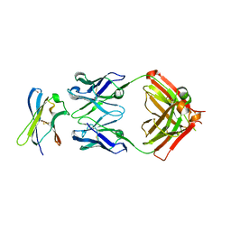
 | | PcdhgB3 EC1-4 in 50 mM HEPES | | 分子名称: | CALCIUM ION, Protocadherin gamma-B3 | | 著者 | Nicoludis, J.M, Gaudet, R. | | 登録日 | 2018-09-06 | | 公開日 | 2019-09-04 | | 最終更新日 | 2023-10-11 | | 実験手法 | X-RAY DIFFRACTION (2.9 Å) | | 主引用文献 | Interaction specificity of clustered protocadherins inferred from sequence covariation and structural analysis.
Proc.Natl.Acad.Sci.USA, 116, 2019
|
|
5IRR
 | | Crystal structure of Septin GTPase domain from Chlamydomonas reinhardtii | | 分子名称: | 5'-GUANOSINE-DIPHOSPHATE-MONOTHIOPHOSPHATE, MAGNESIUM ION, Septin-like protein | | 著者 | Pinto, A.P.A, Pereira, H.M, Navarro, M.V.A.S, Brandao-Neto, J, Garratt, R.C, Araujo, A.P.U. | | 登録日 | 2016-03-14 | | 公開日 | 2017-04-26 | | 最終更新日 | 2020-01-01 | | 実験手法 | X-RAY DIFFRACTION (2.04 Å) | | 主引用文献 | Filaments and fingers: Novel structural aspects of the single septin from Chlamydomonas reinhardtii.
J. Biol. Chem., 292, 2017
|
|
4V7N
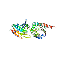
 | | Glycocyamine kinase, beta-beta homodimer from marine worm Namalycastis sp., with transition state analog Mg(II)-ADP-NO3-glycocyamine. | | 分子名称: | ADENOSINE-5'-DIPHOSPHATE, GUANIDINO ACETATE, Glycocyamine kinase beta chain, ... | | 著者 | Lim, K, Pullalarevu, S, Herzberg, O. | | 登録日 | 2009-12-15 | | 公開日 | 2014-07-09 | | 最終更新日 | 2023-09-20 | | 実験手法 | X-RAY DIFFRACTION (2.3 Å) | | 主引用文献 | Structural basis for the mechanism and substrate specificity of glycocyamine kinase, a phosphagen kinase family member.
Biochemistry, 49, 2010
|
|
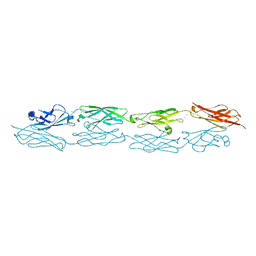
6MER
 | | PcdhgB3 EC1-4 in 50 mM HEPES | | 分子名称: | CALCIUM ION, Protocadherin gamma-B3 | | 著者 | Nicoludis, J.M, Gaudet, R. | | 登録日 | 2018-09-06 | | 公開日 | 2019-09-04 | | 最終更新日 | 2023-10-11 | | 実験手法 | X-RAY DIFFRACTION (3 Å) | | 主引用文献 | Interaction specificity of clustered protocadherins inferred from sequence covariation and structural analysis.
Proc.Natl.Acad.Sci.USA, 116, 2019
|
|
6V88
 | |
6MV3
 | | NMR structure of the cNTnC-cTnI chimera bound to calcium desensitizer W7 | | 分子名称: | CALCIUM ION, N-(6-AMINOHEXYL)-5-CHLORO-1-NAPHTHALENESULFONAMIDE, Troponin C, ... | | 著者 | Cai, F, Hwang, P.M, Sykes, B.D. | | 登録日 | 2018-10-24 | | 公開日 | 2018-11-14 | | 最終更新日 | 2024-05-15 | | 実験手法 | SOLUTION NMR | | 主引用文献 | Structural Changes Induced by the Binding of the Calcium Desensitizer W7 to Cardiac Troponin.
Biochemistry, 57, 2018
|
|
5K2J
 | |
5K1G
 | |


