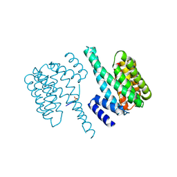
1O9F
 
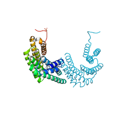
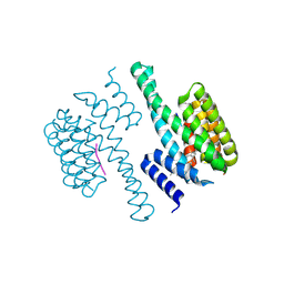 | | Structural view of a fungal toxin acting on a 14-3-3 regulatory complex | | 分子名称: | 14-3-3-LIKE PROTEIN C, FUSICOCCIN, PLASMA MEMBRANE H+ ATPASE | | 著者 | Wurtele, M, Jelich-Ottmann, C, Wittinghofer, A, Oecking, C. | | 登録日 | 2002-12-12 | | 公開日 | 2003-03-06 | | 最終更新日 | 2023-12-13 | | 実験手法 | X-RAY DIFFRACTION (2.7 Å) | | 主引用文献 | Structural View of a Fungal Toxin Acting on a 14-3-3 Regulatory Complex
Embo J., 22, 2003
|
|
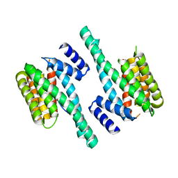
2O98
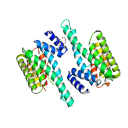
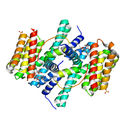 | | Structure of the 14-3-3 / H+-ATPase plant complex | | 分子名称: | 14-3-3-like protein C, FUSICOCCIN, Plasma membrane H+ ATPase, ... | | 著者 | Ottmann, C, Weyand, M, Wittinghofer, A, Oecking, C. | | 登録日 | 2006-12-13 | | 公開日 | 2007-04-03 | | 最終更新日 | 2023-12-27 | | 実験手法 | X-RAY DIFFRACTION (2.7 Å) | | 主引用文献 | Structure of a 14-3-3 coordinated hexamer of the plant plasma membrane H+ -ATPase by combining X-ray crystallography and electron cryomicroscopy
Mol.Cell, 25, 2007
|
|
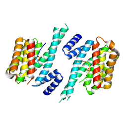
2C74
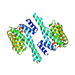
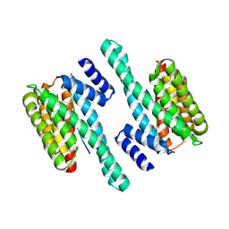
 | | 14-3-3 Protein Eta (Human) Complexed to Peptide | | 分子名称: | 14-3-3 PROTEIN ETA, CITRIC ACID, CONSENSUS PEPTIDE MODE 1 FOR 14-3-3 PROTEINS | | 著者 | Elkins, J.M, Yang, X, Smee, C.E.A, Johansson, C, Sundstrom, M, Edwards, A, Weigelt, J, Arrowsmith, C, Doyle, D.A. | | 登録日 | 2005-11-17 | | 公開日 | 2005-12-02 | | 最終更新日 | 2023-12-13 | | 実験手法 | X-RAY DIFFRACTION (2.7 Å) | | 主引用文献 | Structural basis for protein-protein interactions in the 14-3-3 protein family.
Proc. Natl. Acad. Sci. U.S.A., 103, 2006
|
|
7A6R
 | |
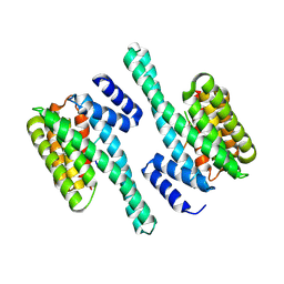
1IB1
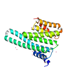
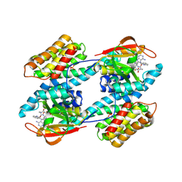 | | CRYSTAL STRUCTURE OF THE 14-3-3 ZETA:SEROTONIN N-ACETYLTRANSFERASE COMPLEX | | 分子名称: | 14-3-3 ZETA ISOFORM, COA-S-ACETYL TRYPTAMINE, SEROTONIN N-ACETYLTRANSFERASE | | 著者 | Obsil, T, Ghirlando, R, Klein, D.C, Ganguly, S, Dyda, F. | | 登録日 | 2001-03-26 | | 公開日 | 2001-05-02 | | 最終更新日 | 2023-08-09 | | 実験手法 | X-RAY DIFFRACTION (2.7 Å) | | 主引用文献 | Crystal structure of the 14-3-3zeta:serotonin N-acetyltransferase complex. a role for scaffolding in enzyme regulation.
Cell(Cambridge,Mass.), 105, 2001
|
|
5D3F
 | |
7EXE
 | |
6YOS
 | | Binary complex of 14-3-3 zeta with Glucocorticoid Receptor (GR) pT524 pS617 peptide | | 分子名称: | 14-3-3 protein zeta/delta, Glucocorticoid receptor,Glucocorticoid receptor | | 著者 | Munier, C.C, Edman, K, Perry, M.W.D, Ottmann, C. | | 登録日 | 2020-04-15 | | 公開日 | 2021-03-24 | | 最終更新日 | 2024-01-24 | | 実験手法 | X-RAY DIFFRACTION (2.75 Å) | | 主引用文献 | Glucocorticoid receptor Thr524 phosphorylation by MINK1 induces interactions with 14-3-3 protein regulators.
J.Biol.Chem., 296, 2021
|
|
5D3E
 | |
6SAD
 | |
1A4O
 | | 14-3-3 PROTEIN ZETA ISOFORM | | 分子名称: | 14-3-3 PROTEIN ZETA | | 著者 | Liu, D, Bienkowska, J, Petosa, C, Collier, R.J, Fu, H, Liddington, R.C. | | 登録日 | 1998-02-01 | | 公開日 | 1999-03-02 | | 最終更新日 | 2024-02-07 | | 実験手法 | X-RAY DIFFRACTION (2.8 Å) | | 主引用文献 | Crystal structure of the zeta isoform of the 14-3-3 protein.
Nature, 376, 1995
|
|
6BQT
 | |
6TWZ
 | | 14-3-3 sigma complexed with a phosphorylated 16E6 peptide | | 分子名称: | 14-3-3 protein sigma, 2-[3-(2-HYDROXY-1,1-DIHYDROXYMETHYL-ETHYLAMINO)-PROPYLAMINO]-2-HYDROXYMETHYL-PROPANE-1,3-DIOL, D(-)-TARTARIC ACID, ... | | 著者 | Gogl, G, Cousido-Siah, A, Sluchanko, N.N, Trave, G. | | 登録日 | 2020-01-13 | | 公開日 | 2020-05-06 | | 最終更新日 | 2024-10-09 | | 実験手法 | X-RAY DIFFRACTION (2.8 Å) | | 主引用文献 | Dual Specificity PDZ- and 14-3-3-Binding Motifs: A Structural and Interactomics Study.
Structure, 28, 2020
|
|
1YZ5
 | |
2BTP
 | | 14-3-3 Protein Theta (Human) Complexed to Peptide | | 分子名称: | 14-3-3 PROTEIN TAU, CONSENSUS PEPTIDE FOR 14-3-3 PROTEINS | | 著者 | Elkins, J.M, Johansson, A.C.E, Smee, C, Yang, X, Sundstrom, M, Edwards, A, Arrowsmith, C, Doyle, D.A, Structural Genomics Consortium (SGC) | | 登録日 | 2005-06-05 | | 公開日 | 2005-06-28 | | 最終更新日 | 2024-10-16 | | 実験手法 | X-RAY DIFFRACTION (2.8 Å) | | 主引用文献 | Structural Basis for Protein-Protein Interactions in the 14-3-3 Protein Family.
Proc.Natl.Acad.Sci.USA, 103, 2006
|
|
6FEL
 | |
6GKG
 | |
4ZDR
 | | Crystal structure of 14-3-3[zeta]-LKB1 fusion protein | | 分子名称: | 14-3-3 protein zeta/delta,GGSGGS linker,Serine/threonine-protein kinase STK11, GLYCEROL, PROPANE, ... | | 著者 | Ding, S, Shi, Z.B. | | 登録日 | 2015-04-18 | | 公開日 | 2015-09-09 | | 最終更新日 | 2023-11-08 | | 実験手法 | X-RAY DIFFRACTION (2.899 Å) | | 主引用文献 | Structure of the 14-3-3 zeta-LKB1 fusion protein provides insight into a novel ligand-binding mode of 14-3-3.
Acta Crystallogr.,Sect.F, 71, 2015
|
|
6TLF
 | |
8AH2
 | | Crystal structure of human 14-3-3 zeta fused to the NPM1 peptide including phosphoserine-48 | | 分子名称: | 14-3-3 protein zeta/delta,Nucleophosmin | | 著者 | Boyko, K.M, Kapitonova, A.A, Tugaeva, K.V, Varfolomeeva, L.A, Sluchanko, N.N. | | 登録日 | 2022-07-20 | | 公開日 | 2022-09-14 | | 最終更新日 | 2024-10-16 | | 実験手法 | X-RAY DIFFRACTION (2.9 Å) | | 主引用文献 | Structural basis for the recognition by 14-3-3 proteins of a conditional binding site within the oligomerization domain of human nucleophosmin.
Biochem.Biophys.Res.Commun., 627, 2022
|
|
6BD2
 | |
6BYJ
 | |
4O46
 | | 14-3-3-gamma in complex with influenza NS1 C-terminal tail phosphorylated at S228 | | 分子名称: | 14-3-3 protein gamma, Nonstructural protein 1, UNKNOWN ATOM OR ION, ... | | 著者 | Qin, S, Liu, Y, Tempel, W, Arrowsmith, C.H, Bountra, C, Edwards, A.M, Min, J, Structural Genomics Consortium (SGC) | | 登録日 | 2013-12-18 | | 公開日 | 2014-04-30 | | 最終更新日 | 2023-09-20 | | 実験手法 | X-RAY DIFFRACTION (2.9 Å) | | 主引用文献 | Structural basis for histone mimicry and hijacking of host proteins by influenza virus protein NS1.
Nat Commun, 5, 2014
|
|
6QK8
 | |
5WXN
 | | Structure of the LKB1 and 14-3-3 complex | | 分子名称: | 14-3-3 protein zeta/delta, Serine/threonine-protein kinase STK11 | | 著者 | Ding, S, Shi, Z.B. | | 登録日 | 2017-01-08 | | 公開日 | 2017-04-19 | | 最終更新日 | 2023-11-22 | | 実験手法 | X-RAY DIFFRACTION (2.93 Å) | | 主引用文献 | Structure of the complex of phosphorylated liver kinase B1 and 14-3-3 zeta
Acta Crystallogr F Struct Biol Commun, 73, 2017
|
|


