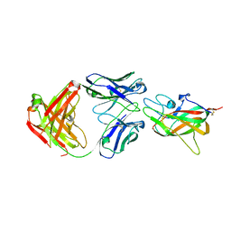
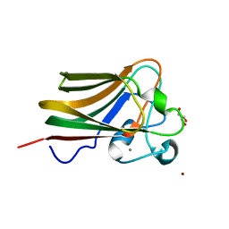
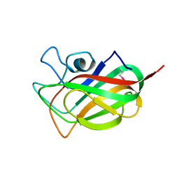
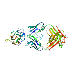
4DEQ
 
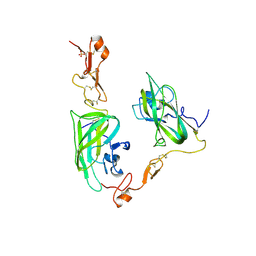 | | Structure of the Neuropilin-1/VEGF-A complex | | 分子名称: | Neuropilin-1, Vascular endothelial growth factor A, PHOSPHATE ION | | 著者 | Vander Kooi, C.W. | | 登録日 | 2012-01-21 | | 公開日 | 2012-02-08 | | 最終更新日 | 2023-09-13 | | 実験手法 | X-RAY DIFFRACTION (2.649 Å) | | 主引用文献 | Structural Basis for Selective Vascular Endothelial Growth Factor-A (VEGF-A) Binding to Neuropilin-1.
J.Biol.Chem., 287, 2012
|
|
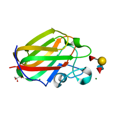
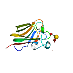
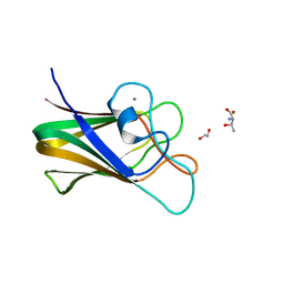
5JGQ
 | | X-ray structure of neuropilin-1 b1 domain complexed with Arg-7 ligand. | | 分子名称: | DIMETHYL SULFOXIDE, Neuropilin-1, N~2~-(benzenecarbonyl)-L-arginine | | 著者 | Fotinou, C, Rana, R, Djordjevic, S, Yelland, T. | | 登録日 | 2016-04-20 | | 公開日 | 2017-05-10 | | 最終更新日 | 2024-01-10 | | 実験手法 | X-RAY DIFFRACTION (1.6 Å) | | 主引用文献 | Architecture and hydration of the arginine-binding site of neuropilin-1.
FEBS J., 285, 2018
|
|
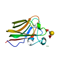
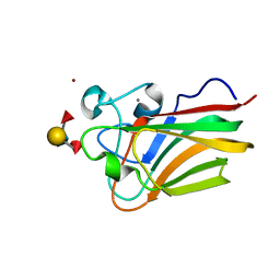
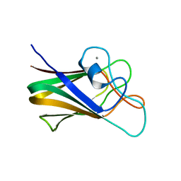
5JGI
 | | X-ray structure of neuropilin-1 b1 domain complexed with M45 compound | | 分子名称: | N-ALPHA-L-ACETYL-ARGININE, Neuropilin-1 | | 著者 | Fotinou, C, Rana, R, Djordjevic, S, Yelland, T. | | 登録日 | 2016-04-20 | | 公開日 | 2017-05-10 | | 最終更新日 | 2024-01-10 | | 実験手法 | X-RAY DIFFRACTION (1.38 Å) | | 主引用文献 | Architecture and hydration of the arginine-binding site of neuropilin-1.
FEBS J., 285, 2018
|
|
2QQI
 | |
2QQJ
 | |
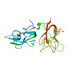
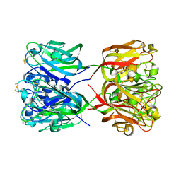
1IQD
 | | Human Factor VIII C2 Domain complexed to human monoclonal BO2C11 Fab. | | 分子名称: | HUMAN FACTOR VIII, HUMAN MONOCLONAL BO2C11 FAB HEAVY CHAIN, HUMAN MONOCLONAL BO2C11 FAB LIGHT CHAIN | | 著者 | Spiegel Jr, P.C, Jacquemin, M, Saint-Remy, J.M, Stoddard, B.L, Pratt, K.P. | | 登録日 | 2001-07-21 | | 公開日 | 2001-08-15 | | 最終更新日 | 2023-12-27 | | 実験手法 | X-RAY DIFFRACTION (2 Å) | | 主引用文献 | Structure of a factor VIII C2 domain-immunoglobulin G4kappa Fab complex: identification of an inhibitory antibody epitope on the surface of factor VIII.
Blood, 98, 2001
|
|
4GWI
 | |
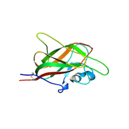
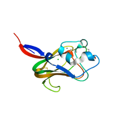
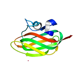
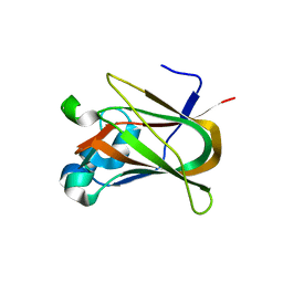
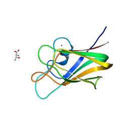
1KEX
 | | Crystal Structure of the b1 Domain of Human Neuropilin-1 | | 分子名称: | Neuropilin-1 | | 著者 | Lee, C.C, Kreusch, A, McMullan, D, Ng, K, Spraggon, G. | | 登録日 | 2001-11-18 | | 公開日 | 2003-01-28 | | 最終更新日 | 2023-08-16 | | 実験手法 | X-RAY DIFFRACTION (1.9 Å) | | 主引用文献 | Crystal Structure of the Human Neuropilin-1 b1 Domain
Structure, 11, 2003
|
|
4GWJ
 | |
1K12
 | | Fucose Binding lectin | | 分子名称: | CALCIUM ION, CHLORIDE ION, LECTIN, ... | | 著者 | Bianchet, M.A, Odom, E.W, Vasta, G.R, Amzel, L.M. | | 登録日 | 2001-09-23 | | 公開日 | 2002-07-31 | | 最終更新日 | 2020-07-29 | | 実験手法 | X-RAY DIFFRACTION (1.9 Å) | | 主引用文献 | A novel fucose recognition fold involved in innate immunity.
Nat.Struct.Biol., 9, 2002
|
|
3LE0
 | | Lectin Domain of Lectinolysin complexed with Glycerol | | 分子名称: | CALCIUM ION, GLYCEROL, NICKEL (II) ION, ... | | 著者 | Feil, S.C. | | 登録日 | 2010-01-13 | | 公開日 | 2010-12-29 | | 最終更新日 | 2023-09-06 | | 実験手法 | X-RAY DIFFRACTION (1.91 Å) | | 主引用文献 | Structure of the lectin regulatory domain of the cholesterol-dependent cytolysin lectinolysin reveals the basis for its lewis antigen specificity.
Structure, 20, 2012
|
|
3LEG
 | | Lectin Domain of Lectinolysin complexed with Lewis Y Antigen | | 分子名称: | CALCIUM ION, NICKEL (II) ION, Platelet aggregation factor Sm-hPAF, ... | | 著者 | Feil, S.C. | | 登録日 | 2010-01-14 | | 公開日 | 2010-12-29 | | 最終更新日 | 2023-09-06 | | 実験手法 | X-RAY DIFFRACTION (2.01 Å) | | 主引用文献 | Structure of the lectin regulatory domain of the cholesterol-dependent cytolysin lectinolysin reveals the basis for its lewis antigen specificity.
Structure, 20, 2012
|
|
3LEK
 | | Lectin Domain of Lectinolysin complexed with Lewis B Antigen | | 分子名称: | CALCIUM ION, NICKEL (II) ION, Platelet aggregation factor Sm-hPAF, ... | | 著者 | Feil, S.C. | | 登録日 | 2010-01-15 | | 公開日 | 2010-12-29 | | 最終更新日 | 2023-09-06 | | 実験手法 | X-RAY DIFFRACTION (2 Å) | | 主引用文献 | Structure of the lectin regulatory domain of the cholesterol-dependent cytolysin lectinolysin reveals the basis for its lewis antigen specificity.
Structure, 20, 2012
|
|
1TVG
 | | X-ray structure of human PP25 gene product, HSPC034. Northeast Structural Genomics Target HR1958. | | 分子名称: | CALCIUM ION, LOC51668 protein, SAMARIUM (III) ION | | 著者 | Kuzin, A.P, Vorobiev, S.M, Lee, I, Acton, T.B, Montelione, G.T, Tong, L, Hunt, J.F, Northeast Structural Genomics Consortium (NESG) | | 登録日 | 2004-06-29 | | 公開日 | 2004-11-09 | | 最終更新日 | 2024-02-14 | | 実験手法 | X-RAY DIFFRACTION (1.6 Å) | | 主引用文献 | Improving NMR protein structure quality by Rosetta refinement: a molecular replacement study.
Proteins, 75, 2009
|
|
3F2Z
 | | Crystal structure of the C-terminal domain of a chitobiase (BF3579) from Bacteroides fragilis, Northeast Structural Genomics Consortium Target BfR260B | | 分子名称: | uncharacterized protein BF3579 | | 著者 | Forouhar, F, Lew, S, Seetharaman, J, Janjua, H, Xiao, R, Foote, E.L, Ciccosanti, C, Lee, D, Nair, R, Everett, J.K, Acton, T.B, Rost, B, Montelione, G.T, Hunt, J.F, Tong, L, Northeast Structural Genomics Consortium (NESG) | | 登録日 | 2008-10-30 | | 公開日 | 2008-11-18 | | 最終更新日 | 2023-12-27 | | 実験手法 | X-RAY DIFFRACTION (1.3 Å) | | 主引用文献 | Crystal structure of the C-terminal domain of a chitobiase (BF3579) from Bacteroides fragilis, Northeast Structural Genomics Consortium Target BfR260B
To be Published
|
|
3NNG
 | | Crystal structure of the F5/8 type C domain of Q5LFR2_BACFN protein from Bacteroides fragilis. Northeast Structural Genomics Consortium Target BfR258E | | 分子名称: | CALCIUM ION, uncharacterized protein | | 著者 | Vorobiev, S, Su, M, Dimaio, F, Baker, D, Seetharaman, J, Janjua, J, Xiao, R, Ciccosanti, C, Foote, E.L, Lee, D, Everett, J.K, Nair, R, Acton, T.B, Rost, B, Montelione, G.T, Hunt, J.F, Tong, L, Northeast Structural Genomics Consortium (NESG) | | 登録日 | 2010-06-23 | | 公開日 | 2010-08-18 | | 最終更新日 | 2023-09-06 | | 実験手法 | X-RAY DIFFRACTION (2.177 Å) | | 主引用文献 | Crystal structure of the F5/8 type C domain of Q5LFR2_BACFN protein from Bacteroides fragilis.
To be Published
|
|
3CQO
 | |
3GGL
 | | X-Ray Structure of the C-terminal domain (277-440) of Putative chitobiase from Bacteroides thetaiotaomicron. Northeast Structural Genomics Consortium Target BtR324A. | | 分子名称: | DI(HYDROXYETHYL)ETHER, Putative chitobiase, ZINC ION | | 著者 | Kuzin, A, Neely, H, Seetharaman, R, Lee, D, Ciccosanti, C, Foote, E.L, Janjua, H, Xiao, R, Nair, R, Rost, B, Acton, T, Everett, J.K, Montelione, G.T, Tong, L, Hunt, J, Northeast Structural Genomics Consortium (NESG) | | 登録日 | 2009-02-28 | | 公開日 | 2009-03-31 | | 最終更新日 | 2023-11-22 | | 実験手法 | X-RAY DIFFRACTION (3 Å) | | 主引用文献 | Northeast Structural Genomics Consortium Target BtR324A
To be Published
|
|
4XZU
 | |
7BLJ
 | |
7BLG
 | |
7BLH
 | |
7BLK
 | | Structure of CBM BT3015C from Bacteroides thetaiotaomicron | | 分子名称: | CALCIUM ION, family 32 carbohydrate-binding module from Bacteroides thetaiotaomicron | | 著者 | Costa, R.L. | | 登録日 | 2021-01-18 | | 公開日 | 2022-03-02 | | 最終更新日 | 2024-01-31 | | 実験手法 | X-RAY DIFFRACTION (1.06 Å) | | 主引用文献 | Structural basis for mucin-type O-glycan recognition by proteins of a Bacteroides thetaiotaomicron polysaccharide utilization loci
To Be Published
|
|
4LKS
 | | Structure of CBM32-3 from a family 31 glycoside hydrolase from Clostridium perfringens in complex with galactose | | 分子名称: | CALCIUM ION, Glycosyl hydrolase, family 31/fibronectin type III domain protein, ... | | 著者 | Grondin, J.M, Duan, D, Kirlin, A.C, Furness, H.S, Allingham, J.S, Smith, S.P. | | 登録日 | 2013-07-08 | | 公開日 | 2014-12-24 | | 最終更新日 | 2023-09-20 | | 実験手法 | X-RAY DIFFRACTION (1.5 Å) | | 主引用文献 | Diverse modes of galacto-specific carbohydrate recognition by a family 31 glycoside hydrolase from Clostridium perfringens.
Plos One, 12, 2017
|
|
4ZXE
 | | X-ray crystal structure of chitosan-binding module 1 derived from chitosanase/glucanase from Paenibacillus sp. IK-5. | | 分子名称: | 1,2-ETHANEDIOL, Glucanase/Chitosanase, SULFATE ION | | 著者 | Shinya, S, Oi, H, Kitaoku, Y, Ohnuma, T, Numata, T, Fukamizo, T. | | 登録日 | 2015-05-20 | | 公開日 | 2016-04-13 | | 最終更新日 | 2024-03-20 | | 実験手法 | X-RAY DIFFRACTION (1.4 Å) | | 主引用文献 | Mechanism of chitosan recognition by CBM32 carbohydrate-binding modules from a Paenibacillus sp. IK-5 chitosanase/glucanase
Biochem.J., 473, 2016
|
|


