1JVP
 
 | |
7UWM
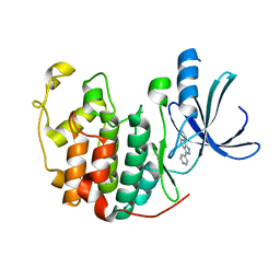
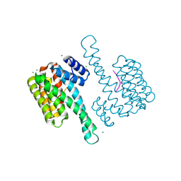
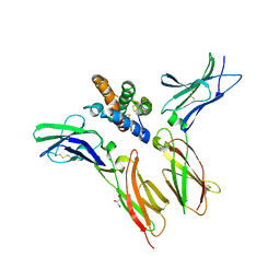
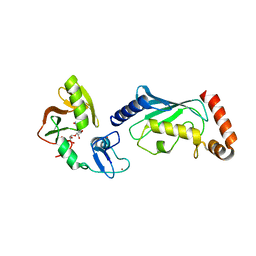
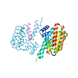
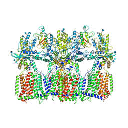
 | | Structure of the IL-17A-IL-17RA binary complex | | 分子名称: | 2-acetamido-2-deoxy-beta-D-glucopyranose, 2-acetamido-2-deoxy-beta-D-glucopyranose-(1-4)-2-acetamido-2-deoxy-beta-D-glucopyranose, Interleukin-17 receptor A, ... | | 著者 | Wilson, S.C, Caveney, N.A, Jude, K.M, Garcia, K.C. | | 登録日 | 2022-05-03 | | 公開日 | 2022-07-27 | | 最終更新日 | 2025-05-28 | | 実験手法 | ELECTRON MICROSCOPY (2.5 Å) | | 主引用文献 | Organizing structural principles of the IL-17 ligand-receptor axis.
Nature, 609, 2022
|
|
3N8B
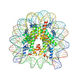
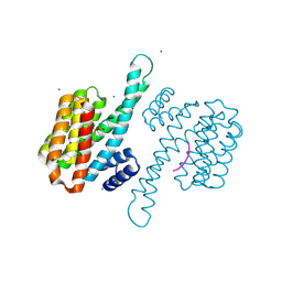
 | | Crystal Structure of Borrelia burgdorferi Pur-alpha | | 分子名称: | 1,2-ETHANEDIOL, MAGNESIUM ION, Uncharacterized protein | | 著者 | Graebsch, A, Roche, S, Kostrewa, D, Niessing, D. | | 登録日 | 2010-05-28 | | 公開日 | 2010-10-06 | | 最終更新日 | 2024-10-09 | | 実験手法 | X-RAY DIFFRACTION (1.9 Å) | | 主引用文献 | Of bits and bugs--on the use of bioinformatics and a bacterial crystal structure to solve a eukaryotic repeat-protein structure.
Plos One, 5, 2010
|
|
1TIV
 | |
8BFC
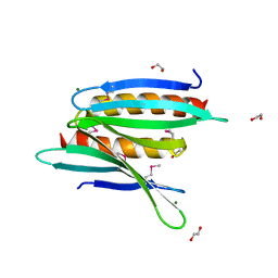
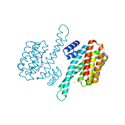
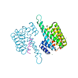
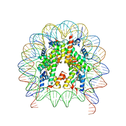
 | | Binary structure of 14-3-3s and RND3 phosphopeptide | | 分子名称: | 14-3-3 protein sigma, CHLORIDE ION, MAGNESIUM ION, ... | | 著者 | Somsen, B.A, Ottmann, C. | | 登録日 | 2022-10-24 | | 公開日 | 2023-03-29 | | 最終更新日 | 2024-11-06 | | 実験手法 | X-RAY DIFFRACTION (1.4 Å) | | 主引用文献 | Reversible Dual-Covalent Molecular Locking of the 14-3-3/ERR gamma Protein-Protein Interaction as a Molecular Glue Drug Discovery Approach.
J.Am.Chem.Soc., 145, 2023
|
|
6IPU
 | |
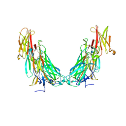
6WDQ
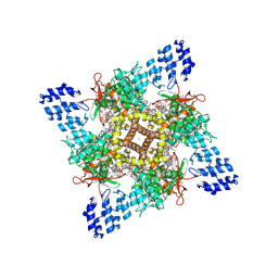
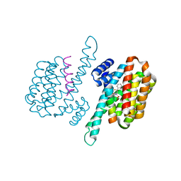
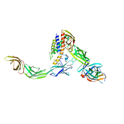 | | IL23/IL23R/IL12Rb1 signaling complex | | 分子名称: | 2-acetamido-2-deoxy-beta-D-glucopyranose, 2-acetamido-2-deoxy-beta-D-glucopyranose-(1-4)-2-acetamido-2-deoxy-beta-D-glucopyranose, Interleukin-12 receptor subunit beta-1, ... | | 著者 | Jude, K.M, Ely, L.K, Glassman, C.R, Thomas, C, Spangler, J.B, Lupardus, P.J, Garcia, K.C. | | 登録日 | 2020-04-01 | | 公開日 | 2021-02-24 | | 最終更新日 | 2024-10-16 | | 実験手法 | X-RAY DIFFRACTION (3.4 Å) | | 主引用文献 | Structural basis for IL-12 and IL-23 receptor sharing reveals a gateway for shaping actions on T versus NK cells.
Cell, 184, 2021
|
|
3DDP
 | |
6WEO
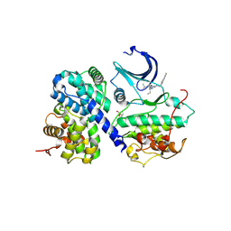
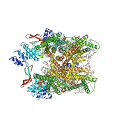
 | | IL-22 Signaling Complex with IL-22R1 and IL-10Rbeta | | 分子名称: | 2-acetamido-2-deoxy-beta-D-glucopyranose, 2-acetamido-2-deoxy-beta-D-glucopyranose-(1-4)-2-acetamido-2-deoxy-beta-D-glucopyranose, 2-acetamido-2-deoxy-beta-D-glucopyranose-(1-4)-[alpha-L-fucopyranose-(1-6)]2-acetamido-2-deoxy-beta-D-glucopyranose, ... | | 著者 | Saxton, R.A, Jude, K.M, Henneberg, L.T, Garcia, K.C. | | 登録日 | 2020-04-02 | | 公開日 | 2021-04-28 | | 最終更新日 | 2024-10-23 | | 実験手法 | X-RAY DIFFRACTION (2.6 Å) | | 主引用文献 | The tissue protective functions of interleukin-22 can be decoupled from pro-inflammatory actions through structure-based design.
Immunity, 54, 2021
|
|
8GF8
 | | Cryo-EM structure of human TRPV1 in cNW11 nanodisc and soybean lipids | | 分子名称: | (2R)-3-{[(R)-hydroxy{[(1S,2R,3R,4S,5S,6R)-2,3,4,5,6-pentahydroxycyclohexyl]oxy}phosphoryl]oxy}propane-1,2-diyl dioctadecanoate, (2S)-3-(hexadecanoyloxy)-2-[(9Z)-octadec-9-enoyloxy]propyl 2-(trimethylammonio)ethyl phosphate, SODIUM ION, ... | | 著者 | Neuberger, A, Nadezhdin, K.D, Sobolevsky, A.I. | | 登録日 | 2023-03-07 | | 公開日 | 2023-05-10 | | 最終更新日 | 2024-10-23 | | 実験手法 | ELECTRON MICROSCOPY (2.9 Å) | | 主引用文献 | Human TRPV1 structure and inhibition by the analgesic SB-366791.
Nat Commun, 14, 2023
|
|
8GF9
 | | Cryo-EM structure of human TRPV1 in cNW11 nanodisc and POPC:POPE:POPG lipids | | 分子名称: | (2R)-3-{[(R)-hydroxy{[(1S,2R,3R,4S,5S,6R)-2,3,4,5,6-pentahydroxycyclohexyl]oxy}phosphoryl]oxy}propane-1,2-diyl dioctadecanoate, (2S)-3-(hexadecanoyloxy)-2-[(9Z)-octadec-9-enoyloxy]propyl 2-(trimethylammonio)ethyl phosphate, SODIUM ION, ... | | 著者 | Neuberger, A, Nadezhdin, K.D, Sobolevsky, A.I. | | 登録日 | 2023-03-07 | | 公開日 | 2023-05-10 | | 最終更新日 | 2024-10-09 | | 実験手法 | ELECTRON MICROSCOPY (2.58 Å) | | 主引用文献 | Human TRPV1 structure and inhibition by the analgesic SB-366791.
Nat Commun, 14, 2023
|
|
8GFA
 | | Cryo-EM structure of human TRPV1 in complex with the analgesic drug SB-366791 | | 分子名称: | (2E)-3-(4-chlorophenyl)-N-(3-methoxyphenyl)prop-2-enamide, (2S)-3-(hexadecanoyloxy)-2-[(9Z)-octadec-9-enoyloxy]propyl 2-(trimethylammonio)ethyl phosphate, 2-[2-[(1~{S},2~{S},4~{S},5'~{R},6~{R},7~{S},8~{R},9~{S},12~{S},13~{R},16~{S})-5',7,9,13-tetramethylspiro[5-oxapentacyclo[10.8.0.0^{2,9}.0^{4,8}.0^{13,18}]icos-18-ene-6,2'-oxane]-16-yl]oxyethyl]propane-1,3-diol, ... | | 著者 | Neuberger, A, Nadezhdin, K.D, Sobolevsky, A.I. | | 登録日 | 2023-03-07 | | 公開日 | 2023-05-10 | | 最終更新日 | 2024-10-23 | | 実験手法 | ELECTRON MICROSCOPY (2.29 Å) | | 主引用文献 | Human TRPV1 structure and inhibition by the analgesic SB-366791.
Nat Commun, 14, 2023
|
|
4QPL
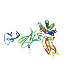
 | | Crystal structure of RNF146(RING-WWE)/UbcH5a/iso-ADPr complex | | 分子名称: | 2'-O-(5-O-phosphono-alpha-D-ribofuranosyl)adenosine 5'-(dihydrogen phosphate), E3 ubiquitin-protein ligase RNF146, Ubiquitin-conjugating enzyme E2 D1, ... | | 著者 | Wang, Z, DaRosa, P.A, Klevit, R.E, Xu, W. | | 登録日 | 2014-06-23 | | 公開日 | 2014-10-15 | | 最終更新日 | 2024-02-28 | | 実験手法 | X-RAY DIFFRACTION (1.9 Å) | | 主引用文献 | Allosteric activation of the RNF146 ubiquitin ligase by a poly(ADP-ribosyl)ation signal.
Nature, 517, 2015
|
|
8C40
 | |
8C3Z
 | |
8C1Y
 | |
8BTQ
 | |
8BWH
 | |
8BWE
 | |
2C6B
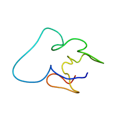
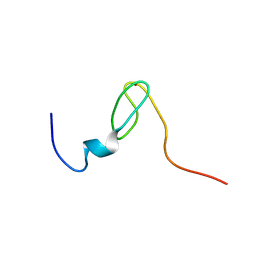
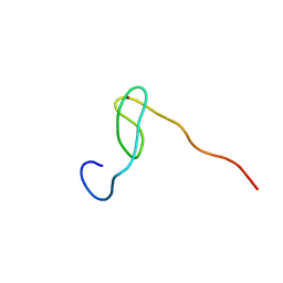 | | Solution structure of the C4 zinc-finger domain of HDM2 | | 分子名称: | UBIQUITIN-PROTEIN LIGASE E3 MDM2, ZINC ION | | 著者 | Yu, G.W, Allen, M.D, Andreeva, A, Fersht, A.R, Bycroft, M. | | 登録日 | 2005-11-08 | | 公開日 | 2006-01-04 | | 最終更新日 | 2024-05-15 | | 実験手法 | SOLUTION NMR | | 主引用文献 | Solution Structure of the C4 Zinc Finger Domain of Hdm2.
Protein Sci., 15, 2006
|
|
7DWX
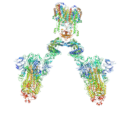 | | Conformation 1 of S-ACE2-B0AT1 ternary complex | | 分子名称: | 2-acetamido-2-deoxy-beta-D-glucopyranose, 2-acetamido-2-deoxy-beta-D-glucopyranose-(1-4)-2-acetamido-2-deoxy-beta-D-glucopyranose, Angiotensin-converting enzyme 2, ... | | 著者 | Yan, R.H, Zhang, Y.Y, Li, Y.N, Ye, F.F, Guo, Y.Y, Xia, L, Zhong, X.Y, Chi, X.M, Zhou, Q. | | 登録日 | 2021-01-18 | | 公開日 | 2021-03-31 | | 最終更新日 | 2024-10-23 | | 実験手法 | ELECTRON MICROSCOPY (8.3 Å) | | 主引用文献 | Structural basis for the different states of the spike protein of SARS-CoV-2 in complex with ACE2.
Cell Res., 31, 2021
|
|
6JXD
 | |
2C6A
 | | Solution structure of the C4 zinc-finger domain of HDM2 | | 分子名称: | UBIQUITIN-PROTEIN LIGASE E3 MDM2, ZINC ION | | 著者 | Yu, G.W, Allen, M.D, Andreeva, A, Fersht, A.R, Bycroft, M. | | 登録日 | 2005-11-08 | | 公開日 | 2006-01-04 | | 最終更新日 | 2024-05-15 | | 実験手法 | SOLUTION NMR | | 主引用文献 | Solution Structure of the C4 Zinc Finger Domain of Hdm2.
Protein Sci., 15, 2006
|
|
6N7K
 | | Cryo-EM structure of tetrameric Ptch1 in complex with ShhNp (form II) | | 分子名称: | 2-acetamido-2-deoxy-beta-D-glucopyranose, 2-acetamido-2-deoxy-beta-D-glucopyranose-(1-4)-2-acetamido-2-deoxy-beta-D-glucopyranose, CALCIUM ION, ... | | 著者 | Yan, N, Gong, X, Qian, H.W. | | 登録日 | 2018-11-27 | | 公開日 | 2019-05-29 | | 最終更新日 | 2025-05-28 | | 実験手法 | ELECTRON MICROSCOPY (6.5 Å) | | 主引用文献 | Inhibition of tetrameric Patched1 by Sonic Hedgehog through an asymmetric paradigm.
Nat Commun, 10, 2019
|
|
6UIB
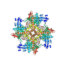
 | | Crystal structure of IL23 bound to peptide 23-652 | | 分子名称: | 2-acetamido-2-deoxy-beta-D-glucopyranose-(1-4)-2-acetamido-2-deoxy-beta-D-glucopyranose, Interleukin-12 subunit beta, Interleukin-23 subunit alpha, ... | | 著者 | Durbin, J.D, Wang, J, Afshar, S. | | 登録日 | 2019-09-30 | | 公開日 | 2020-07-15 | | 最終更新日 | 2024-11-06 | | 実験手法 | X-RAY DIFFRACTION (2.74 Å) | | 主引用文献 | Integration of phage and yeast display platforms: A reliable and cost effective approach for binning of peptides as displayed on-phage.
Plos One, 15, 2020
|
|


