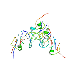
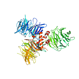
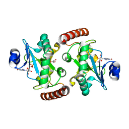
7ZVT
 
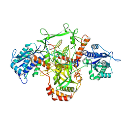 | | CryoEM structure of Ku heterodimer bound to DNA | | 分子名称: | DNA (5'-D(P*CP*GP*AP*TP*AP*TP*CP*TP*AP*GP*AP*GP*GP*GP*AP*T)-3'), DNA (5'-D(P*TP*CP*CP*CP*TP*CP*TP*AP*GP*AP*TP*AP*TP*C)-3'), INOSITOL HEXAKISPHOSPHATE, ... | | 著者 | Hardwick, S.W, Kefala-Stavridi, A, Chirgadze, D.Y, Blundell, T.L, Chaplin, A.K. | | 登録日 | 2022-05-17 | | 公開日 | 2023-05-24 | | 最終更新日 | 2023-12-06 | | 実験手法 | ELECTRON MICROSCOPY (2.74 Å) | | 主引用文献 | Structural and functional basis of inositol hexaphosphate stimulation of NHEJ through stabilization of Ku-XLF interaction.
Nucleic Acids Res., 51, 2023
|
|
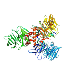
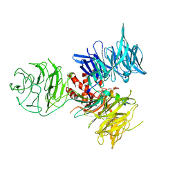
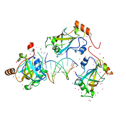
7ZWA
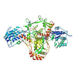 | | CryoEM structure of Ku heterodimer bound to DNA and PAXX | | 分子名称: | DNA (5'-D(P*CP*GP*AP*TP*AP*TP*CP*TP*AP*GP*AP*GP*GP*GP*AP*TP*C)-3'), DNA (5'-D(P*GP*AP*TP*CP*CP*CP*TP*CP*TP*AP*GP*AP*TP*AP*T)-3'), PHOSPHATE ION, ... | | 著者 | Hardwick, S.W, Kefala-Stavridi, A, Chirgadze, D.Y, Blundell, T.L, Chaplin, A.K. | | 登録日 | 2022-05-19 | | 公開日 | 2023-05-31 | | 最終更新日 | 2024-07-24 | | 実験手法 | ELECTRON MICROSCOPY (2.8 Å) | | 主引用文献 | PAXX binding to the NHEJ machinery explains functional redundancy with XLF.
Sci Adv, 9, 2023
|
|
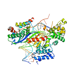
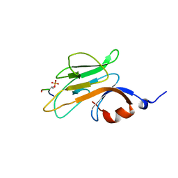
6CIK
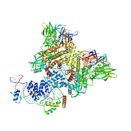 | | Pre-Reaction Complex, RAG1(E962Q)/2-intact/nicked 12/23RSS complex in Mn2+ | | 分子名称: | DNA (5'-D(*AP*TP*CP*TP*GP*GP*CP*CP*TP*GP*TP*CP*TP*TP*A)-3'), High mobility group protein B1, Intact 12RSS substrate forward strand, ... | | 著者 | Chuenchor, W, Chen, X, Kim, M.S, Gellert, M, Yang, W. | | 登録日 | 2018-02-24 | | 公開日 | 2018-04-25 | | 最終更新日 | 2023-10-04 | | 実験手法 | X-RAY DIFFRACTION (3.15 Å) | | 主引用文献 | Cracking the DNA Code for V(D)J Recombination.
Mol. Cell, 70, 2018
|
|
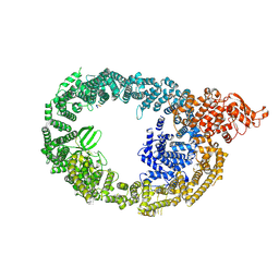
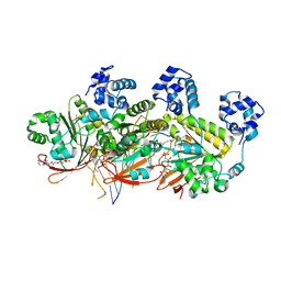
7OOP
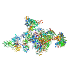 | | Pol II-CSB-CSA-DDB1-UVSSA-PAF-SPT6 (Structure 3) | | 分子名称: | DNA damage-binding protein 1, DNA excision repair protein ERCC-6, DNA excision repair protein ERCC-8, ... | | 著者 | Kokic, G, Cramer, P. | | 登録日 | 2021-05-28 | | 公開日 | 2021-10-06 | | 最終更新日 | 2024-07-17 | | 実験手法 | ELECTRON MICROSCOPY (2.9 Å) | | 主引用文献 | Structural basis of human transcription-DNA repair coupling.
Nature, 598, 2021
|
|
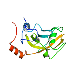
4RKH
 | | Structure of the MSL2 CXC domain bound with a specific MRE sequence | | 分子名称: | DNA (5'-D(*AP*TP*CP*CP*AP*TP*CP*TP*CP*GP*CP*TP*CP*AP*T)-3'), DNA (5'-D(*AP*TP*GP*AP*GP*CP*GP*AP*GP*AP*TP*GP*GP*AP*T)-3'), E3 ubiquitin-protein ligase msl-2, ... | | 著者 | Zheng, S, Ye, K. | | 登録日 | 2014-10-13 | | 公開日 | 2015-01-21 | | 最終更新日 | 2024-03-20 | | 実験手法 | X-RAY DIFFRACTION (2 Å) | | 主引用文献 | Structural basis of X chromosome DNA recognition by the MSL2 CXC domain during Drosophila dosage compensation.
Genes Dev., 28, 2014
|
|
7UKN
 | | Crystal Structure of DDB1 in Complex with the H-Box Motif of pUL145 | | 分子名称: | DNA damage-binding protein 1, H-Box Motif of pUL145 | | 著者 | Wick, E.T, Treadway, C.J, Nicely, N.I, Li, Z, Ren, Z, Baldwin, A.S, Xiong, Y, Harrison, J.S, Brown, N.G. | | 登録日 | 2022-04-01 | | 公開日 | 2022-08-10 | | 最終更新日 | 2023-10-18 | | 実験手法 | X-RAY DIFFRACTION (2.9 Å) | | 主引用文献 | Insight into Viral Hijacking of CRL4 Ubiquitin Ligase through Structural Analysis of the pUL145-DDB1 Complex.
J.Virol., 96, 2022
|
|
5N9G
 | | TFIIIB -TBP/Brf2/DNA and SANT domain of Bdp1- | | 分子名称: | DNA/RNA (25-MER), DNA/RNA (27-MER), SODIUM ION, ... | | 著者 | Gouge, J, Vannini, A, Guthertz, N. | | 登録日 | 2017-02-24 | | 公開日 | 2017-06-14 | | 最終更新日 | 2024-01-17 | | 実験手法 | X-RAY DIFFRACTION (2.7 Å) | | 主引用文献 | Molecular mechanisms of Bdp1 in TFIIIB assembly and RNA polymerase III transcription initiation.
Nat Commun, 8, 2017
|
|
6QCG
 | |
3I7L
 | | Crystal Structure of DDB1 in Complex with the H-Box Motif of DDB2 | | 分子名称: | DNA damage-binding protein 1, DNA damage-binding protein 2 | | 著者 | Li, T, Robert, E.I, Breugel, P.C.V, Strubin, M, Zheng, N. | | 登録日 | 2009-07-08 | | 公開日 | 2009-12-08 | | 最終更新日 | 2023-09-06 | | 実験手法 | X-RAY DIFFRACTION (2.8 Å) | | 主引用文献 | A promiscuous alpha-helical motif anchors viral hijackers and substrate receptors to the CUL4-DDB1 ubiquitin ligase machinery.
Nat.Struct.Mol.Biol., 17, 2010
|
|
4A11
 | | Structure of the hsDDB1-hsCSA complex | | 分子名称: | DNA DAMAGE-BINDING PROTEIN 1, DNA EXCISION REPAIR PROTEIN ERCC-8 | | 著者 | Bohm, K, Scrima, A, Fischer, E.S, Gut, H, Thomae, N.H. | | 登録日 | 2011-09-13 | | 公開日 | 2011-12-07 | | 最終更新日 | 2023-12-20 | | 実験手法 | X-RAY DIFFRACTION (3.31 Å) | | 主引用文献 | The Molecular Basis of Crl4(Ddb2/Csa) Ubiquitin Ligase Architecture, Targeting, and Activation.
Cell(Cambridge,Mass.), 147, 2011
|
|
3I7P
 | | Crystal Structure of DDB1 in Complex with the H-Box Motif of WDR40A | | 分子名称: | DNA damage-binding protein 1, WD repeat-containing protein 40A | | 著者 | Li, T, Robert, E.I, Breugel, P.C.V, Strubin, M, Zheng, N. | | 登録日 | 2009-07-08 | | 公開日 | 2009-12-08 | | 最終更新日 | 2023-09-06 | | 実験手法 | X-RAY DIFFRACTION (3 Å) | | 主引用文献 | A promiscuous alpha-helical motif anchors viral hijackers and substrate receptors to the CUL4-DDB1 ubiquitin ligase machinery.
Nat.Struct.Mol.Biol., 17, 2010
|
|
3I7O
 | | Crystal Structure of DDB1 in Complex with the H-Box Motif of IQWD1 | | 分子名称: | DNA damage-binding protein 1, IQ motif and WD repeat-containing protein 1 | | 著者 | Li, T, Robert, E.I, Breugel, P.C.V, Strubin, M, Zheng, N. | | 登録日 | 2009-07-08 | | 公開日 | 2009-12-08 | | 最終更新日 | 2023-09-06 | | 実験手法 | X-RAY DIFFRACTION (2.8 Å) | | 主引用文献 | A promiscuous alpha-helical motif anchors viral hijackers and substrate receptors to the CUL4-DDB1 ubiquitin ligase machinery.
Nat.Struct.Mol.Biol., 17, 2010
|
|
3I7N
 | | Crystal Structure of DDB1 in Complex with the H-Box Motif of WDTC1 | | 分子名称: | DNA damage-binding protein 1, WD and tetratricopeptide repeats protein 1 | | 著者 | Li, T, Robert, E.I, Breugel, P.C.V, Strubin, M, Zheng, N. | | 登録日 | 2009-07-08 | | 公開日 | 2009-12-08 | | 最終更新日 | 2023-09-06 | | 実験手法 | X-RAY DIFFRACTION (2.8 Å) | | 主引用文献 | A promiscuous alpha-helical motif anchors viral hijackers and substrate receptors to the CUL4-DDB1 ubiquitin ligase machinery.
Nat.Struct.Mol.Biol., 17, 2010
|
|
3I7H
 | | Crystal Structure of DDB1 in Complex with the H-Box Motif of HBX | | 分子名称: | DNA damage-binding protein 1, X protein | | 著者 | Li, T, Robert, E.I, Breugel, P.C.V, Strubin, M, Zheng, N. | | 登録日 | 2009-07-08 | | 公開日 | 2009-12-08 | | 最終更新日 | 2023-09-06 | | 実験手法 | X-RAY DIFFRACTION (2.9 Å) | | 主引用文献 | A promiscuous alpha-helical motif anchors viral hijackers and substrate receptors to the CUL4-DDB1 ubiquitin ligase machinery.
Nat.Struct.Mol.Biol., 17, 2010
|
|
3I7K
 | | Crystal Structure of DDB1 in Complex with the H-Box Motif of WHX | | 分子名称: | DNA damage-binding protein 1, X protein | | 著者 | Li, T, Robert, E.I, Breugel, P.C.V, Strubin, M, Zheng, N. | | 登録日 | 2009-07-08 | | 公開日 | 2009-12-08 | | 最終更新日 | 2023-09-06 | | 実験手法 | X-RAY DIFFRACTION (2.8 Å) | | 主引用文献 | A promiscuous alpha-helical motif anchors viral hijackers and substrate receptors to the CUL4-DDB1 ubiquitin ligase machinery.
Nat.Struct.Mol.Biol., 17, 2010
|
|
3I8E
 | | Crystal Structure of DDB1 in Complex with the H-Box Motif of WDR42A | | 分子名称: | DNA damage-binding protein 1, WD repeat-containing protein 42A | | 著者 | Li, T, Robert, E.I, Breugel, P.C.V, Strubin, M, Zheng, N. | | 登録日 | 2009-07-09 | | 公開日 | 2009-12-08 | | 最終更新日 | 2023-09-06 | | 実験手法 | X-RAY DIFFRACTION (3.4 Å) | | 主引用文献 | A promiscuous alpha-helical motif anchors viral hijackers and substrate receptors to the CUL4-DDB1 ubiquitin ligase machinery.
Nat.Struct.Mol.Biol., 17, 2010
|
|
3I89
 | | Crystal Structure of DDB1 in Complex with the H-Box Motif of WDR22 | | 分子名称: | DNA damage-binding protein 1, WD repeat-containing protein 22 | | 著者 | Li, T, Robert, E.I, Breugel, P.C.V, Strubin, M, Zheng, N. | | 登録日 | 2009-07-09 | | 公開日 | 2009-12-08 | | 最終更新日 | 2023-09-06 | | 実験手法 | X-RAY DIFFRACTION (3 Å) | | 主引用文献 | A promiscuous alpha-helical motif anchors viral hijackers and substrate receptors to the CUL4-DDB1 ubiquitin ligase machinery.
Nat.Struct.Mol.Biol., 17, 2010
|
|
3I8C
 | | Crystal Structure of DDB1 in Complex with the H-Box Motif of WDR21A | | 分子名称: | DNA damage-binding protein 1, WD repeat-containing protein 21A | | 著者 | Li, T, Robert, E.I, Breugel, P.C.V, Strubin, M, Zheng, N. | | 登録日 | 2009-07-09 | | 公開日 | 2009-12-08 | | 最終更新日 | 2023-09-06 | | 実験手法 | X-RAY DIFFRACTION (2.8 Å) | | 主引用文献 | A promiscuous alpha-helical motif anchors viral hijackers and substrate receptors to the CUL4-DDB1 ubiquitin ligase machinery.
Nat.Struct.Mol.Biol., 17, 2010
|
|
7MOP
 | |
2ZKG
 | | Crystal structure of unliganded SRA domain of mouse Np95 | | 分子名称: | 1,2-ETHANEDIOL, E3 ubiquitin-protein ligase UHRF1 | | 著者 | Arita, K, Ariyoshi, M, Tochio, H, Nakamura, Y, Shirakawa, M. | | 登録日 | 2008-03-19 | | 公開日 | 2008-09-09 | | 最終更新日 | 2023-11-01 | | 実験手法 | X-RAY DIFFRACTION (1.77 Å) | | 主引用文献 | Recognition of hemi-methylated DNA by the SRA protein UHRF1 by a base-flipping mechanism
Nature, 455, 2008
|
|
4XBA
 | | Hnt3 | | 分子名称: | Aprataxin-like protein, GLYCEROL, GUANOSINE, ... | | 著者 | Jacewicz, A, Chauleau, M, Shuman, S. | | 登録日 | 2014-12-16 | | 公開日 | 2015-06-03 | | 最終更新日 | 2023-09-27 | | 実験手法 | X-RAY DIFFRACTION (1.5 Å) | | 主引用文献 | DNA3'pp5'G de-capping activity of aprataxin: effect of cap nucleoside analogs and structural basis for guanosine recognition.
Nucleic Acids Res., 43, 2015
|
|
6VCS
 | | SRA domain of UHRF1 in complex with DNA | | 分子名称: | DNA (5'-D(*GP*CP*CP*TP*GP*TP*AP*CP*AP*GP*GP*C)-3'), E3 ubiquitin-protein ligase UHRF1, UNK-UNK-UNK-UNK, ... | | 著者 | Dong, C, Tempel, W, Bountra, C, Edwards, A.M, Arrowsmith, C.H, Min, J, Structural Genomics Consortium (SGC) | | 登録日 | 2019-12-22 | | 公開日 | 2020-03-04 | | 最終更新日 | 2023-10-11 | | 実験手法 | X-RAY DIFFRACTION (1.7 Å) | | 主引用文献 | SRA domain of UHRF1 in complex with DNA
To Be Published
|
|
2PIE
 | |
5H1C
 | | Human RAD51 post-synaptic complexes | | 分子名称: | DNA (5'-D(P*AP*AP*AP*AP*AP*AP*AP*AP*A)-3'), DNA (5'-D(P*TP*TP*TP*TP*TP*TP*TP*TP*T)-3'), DNA repair protein RAD51 homolog 1, ... | | 著者 | Xu, J, Zhao, L, Xu, Y, Zhao, W, Sung, P, Wang, H.W. | | 登録日 | 2016-10-08 | | 公開日 | 2016-12-21 | | 最終更新日 | 2022-03-23 | | 実験手法 | ELECTRON MICROSCOPY (4.5 Å) | | 主引用文献 | Cryo-EM structures of human RAD51 recombinase filaments during catalysis of DNA-strand exchange
Nat. Struct. Mol. Biol., 24, 2017
|
|
5JZC
 | | helical filament | | 分子名称: | DNA repair protein RAD51 homolog 1 | | 著者 | Short, J, Liu, Y. | | 登録日 | 2016-05-16 | | 公開日 | 2016-09-21 | | 最終更新日 | 2024-05-15 | | 実験手法 | ELECTRON MICROSCOPY (4.2 Å) | | 主引用文献 | High-resolution structure of the presynaptic RAD51 filament on single-stranded DNA by electron cryo-microscopy.
Nucleic Acids Res., 44, 2016
|
|


