Loading
PDBj
メニューPDBj@FacebookPDBj@TwitterPDBj@YouTubewwPDB FoundationwwPDB
RCSB PDBPDBeBMRBAdv. SearchSearch help

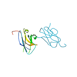
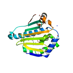
5AJK
DownloadVisualize
BU of 5ajk by Molmil
Crystal structure of variola virus virulence factor F1L in complex with human Bak BH3 domain
分子名称: ACETATE ION, BCL-2 HOMOLOGOUS ANTAGONIST/KILLER, CHLORIDE ION, ...
著者Kvansakul, M, Colman, P.M.
登録日2015-02-25
公開日2015-03-25
最終更新日2024-01-10
実験手法X-RAY DIFFRACTION (2.55 Å)
主引用文献Variola Virus F1L is a Bcl-2-Like Protein that Unlike its Vaccinia Virus Counterpart Inhibits Apoptosis Independent of Bim.
Cell Death Dis., 6, 2015
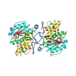
3BMB
DownloadVisualize
BU of 3bmb by Molmil
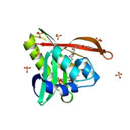
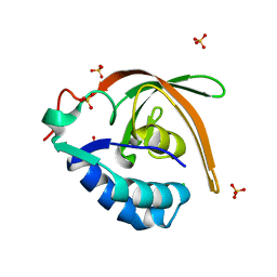
Crystal structure of a new RNA polymerase interacting protein
分子名称: CHLORIDE ION, Regulator of nucleoside diphosphate kinase, SULFATE ION
著者Darst, S.A, Lamour, V.
登録日2007-12-12
公開日2008-10-28
最終更新日2024-02-21
実験手法X-RAY DIFFRACTION (1.91 Å)
主引用文献Crystal structure of Escherichia coli Rnk, a new RNA polymerase-interacting protein.
J.Mol.Biol., 383, 2008
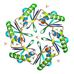
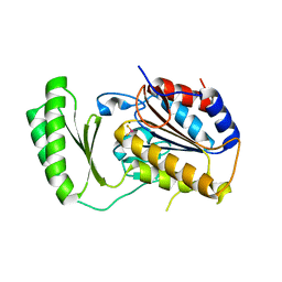
3BN4
DownloadVisualize
BU of 3bn4 by Molmil
Carboxysome Subunit, CcmK1
分子名称: Carbon dioxide-concentrating mechanism protein ccmK homolog 1, SULFATE ION
著者Tanaka, S, Sawaya, M.R, Yeates, T.O.
登録日2007-12-13
公開日2008-03-04
最終更新日2024-02-21
実験手法X-RAY DIFFRACTION (2 Å)
主引用文献Atomic-level models of the bacterial carboxysome shell.
Science, 319, 2008
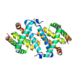
3VVV
DownloadVisualize
BU of 3vvv by Molmil
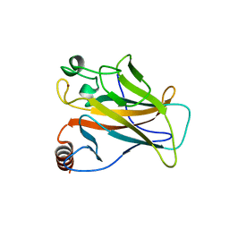
Skich domain of NDP52
分子名称: Calcium-binding and coiled-coil domain-containing protein 2
著者Akutsu, M, Muhlinen, N.V, Randow, F, Komander, D.
登録日2012-07-28
公開日2013-02-27
最終更新日2024-03-20
実験手法X-RAY DIFFRACTION (1.35 Å)
主引用文献LC3C, bound selectively by a noncanonical LIR motif in NDP52, is required for antibacterial autophagy
Mol.Cell, 48, 2012
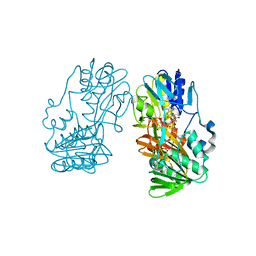
5THL
DownloadVisualize
BU of 5thl by Molmil
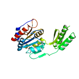
Crystal structure of the human tyrosyl-tRNA synthetase mutant G41R
分子名称: Tyrosine--tRNA ligase, cytoplasmic
著者Blocquel, D, Sajish, M, Yang, X.L.
登録日2016-09-29
公開日2017-06-07
最終更新日2023-10-04
実験手法X-RAY DIFFRACTION (1.6 Å)
主引用文献Alternative stable conformation capable of protein misinteraction links tRNA synthetase to peripheral neuropathy.
Nucleic Acids Res., 45, 2017
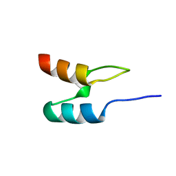
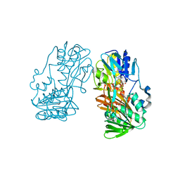
1W4H
DownloadVisualize
BU of 1w4h by Molmil
Peripheral-subunit from mesophilic, thermophilic and hyperthermophilic bacteria fold by ultrafast, apparently two-state transitions
分子名称: DIHYDROLIPOYLLYSINE-RESIDUE ACETYLTRANSFERASE
著者Ferguson, N, Sharpe, T.D, Schartau, P.J, Allen, M.D, Johnson, C.M, Fersht, A.R.
登録日2004-07-23
公開日2005-07-20
最終更新日2024-05-15
実験手法SOLUTION NMR
主引用文献Ultra-Fast Barrier-Limited Folding in the Peripheral Subunit-Binding Domain Family.
J.Mol.Biol., 353, 2005
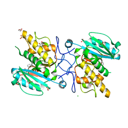
5DXN
DownloadVisualize
BU of 5dxn by Molmil
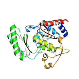
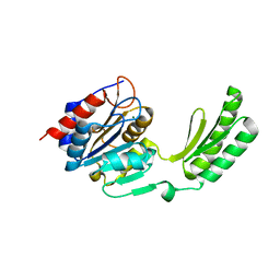
Structure of Aspergillus fumigatus trehalose-6-phosphate phosphatase crystal form 2
分子名称: MAGNESIUM ION, trehalose-6-phosphate phosphatase
著者Miao, Y, Brennan, R.G.
登録日2015-09-23
公開日2016-06-15
最終更新日2023-09-27
実験手法X-RAY DIFFRACTION (1.65 Å)
主引用文献Structures of trehalose-6-phosphate phosphatase from pathogenic fungi reveal the mechanisms of substrate recognition and catalysis.
Proc.Natl.Acad.Sci.USA, 113, 2016
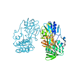
5THH
DownloadVisualize
BU of 5thh by Molmil
Crystal structure of a human tyrosyl-tRNA synthetase mutant
分子名称: TYROSINE, Tyrosine--tRNA ligase, cytoplasmic
著者Blocquel, D, Yang, X.L.
登録日2016-09-29
公開日2017-05-17
最終更新日2023-10-04
実験手法X-RAY DIFFRACTION (1.959 Å)
主引用文献Alternative stable conformation capable of protein misinteraction links tRNA synthetase to peripheral neuropathy.
Nucleic Acids Res., 45, 2017
4OKV
DownloadVisualize
BU of 4okv by Molmil
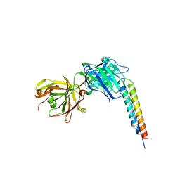
Crystal structure of anopheline anti-platelet protein with Fab antibody
分子名称: Anti-platelet aggregation protein, heavy chain of 8H7 mAb, light chain of 8H7 mAb
著者Park, S.Y, Sugiyama, K.
登録日2014-01-23
公開日2014-04-30
実験手法X-RAY DIFFRACTION (1.802 Å)
主引用文献The crystal structure of anopheles anti-platelet protein, a powerful anti-coagulant, in complex with an antibody
To be Published
2X0U
DownloadVisualize
BU of 2x0u by Molmil
STRUCTURE OF THE P53 CORE DOMAIN MUTANT Y220C BOUND TO A 2-amino substituted benzothiazole scaffold
分子名称: 6,7-DIHYDRO[1,4]DIOXINO[2,3-F][1,3]BENZOTHIAZOL-2-AMINE, CELLULAR TUMOR ANTIGEN P53, ZINC ION
著者Joerger, A.C, Kaar, J.L, Basse, N, Fersht, A.R.
登録日2009-12-17
公開日2010-01-26
最終更新日2023-12-20
実験手法X-RAY DIFFRACTION (1.6 Å)
主引用文献Toward the Rational Design of P53-Stabilizing Drugs: Probing the Surface of the Oncogenic Y220C Mutant.
Chem.Biol., 17, 2010
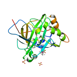
5BRV
DownloadVisualize
BU of 5brv by Molmil
Catalytic Improvement of an Artificial Metalloenzyme by Computational Design
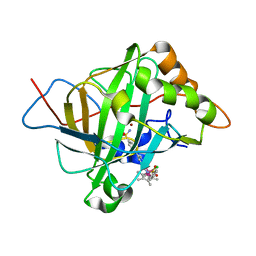
分子名称: Carbonic anhydrase 2, ZINC ION, pentamethylcyclopentadienyl iridium [N-benzensulfonamide-(2-pyridylmethyl-4-benzensulfonamide)amin] chloride
著者Heinisch, T, Pellizzoni, M, Duerrenberger, M, Tinberg, C.E, Koehler, V, Klehr, J, Haeussinger, D, Baker, D, Ward, T.R.
登録日2015-06-01
公開日2015-06-24
最終更新日2024-01-10
実験手法X-RAY DIFFRACTION (1.6 Å)
主引用文献Improving the Catalytic Performance of an Artificial Metalloenzyme by Computational Design.
J.Am.Chem.Soc., 137, 2015
5VD6
DownloadVisualize
BU of 5vd6 by Molmil
Crystal structure of a GNAT superfamily acetyltransferase PA4794 in complex with bisubstrate analog 6
分子名称: (3R,5S,9R,23S)-1-[(2R,3S,4R,5R)-5-(6-amino-9H-purin-9-yl)-4-hydroxy-3-(phosphonooxy)tetrahydrofuran-2-yl]-3,5,9-trihydroxy-8,8-dimethyl-10,14-dioxo-23-({[(phenylacetyl)amino]acetyl}amino)-2,4,6-trioxa-18-thia-11,15-diaza-3,5-diphosphatetracosan-24-oic acid 3,5-dioxide (non-preferred name), SULFATE ION, acetyltransferase PA4794
著者Majorek, K.A, Joachimiak, A, Minor, W, Midwest Center for Structural Genomics (MCSG)
登録日2017-04-01
公開日2017-07-26
最終更新日2023-10-04
実験手法X-RAY DIFFRACTION (1.2 Å)
主引用文献Generating enzyme and radical-mediated bisubstrates as tools for investigating Gcn5-related N-acetyltransferases.
FEBS Lett., 591, 2017
5DX9
DownloadVisualize
BU of 5dx9 by Molmil
Structure of trehalose-6-phosphate phosphatase from Cryptococcus neoformans
分子名称: 6-O-phosphono-alpha-D-glucopyranose-(1-1)-alpha-D-glucopyranose, BETA-MERCAPTOETHANOL, MAGNESIUM ION, ...
著者Miao, Y, Brennan, R.G.
登録日2015-09-23
公開日2016-06-15
最終更新日2020-07-29
実験手法X-RAY DIFFRACTION (2.15 Å)
主引用文献Structures of trehalose-6-phosphate phosphatase from pathogenic fungi reveal the mechanisms of substrate recognition and catalysis.
Proc.Natl.Acad.Sci.USA, 113, 2016
5DXO
DownloadVisualize
BU of 5dxo by Molmil
Structure of Aspergillus fumigatus trehalose-6-phosphate phosphatase crystal form 3
分子名称: MAGNESIUM ION, trehalose-6-phosphate phosphatase
著者Miao, Y, Brennan, R.G.
登録日2015-09-23
公開日2016-06-15
最終更新日2023-09-27
実験手法X-RAY DIFFRACTION (1.9 Å)
主引用文献Structures of trehalose-6-phosphate phosphatase from pathogenic fungi reveal the mechanisms of substrate recognition and catalysis.
Proc.Natl.Acad.Sci.USA, 113, 2016
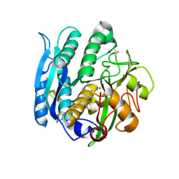
1GA1
DownloadVisualize
BU of 1ga1 by Molmil
CRYSTAL STRUCTURE ANALYSIS OF PSCP (PSEUDOMONAS SERINE-CARBOXYL PROTEINASE) COMPLEXED WITH A FRAGMENT OF IODOTYROSTATIN (THIS ENZYME RENAMED "SEDOLISIN" IN 2003)
分子名称: CALCIUM ION, CHLORIDE ION, FRAGMENT OF IODOTYROSTATIN, ...
著者Dauter, Z, Li, M, Wlodawer, A.
登録日2000-11-29
公開日2000-12-13
最終更新日2011-07-13
実験手法X-RAY DIFFRACTION (1.4 Å)
主引用文献Practical experience with the use of halides for phasing macromolecular structures: a powerful tool for structural genomics.
Acta Crystallogr.,Sect.D, 57, 2001
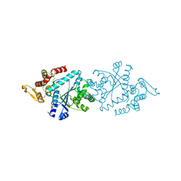
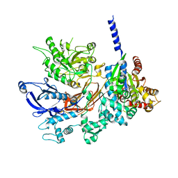
4GNK
DownloadVisualize
BU of 4gnk by Molmil
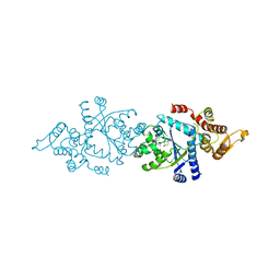
Crystal structure of Galphaq in complex with full-length human PLCbeta3
分子名称: 1-phosphatidylinositol 4,5-bisphosphate phosphodiesterase beta-3, CALCIUM ION, GUANOSINE-5'-DIPHOSPHATE, ...
著者Lyon, A.M, Tesmer, J.J.G.
登録日2012-08-17
公開日2013-02-06
最終更新日2023-09-13
実験手法X-RAY DIFFRACTION (4 Å)
主引用文献Full-length G alpha (q)-phospholipase C-beta 3 structure reveals interfaces of the C-terminal coiled-coil domain.
Nat.Struct.Mol.Biol., 20, 2013
3U0X
DownloadVisualize
BU of 3u0x by Molmil
Crystal structure of the B-specific-1,3-galactosyltransferase (GTB) in complex with compound 382
分子名称: 1-(3-phenyl-1,2,4-thiadiazol-5-yl)piperazine, CHLORIDE ION, GLYCEROL, ...
著者Palcic, M.M, Jorgensen, R.
登録日2011-09-29
公開日2012-10-24
最終更新日2023-09-13
実験手法X-RAY DIFFRACTION (1.85 Å)
主引用文献A novel compound from a molecular fragment library screen inhibits glycosyltransferases by displacing the metal ion and interfering with substrate binding
To be Published
5BRU
DownloadVisualize
BU of 5bru by Molmil
Catalytic Improvement of an Artificial Metalloenzyme by Computational Design
分子名称: Carbonic anhydrase 2, SULFATE ION, ZINC ION, ...
著者Heinisch, T, Pellizzoni, M, Duerrenberger, M, Tinberg, C.E, Koehler, V, Klehr, J, Haeussinger, D, Baker, D, Ward, T.R.
登録日2015-06-01
公開日2015-06-24
最終更新日2024-01-10
実験手法X-RAY DIFFRACTION (1.6 Å)
主引用文献Improving the Catalytic Performance of an Artificial Metalloenzyme by Computational Design.
J.Am.Chem.Soc., 137, 2015
2GR3
DownloadVisualize
BU of 2gr3 by Molmil
Crystal structure of Ferredoxin reductase, BphA4 (oxidized form)
分子名称: FLAVIN-ADENINE DINUCLEOTIDE, FORMIC ACID, GLYCEROL, ...
著者Senda, T, Senda, M.
登録日2006-04-22
公開日2007-05-22
最終更新日2024-05-29
実験手法X-RAY DIFFRACTION (1.5 Å)
主引用文献A ferredoxin reductase BphA4 uses a butterfly motion of FAD to regulate affinity for ferredoxin
To be Published
2GR1
DownloadVisualize
BU of 2gr1 by Molmil
Crystal structure of Ferredoxin reductase, BphA4 (hydroquinone)
分子名称: FLAVIN-ADENINE DINUCLEOTIDE, ferredoxin reductase
著者Senda, T, Senda, M.
登録日2006-04-22
公開日2007-05-22
最終更新日2024-03-13
実験手法X-RAY DIFFRACTION (1.8 Å)
主引用文献A ferredoxin reductase BphA4 uses a butterfly motion of FAD to regulate affinity for ferredoxin
To be Published
2GR2
DownloadVisualize
BU of 2gr2 by Molmil
Crystal structure of Ferredoxin reductase, BphA4 (oxidized form)
分子名称: ADENOSINE-5-DIPHOSPHORIBOSE, FLAVIN-ADENINE DINUCLEOTIDE, ferredoxin reductase
著者Senda, T, Senda, M.
登録日2006-04-22
公開日2007-05-22
最終更新日2024-03-13
実験手法X-RAY DIFFRACTION (1.85 Å)
主引用文献A ferredoxin reductase BphA4 uses a butterfly motion of FAD to regulate affinity for ferredoxin
To be Published
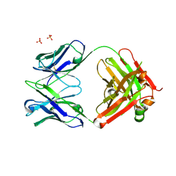
1XF4
DownloadVisualize
BU of 1xf4 by Molmil
Structure of ligand-free Fab DNA-1 in space group P321 solved from crystals with perfect hemihedral twinning
分子名称: Fab heavy chain, Fab light chain, SULFATE ION
著者Schuermann, J.P, Prewitt, S.P, Deutscher, S.L, Tanner, J.J.
登録日2004-09-13
公開日2005-04-12
最終更新日2023-08-23
実験手法X-RAY DIFFRACTION (2.5 Å)
主引用文献Evidence for Structural Plasticity of Heavy Chain Complementarity-determining Region 3 in Antibody-ssDNA Recognition
J.Mol.Biol., 347, 2005
5VYY
DownloadVisualize
BU of 5vyy by Molmil
Structure of human Hsp90-alpha bound to resorcinylic inhibitor BnIm
分子名称: 1,2-ETHANEDIOL, FORMIC ACID, Heat shock protein HSP 90-alpha, ...
著者Que, N.S, Gewirth, D.T.
登録日2017-05-26
公開日2018-04-18
最終更新日2024-03-13
実験手法X-RAY DIFFRACTION (1.792 Å)
主引用文献Structure Based Design of a Grp94-Selective Inhibitor: Exploiting a Key Residue in Grp94 To Optimize Paralog-Selective Binding.
J. Med. Chem., 61, 2018
3U0Y
DownloadVisualize
BU of 3u0y by Molmil
Crystal structure of the Fucosylgalactoside alpha N-acetylgalactosaminyltransferase (GTA, cisAB mutant L266G, G268A) in complex with compound 382 and UDP
分子名称: 1-(3-phenyl-1,2,4-thiadiazol-5-yl)piperazine, GLYCEROL, Histo-blood group ABO system transferase, ...
著者Palcic, M.M, Jorgensen, R.
登録日2011-09-29
公開日2012-10-24
最終更新日2023-09-13
実験手法X-RAY DIFFRACTION (1.6 Å)
主引用文献A novel compound from a molecular fragment library screen inhibits glycosyltransferases by displacing the metal ion and interfering with substrate binding
To be Published
5DXI
DownloadVisualize
BU of 5dxi by Molmil
Structure of C. albicans Trehalose-6-phosphate phosphatase C-terminal domain
分子名称: BERYLLIUM TRIFLUORIDE ION, MAGNESIUM ION, alpha-D-glucopyranose-(1-1)-alpha-D-glucopyranose, ...
著者Miao, Y, Brennan, R.G.
登録日2015-09-23
公開日2016-06-15
最終更新日2024-03-06
実験手法X-RAY DIFFRACTION (2 Å)
主引用文献Structures of trehalose-6-phosphate phosphatase from pathogenic fungi reveal the mechanisms of substrate recognition and catalysis.
Proc.Natl.Acad.Sci.USA, 113, 2016

224931

件を2024-09-11に公開中

PDB statisticsPDBj update infoContact PDBjnumon