6GWP
 
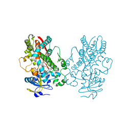
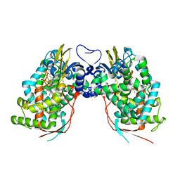
 | | Crystal Structure of Stabilized Active Plasminogen Activator Inhibitor-1 (PAI-1-stab) in Complex with Two Inhibitory Nanobodies (VHH-2g-42, VHH-2w-64) | | 分子名称: | Plasminogen Activator Inhibitor-1, VHH-2g-42, VHH-2w-64 | | 著者 | Sillen, M, Weeks, S.D, Strelkov, S.V, Declerck, P.J. | | 登録日 | 2018-06-25 | | 公開日 | 2020-01-01 | | 最終更新日 | 2024-01-17 | | 実験手法 | X-RAY DIFFRACTION (2.28 Å) | | 主引用文献 | Molecular mechanism of two nanobodies that inhibit PAI-1 activity reveals a modulation at distinct stages of the PAI-1/plasminogen activator interaction.
J.Thromb.Haemost., 18, 2020
|
|
8ACF
 | |
8ACI
 | |
6UNA
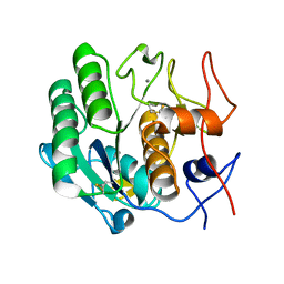
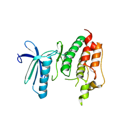
 | | Crystal structure of inactive p38gamma | | 分子名称: | GLYCEROL, Mitogen-activated protein kinase 12, SULFATE ION | | 著者 | Aoto, P.C, Stanfield, R.L, Wilson, I.A, Dyson, H.J, Wright, P.E. | | 登録日 | 2019-10-11 | | 公開日 | 2019-12-18 | | 最終更新日 | 2023-10-11 | | 実験手法 | X-RAY DIFFRACTION (2.554 Å) | | 主引用文献 | A Dynamic Switch in Inactive p38 gamma Leads to an Excited State on the Pathway to an Active Kinase.
Biochemistry, 58, 2019
|
|
6UNM
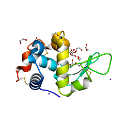
 | | CYP3A4 bound to an inhibitor | | 分子名称: | Cytochrome P450 3A4, PROTOPORPHYRIN IX CONTAINING FE, tert-butyl [(2S)-1-(naphthalen-1-yl)-3-{[(2S)-3-(naphthalen-1-yl)-1-oxo-1-{(E)-[2-(pyridin-3-yl)ethylidene]amino}propan-2-yl]sulfanyl}propan-2-yl]carbamate | | 著者 | Sevrioukova, I. | | 登録日 | 2019-10-12 | | 公開日 | 2020-02-05 | | 最終更新日 | 2023-10-11 | | 実験手法 | X-RAY DIFFRACTION (2.83 Å) | | 主引用文献 | An increase in side-group hydrophobicity largely improves the potency of ritonavir-like inhibitors of CYP3A4.
Bioorg.Med.Chem., 28, 2020
|
|
3DVR
 | |
3DWE
 | |
5DL9
 | | Structure of Tetragonal Lysozyme in complex with Iodine solved by UWO Students | | 分子名称: | 1,2-ETHANEDIOL, ACETATE ION, IODIDE ION, ... | | 著者 | Bednarski, R, Cirricione, N, Greco, A, Hodgson, R, Kent, S, McGowan, J, Notherm, B, Patt, M, Vue, L, Bianchetti, C.M. | | 登録日 | 2015-09-04 | | 公開日 | 2015-09-16 | | 実験手法 | X-RAY DIFFRACTION (1.38 Å) | | 主引用文献 | Structure of Tetragonal Lysozyme in complex with Iodine solved by UWO Students
To Be Published
|
|
3DI8
 | | Crystal structure of bovine pancreatic ribonuclease A variant (V57A) | | 分子名称: | CHLORIDE ION, Ribonuclease pancreatic, SULFATE ION | | 著者 | Kurpiewska, K, Font, J, Ribo, M, Vilanova, M, Lewinski, K. | | 登録日 | 2008-06-20 | | 公開日 | 2008-07-15 | | 最終更新日 | 2023-11-01 | | 実験手法 | X-RAY DIFFRACTION (1.6 Å) | | 主引用文献 | X-ray crystallographic studies of RNase A variants engineered at the most destabilizing positions of the main hydrophobic core: further insight into protein stability
Proteins, 77, 2009
|
|
6SUV
 | |
6GZM
 | | Crystal Structure of Human CKIdelta with A86 | | 分子名称: | CITRIC ACID, Casein kinase I isoform delta, GLYCEROL, ... | | 著者 | Ben-neriah, Y, Venkatachalam, A, Minzel, W, Fink, A, Snir-Alkalay, I, Vacca, J. | | 登録日 | 2018-07-04 | | 公開日 | 2018-08-29 | | 最終更新日 | 2024-01-17 | | 実験手法 | X-RAY DIFFRACTION (1.59 Å) | | 主引用文献 | Small Molecules Co-targeting CKI alpha and the Transcriptional Kinases CDK7/9 Control AML in Preclinical Models.
Cell, 175, 2018
|
|
3DIH
 | |
3DZ8
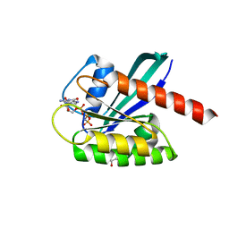
 | | Crystal structure of human Rab3B GTPase bound with GDP | | 分子名称: | GUANOSINE-5'-DIPHOSPHATE, Ras-related protein Rab-3B, UNKNOWN ATOM OR ION | | 著者 | Shen, Y, Tong, Y, Sukumar, D, Tempel, W, Loppnau, P, Arrowsmith, C.H, Edwards, A.M, Bountra, C, Wilkstrom, M, Bochkarev, A, Park, H, Structural Genomics Consortium (SGC) | | 登録日 | 2008-07-29 | | 公開日 | 2008-08-12 | | 最終更新日 | 2023-08-30 | | 実験手法 | X-RAY DIFFRACTION (1.9 Å) | | 主引用文献 | Crystal structure of human Rab3B GTPase bound with GDP
To be Published
|
|
6GHV
 | | Structure of a DC-SIGN CRD in complex with high affinity glycomimetic. | | 分子名称: | CALCIUM ION, CD209 antigen, CHLORIDE ION, ... | | 著者 | Thepaut, M, Achilli, S, Medve, L, Bernardi, A, Fieschi, F. | | 登録日 | 2018-05-09 | | 公開日 | 2019-09-11 | | 最終更新日 | 2024-01-17 | | 実験手法 | X-RAY DIFFRACTION (2.1 Å) | | 主引用文献 | Enhancing Potency and Selectivity of a DC-SIGN Glycomimetic Ligand by Fragment-Based Design: Structural Basis.
Chemistry, 25, 2019
|
|
6GII
 | |
6GJ7
 | | CRYSTAL STRUCTURE OF KRAS G12D (GPPCP) IN COMPLEX WITH 22 | | 分子名称: | (3~{S})-5-oxidanyl-3-[2-[[[1-(phenylmethyl)indol-6-yl]methylamino]methyl]-1~{H}-indol-3-yl]-2,3-dihydroisoindol-1-one, GTPase KRas, MAGNESIUM ION, ... | | 著者 | Kessler, D, Mcconnell, D.M, Mantoulidis, A. | | 登録日 | 2018-05-16 | | 公開日 | 2019-07-31 | | 最終更新日 | 2024-01-17 | | 実験手法 | X-RAY DIFFRACTION (1.67 Å) | | 主引用文献 | Drugging an undruggable pocket on KRAS.
Proc.Natl.Acad.Sci.USA, 116, 2019
|
|
6T0F
 | | Crystal structure of CYP124 in complex with cholest-4-en-3-one | | 分子名称: | (8ALPHA,9BETA)-CHOLEST-4-EN-3-ONE, 3[N-MORPHOLINO]PROPANE SULFONIC ACID, GLYCEROL, ... | | 著者 | Bukhdruker, S, Marin, E, Varaksa, T, Gilep, A, Strushkevich, N, Borshchevskiy, V. | | 登録日 | 2019-10-03 | | 公開日 | 2020-10-14 | | 最終更新日 | 2024-01-24 | | 実験手法 | X-RAY DIFFRACTION (1.65 Å) | | 主引用文献 | Metabolic Fate of Human Immunoactive Sterols in Mycobacterium tuberculosis.
J.Mol.Biol., 433, 2021
|
|
3D97
 | |
6SKC
 | |
5DQ1
 | | Endothiapepsin in complex with fragment 34 | | 分子名称: | 1,2-ETHANEDIOL, 3-[(cyclopropylamino)methyl]-8-ethylquinolin-2(1H)-one, Endothiapepsin, ... | | 著者 | Radeva, N, Uehlein, M, Weiss, M, Heine, A, Klebe, G. | | 登録日 | 2015-09-14 | | 公開日 | 2016-09-28 | | 最終更新日 | 2024-01-10 | | 実験手法 | X-RAY DIFFRACTION (1.489 Å) | | 主引用文献 | Crystallographic Fragment Screening of an Entire Library
To Be Published
|
|
5DQC
 | | Co-crystal of BACE1 with compound 0211 | | 分子名称: | Beta-secretase 1, N-[(2S,3R)-3-hydroxy-4-({(2S,3S)-3-hydroxy-1-[(2-methylpropyl)amino]-1-oxobutan-2-yl}amino)-1-phenylbutan-2-yl]-5-[methyl(methylsulfonyl)amino]-N'-[(1R)-1-phenylethyl]benzene-1,3-dicarboxamide | | 著者 | Ghosh, A.K, Bhavanam, S.R, Yen, T.-C, Cardenas, E.L, Rao, K.V, Downs, D, Huang, X, Tang, J, Mescar, A.D. | | 登録日 | 2015-09-14 | | 公開日 | 2016-02-17 | | 最終更新日 | 2016-07-13 | | 実験手法 | X-RAY DIFFRACTION (2.4651 Å) | | 主引用文献 | Design of Potent and Highly Selective Inhibitors for Human beta-Secretase 2 (Memapsin 1), a Target for Type 2 Diabetes.
Chem Sci, 7, 2016
|
|
3DPQ
 | |
3DAX
 | | Crystal structure of human CYP7A1 | | 分子名称: | Cytochrome P450 7A1, PROTOPORPHYRIN IX CONTAINING FE, UNKNOWN ATOM OR ION | | 著者 | Strushkevich, N.V, Tempel, W, Dombrovski, L, Dong, A, Loppnau, P, Arrowsmith, C.H, Edwards, A.M, Bountra, C, Wilkstrom, M, Bochkarev, A, Park, H, Structural Genomics Consortium (SGC) | | 登録日 | 2008-05-30 | | 公開日 | 2008-08-05 | | 最終更新日 | 2023-08-30 | | 実験手法 | X-RAY DIFFRACTION (2.15 Å) | | 主引用文献 | Crystal structure of human CYP7A1
To be Published
|
|
3DBQ
 | | Crystal structure of TTK kinase domain | | 分子名称: | Dual specificity protein kinase TTK | | 著者 | Wang, W, Yang, Y.T, Gao, Y.F, Zhu, S.C, Wang, F, Old, W, Xu, Q.B, Resing, K, Ahn, N, Lei, M, Liu, X.D. | | 登録日 | 2008-06-02 | | 公開日 | 2009-02-10 | | 最終更新日 | 2011-07-13 | | 実験手法 | X-RAY DIFFRACTION (2.7 Å) | | 主引用文献 | Structural and Mechanistic Insights into Mps1 Kinase Activation
J.CELL.MOL.MED., 13, 2008
|
|
6GOD
 | | KRAS full length wild-type GPPNHP | | 分子名称: | GLYCEROL, GTPase KRas, MAGNESIUM ION, ... | | 著者 | Cruz-Migoni, A, Quevedo, C.E, Carr, S.B, Ehebauer, M.T, Phillips, S.V.E, Rabbitts, T.H. | | 登録日 | 2018-06-01 | | 公開日 | 2019-02-06 | | 最終更新日 | 2024-01-17 | | 実験手法 | X-RAY DIFFRACTION (1.71 Å) | | 主引用文献 | Structure-based development of new RAS-effector inhibitors from a combination of active and inactive RAS-binding compounds.
Proc. Natl. Acad. Sci. U.S.A., 116, 2019
|
|


