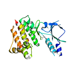
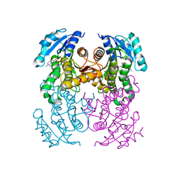
8OWG
 
 | | Crystal structure of D1228V c-MET bound by compound 2 | | 分子名称: | 5-[3,5-bis(fluoranyl)phenyl]-1-[(1S)-1-phenylethyl]pyrimidine-2,4-dione, Hepatocyte growth factor receptor | | 著者 | Collie, G.W. | | 登録日 | 2023-04-27 | | 公開日 | 2023-07-05 | | 最終更新日 | 2024-06-19 | | 実験手法 | X-RAY DIFFRACTION (2.631 Å) | | 主引用文献 | Discovery and Optimization of the First ATP Competitive Type-III c-MET Inhibitor.
J.Med.Chem., 66, 2023
|
|
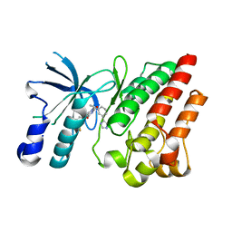
8OVZ
 | | Crystal structure of D1228V c-MET bound by compound 16 | | 分子名称: | 1-[(1S)-1-[3-(1H-imidazol-4-yl)phenyl]ethyl]-5-(1H-indazol-7-yl)pyrimidine-2,4-dione, Hepatocyte growth factor receptor, IODIDE ION | | 著者 | Collie, G.W. | | 登録日 | 2023-04-26 | | 公開日 | 2023-07-05 | | 最終更新日 | 2023-09-20 | | 実験手法 | X-RAY DIFFRACTION (2.206 Å) | | 主引用文献 | Discovery and Optimization of the First ATP Competitive Type-III c-MET Inhibitor.
J.Med.Chem., 66, 2023
|
|
8OUU
 | | Crystal structure of D1228V c-MET bound by compound 29 | | 分子名称: | 1,2-ETHANEDIOL, 5-(3-ethynyl-5-fluoranyl-1H-indazol-7-yl)-1-[(1S)-1-phenylethyl]pyrimidine-2,4-dione, FORMIC ACID, ... | | 著者 | Collie, G.W. | | 登録日 | 2023-04-24 | | 公開日 | 2023-07-05 | | 最終更新日 | 2024-06-19 | | 実験手法 | X-RAY DIFFRACTION (1.77 Å) | | 主引用文献 | Discovery and Optimization of the First ATP Competitive Type-III c-MET Inhibitor.
J.Med.Chem., 66, 2023
|
|
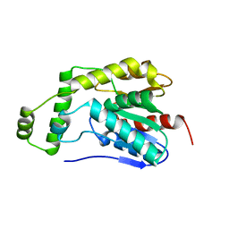
8OUV
 | | Crystal structure of D1228V c-MET bound by compound 15 | | 分子名称: | 5-(1H-indazol-7-yl)-1-[(1S)-1-phenylethyl]pyrimidine-2,4-dione, CHLORIDE ION, Hepatocyte growth factor receptor | | 著者 | Collie, G.W. | | 登録日 | 2023-04-24 | | 公開日 | 2023-07-05 | | 最終更新日 | 2024-06-19 | | 実験手法 | X-RAY DIFFRACTION (1.783 Å) | | 主引用文献 | Discovery and Optimization of the First ATP Competitive Type-III c-MET Inhibitor.
J.Med.Chem., 66, 2023
|
|
8OW3
 | | Crystal structure of wild-type c-MET bound by compound 2 | | 分子名称: | 5-[3,5-bis(fluoranyl)phenyl]-1-[(1S)-1-phenylethyl]pyrimidine-2,4-dione, Hepatocyte growth factor receptor | | 著者 | Collie, G.W. | | 登録日 | 2023-04-26 | | 公開日 | 2023-07-05 | | 最終更新日 | 2024-06-19 | | 実験手法 | X-RAY DIFFRACTION (2.27 Å) | | 主引用文献 | Discovery and Optimization of the First ATP Competitive Type-III c-MET Inhibitor.
J.Med.Chem., 66, 2023
|
|
8OV7
 | | Crystal structure of D1228V c-MET bound by compound 10 | | 分子名称: | 5-[3,5-bis(fluoranyl)phenyl]-1-[(1S)-1-[3-(1H-imidazol-5-yl)phenyl]ethyl]pyrimidine-2,4-dione, Hepatocyte growth factor receptor | | 著者 | Collie, G.W. | | 登録日 | 2023-04-25 | | 公開日 | 2023-07-05 | | 最終更新日 | 2024-06-19 | | 実験手法 | X-RAY DIFFRACTION (1.95 Å) | | 主引用文献 | Discovery and Optimization of the First ATP Competitive Type-III c-MET Inhibitor.
J.Med.Chem., 66, 2023
|
|
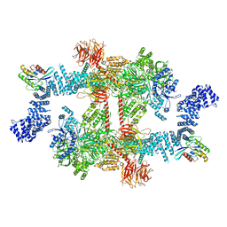
8H38
 | | Cryo-EM Structure of the KBTBD2-CRL3~N8-CSN(mutate) complex | | 分子名称: | COP9 signalosome complex subunit 1, COP9 signalosome complex subunit 2, COP9 signalosome complex subunit 3, ... | | 著者 | Hu, Y, Mao, Q, Chen, Z, Sun, L. | | 登録日 | 2022-10-08 | | 公開日 | 2023-10-11 | | 最終更新日 | 2024-03-20 | | 実験手法 | ELECTRON MICROSCOPY (4.25 Å) | | 主引用文献 | Dynamic molecular architecture and substrate recruitment of cullin3-RING E3 ligase CRL3 KBTBD2.
Nat.Struct.Mol.Biol., 31, 2024
|
|
8HEA
 | | Esterase2 (EaEst2) from Exiguobacterium antarcticum | | 分子名称: | Thermostable carboxylesterase Est30 | | 著者 | Hwang, J, Lee, J.H. | | 登録日 | 2022-11-08 | | 公開日 | 2023-09-20 | | 実験手法 | X-RAY DIFFRACTION (1.74 Å) | | 主引用文献 | Structural and Biochemical Insights into Bis(2-hydroxyethyl) Terephthalate Degrading Carboxylesterase Isolated from Psychrotrophic Bacterium Exiguobacterium antarcticum.
Int J Mol Sci, 24, 2023
|
|
8OTM
 | | structure of InhA from mycobacterium tuberculosis in complex with N-((1-(3-hydroxy-4-phenoxybenzyl)-1H-1,2,3-triazol-4-yl)methyl)-2-oxo-2H-chromene-3-carboxamide | | 分子名称: | 1,2-ETHANEDIOL, 2-oxidanylidene-~{N}-[[1-[(3-oxidanyl-4-phenoxy-phenyl)methyl]-1,2,3-triazol-4-yl]methyl]chromene-3-carboxamide, ACETATE ION, ... | | 著者 | Chebaiki, M, Maveyraud, L, Tamhaev, R, Lherbet, C, Mourey, L. | | 登録日 | 2023-04-21 | | 公開日 | 2023-08-16 | | 実験手法 | X-RAY DIFFRACTION (1.6 Å) | | 主引用文献 | Discovery of new diaryl ether inhibitors against Mycobacterium tuberculosis targeting the minor portal of InhA.
Eur.J.Med.Chem., 259, 2023
|
|
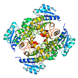
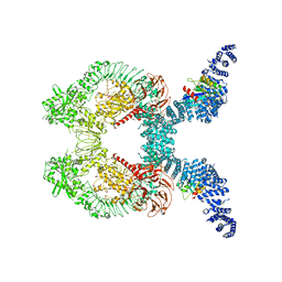
8P5R
 | | Crystal structure of full-length, homohexameric 2-oxoglutarate dehydrogenase KGD from Mycobacterium smegmatis in complex with GarA | | 分子名称: | CALCIUM ION, Glycogen accumulation regulator GarA, MAGNESIUM ION, ... | | 著者 | Wagner, T, Mechaly, A.M, Alzari, P.M, Bellinzoni, M. | | 登録日 | 2023-05-24 | | 公開日 | 2023-08-16 | | 最終更新日 | 2023-08-23 | | 実験手法 | X-RAY DIFFRACTION (4.562 Å) | | 主引用文献 | High resolution cryo-EM and crystallographic snapshots of the actinobacterial two-in-one 2-oxoglutarate dehydrogenase.
Nat Commun, 14, 2023
|
|
8OTN
 | | structure of InhA from mycobacterium tuberculosis in complex with inhibitor 7-((1-(3-Hydroxy-4-phenoxybenzyl)-1H-1,2,3-triazol-4-yl)methoxy)-4-methyl-2H-chromen-2-one | | 分子名称: | 4-methyl-7-[[1-[(3-oxidanyl-4-phenoxy-phenyl)methyl]-1,2,3-triazol-4-yl]methoxy]chromen-2-one, Enoyl-[acyl-carrier-protein] reductase [NADH], NICOTINAMIDE-ADENINE-DINUCLEOTIDE | | 著者 | Chebaiki, M, Maveyraud, L, Tamhaev, R, Lherbet, C, Mourey, L. | | 登録日 | 2023-04-21 | | 公開日 | 2023-08-16 | | 実験手法 | X-RAY DIFFRACTION (1.962 Å) | | 主引用文献 | Discovery of new diaryl ether inhibitors against Mycobacterium tuberculosis targeting the minor portal of InhA.
Eur.J.Med.Chem., 259, 2023
|
|
8HGU
 | |
8HM5
 | |
6QGC
 | | PETase from Ideonella sakaiensis without ligand | | 分子名称: | CHLORIDE ION, Poly(ethylene terephthalate) hydrolase, SULFATE ION | | 著者 | Palm, G.J, Reisky, L, Boettcher, D, Mueller, H, Michels, E.A.P, Walczak, C, Berndt, L, Weiss, M.S, Bornscheuer, U.T, Weber, G. | | 登録日 | 2019-01-10 | | 公開日 | 2019-04-03 | | 最終更新日 | 2024-01-24 | | 実験手法 | X-RAY DIFFRACTION (2 Å) | | 主引用文献 | Structure of the plastic-degrading Ideonella sakaiensis MHETase bound to a substrate.
Nat Commun, 10, 2019
|
|
8OTL
 | | structure of InhA from Mycobacterium tuberculosis in complex with 5-(((4-(2-hydroxyphenoxy)benzyl)(octyl)amino)methyl)-2-phenoxyphenol | | 分子名称: | 1,2-ETHANEDIOL, 5-[[octyl-[[4-(2-oxidanylphenoxy)phenyl]methyl]amino]methyl]-2-phenoxy-phenol, ACETATE ION, ... | | 著者 | Tamhaev, R, Maveyraud, L, Chebaiki, M, Lherbet, C, Mourey, L. | | 登録日 | 2023-04-21 | | 公開日 | 2024-01-24 | | 実験手法 | X-RAY DIFFRACTION (2.108 Å) | | 主引用文献 | Exploring the plasticity of the InhA substrate-binding site using new diaryl ether inhibitors.
Bioorg.Chem., 143, 2023
|
|
5CXT
 | |
8FO2
 | |
8FO8
 | |
8FO9
 | |
8P0F
 | | Crystal structure of the VCB complex with compound 1. | | 分子名称: | (3~{R},5~{R})-~{N}-[[4-(4-methyl-1,3-thiazol-5-yl)phenyl]methyl]-5-oxidanyl-2-oxidanylidene-1-pyridin-2-yl-piperidine-3-carboxamide, CHLORIDE ION, Elongin-B, ... | | 著者 | Bader, G, Boettcher, J, Wolkerstorfer, B. | | 登録日 | 2023-05-10 | | 公開日 | 2023-05-31 | | 最終更新日 | 2023-12-20 | | 実験手法 | X-RAY DIFFRACTION (1.98 Å) | | 主引用文献 | Drugit: Crowd-sourcing molecular design of non-peptidic VHL binders
Chemrxiv, 2023
|
|
6QYX
 | | p38(alpha) MAP kinase with the activation loop of ERK2 | | 分子名称: | 4-(2-HYDROXYETHYL)-1-PIPERAZINE ETHANESULFONIC ACID, Mitogen-activated protein kinase 14,Mitogen-activated protein kinase 1,Mitogen-activated protein kinase 14, octyl beta-D-glucopyranoside | | 著者 | Livnah, O, Eitan-Wexler, M, Vinograd, N. | | 登録日 | 2019-03-10 | | 公開日 | 2020-04-01 | | 最終更新日 | 2024-05-01 | | 実験手法 | X-RAY DIFFRACTION (1.66 Å) | | 主引用文献 | The bacterial metalloprotease NleD selectively cleaves mitogen-activated protein kinases that have high flexibility in their activation loop.
J.Biol.Chem., 295, 2020
|
|
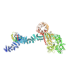
6QZP
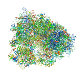 | | High-resolution cryo-EM structure of the human 80S ribosome | | 分子名称: | (3beta)-O~3~-[(2R)-2,6-dihydroxy-2-(2-methoxy-2-oxoethyl)-6-methylheptanoyl]cephalotaxine, 18S rRNA (1740-MER), 28S rRNA (3773-MER), ... | | 著者 | Natchiar, S.K, Myasnikov, A.G, Kratzat, H, Hazemann, I, Klaholz, B.P. | | 登録日 | 2019-03-12 | | 公開日 | 2019-04-24 | | 最終更新日 | 2024-04-24 | | 実験手法 | ELECTRON MICROSCOPY (2.9 Å) | | 主引用文献 | Visualization of chemical modifications in the human 80S ribosome structure.
Nature, 551, 2017
|
|
8POA
 | | Structure of Coxsackievirus A16 (G-10) 2A protease | | 分子名称: | GLYCEROL, Protease 2A, ZINC ION | | 著者 | Lithgo, R.M, Fairhead, M, Koekemoer, L, Aschenbrenner, J.C, Balcomb, B.H, Godoy, A.S, Marples, P.G, Ni, X, Tomlinson, C.W.E, Thompson, W, Wild, C, Fearon, D, Walsh, M.A, von Delft, F. | | 登録日 | 2023-07-04 | | 公開日 | 2023-08-02 | | 最終更新日 | 2024-07-03 | | 実験手法 | X-RAY DIFFRACTION (1.6 Å) | | 主引用文献 | Structure of EV A71 2A protease - to be published
To Be Published
|
|
7ABY
 | |
6ZTB
 | | Crystal Structure of human P-Cadherin EC1_EC2 | | 分子名称: | CALCIUM ION, Cadherin-3, SODIUM ION | | 著者 | Rondeau, J.M, Lehmann, S. | | 登録日 | 2020-07-17 | | 公開日 | 2021-05-05 | | 最終更新日 | 2024-01-31 | | 実験手法 | X-RAY DIFFRACTION (1.4 Å) | | 主引用文献 | PCA062, a P-cadherin Targeting Antibody-Drug Conjugate, Displays Potent Antitumor Activity Against P-cadherin-expressing Malignancies.
Mol.Cancer Ther., 20, 2021
|
|


