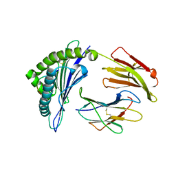
4FIA
 
 | | Crystal Structure of Human CYP46A1 P450 with bicalutamide Bound | | 分子名称: | Cholesterol 24-hydroxylase, GLYCEROL, PROTOPORPHYRIN IX CONTAINING FE, ... | | 著者 | Stout, C.D, Pikuleva, I.A, Mast, N. | | 登録日 | 2012-06-08 | | 公開日 | 2013-01-09 | | 最終更新日 | 2024-02-28 | | 実験手法 | X-RAY DIFFRACTION (2.1 Å) | | 主引用文献 | Binding of a cyano- and fluoro-containing drug bicalutamide to cytochrome P450 46A1: unusual features and spectral response.
J.Biol.Chem., 288, 2013
|
|
3LNK
 | | Structure of BACE bound to SCH743813 | | 分子名称: | Beta-secretase 1, L(+)-TARTARIC ACID, N'-{(1S,2S)-1-(3,5-difluorobenzyl)-2-hydroxy-2-[(2R)-4-(phenylcarbonyl)piperazin-2-yl]ethyl}-5-methyl-N,N-dipropylbenzene-1,3-dicarboxamide | | 著者 | Orth, P, Cumming, J. | | 登録日 | 2010-02-02 | | 公開日 | 2010-04-14 | | 最終更新日 | 2023-09-06 | | 実験手法 | X-RAY DIFFRACTION (1.8 Å) | | 主引用文献 | Piperazine sulfonamide BACE1 inhibitors: design, synthesis, and in vivo characterization.
Bioorg.Med.Chem.Lett., 20, 2010
|
|
3L5D
 | | Structure of BACE Bound to SCH723873 | | 分子名称: | 1-butyl-3-(4-{[(2Z,4R)-2-imino-4-methyl-4-(2-methylpropyl)-5-oxoimidazolidin-1-yl]methyl}benzyl)urea, Beta-secretase 1, D(-)-TARTARIC ACID | | 著者 | Strickland, C, Zhu, Z. | | 登録日 | 2009-12-21 | | 公開日 | 2010-02-16 | | 最終更新日 | 2017-11-01 | | 実験手法 | X-RAY DIFFRACTION (1.75 Å) | | 主引用文献 | Discovery of Cyclic Acylguanidines as Highly Potent and Selective beta-Site Amyloid Cleaving Enzyme (BACE) Inhibitors: Part I-Inhibitor Design and Validation
J.Med.Chem., 53, 2010
|
|
4COK
 | | Functional and Structural Characterization of Pyruvate decarboxylase from Gluconoacetobacter diazotrophicus | | 分子名称: | MAGNESIUM ION, PYRUVATE DECARBOXYLASE, SULFATE ION, ... | | 著者 | vanZyl, L.J, Schubert, W.-D, Tuffin, M, Cowan, D.A. | | 登録日 | 2014-01-29 | | 公開日 | 2014-10-01 | | 最終更新日 | 2023-12-20 | | 実験手法 | X-RAY DIFFRACTION (1.69 Å) | | 主引用文献 | Structure and Functional Characterization of Pyruvate Decarboxylase from Gluconacetobacter Diazotrophicus.
Bmc Struct.Biol., 14, 2014
|
|
3LC5
 | | Selective Benzothiophine Inhibitors of Factor IXa | | 分子名称: | 1-{4-[(R)-phenyl(3-phenyl-1,2,4-oxadiazol-5-yl)methoxy]-1-benzothiophen-2-yl}methanediamine, CALCIUM ION, Coagulation factor IX | | 著者 | Wang, S, Beck, R. | | 登録日 | 2010-01-09 | | 公開日 | 2010-02-23 | | 最終更新日 | 2011-07-13 | | 実験手法 | X-RAY DIFFRACTION (2.62 Å) | | 主引用文献 | Structure Based Drug Design: Development of Potent and Selective Factor IXa (FIXa) Inhibitors.
J.Med.Chem., 53, 2010
|
|
3L5C
 | | Structure of BACE Bound to SCH723871 | | 分子名称: | 1-(4-cyanophenyl)-3-(4-{[(2Z,4R)-2-imino-4-methyl-4-(2-methylpropyl)-5-oxoimidazolidin-1-yl]methyl}benzyl)urea, Beta-secretase 1, D(-)-TARTARIC ACID | | 著者 | Strickland, C, Zhu, Z. | | 登録日 | 2009-12-21 | | 公開日 | 2010-02-16 | | 最終更新日 | 2017-11-01 | | 実験手法 | X-RAY DIFFRACTION (1.8 Å) | | 主引用文献 | Discovery of Cyclic Acylguanidines as Highly Potent and Selective beta-Site Amyloid Cleaving Enzyme (BACE) Inhibitors: Part I-Inhibitor Design and Validation
J.Med.Chem., 53, 2010
|
|
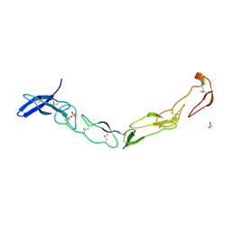
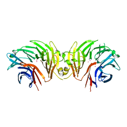
4HPY
 | | Crystal structure of RV144-elicited antibody CH59 in complex with V2 peptide | | 分子名称: | CH59 Fab heavy chain, CH59 Fab light chain, Envelope glycoprotein gp160, ... | | 著者 | McLellan, J.S, Gorman, J, Haynes, B.F, Kwong, P.D. | | 登録日 | 2012-10-24 | | 公開日 | 2013-02-06 | | 最終更新日 | 2023-09-20 | | 実験手法 | X-RAY DIFFRACTION (1.5 Å) | | 主引用文献 | Vaccine Induction of Antibodies against a Structurally Heterogeneous Site of Immune Pressure within HIV-1 Envelope Protein Variable Regions 1 and 2.
Immunity, 38, 2013
|
|
3QHQ
 | | Structure of CRISPR-associated protein Csn2 | | 分子名称: | 1,2-ETHANEDIOL, CALCIUM ION, Sag0897 family CRISPR-associated protein | | 著者 | Ellinger, P, Arslan, Z, Wurm, R, Tschapek, B, Pfeffer, K, Wagner, R, Schmitt, L, Pul, U, Smits, S.H. | | 登録日 | 2011-01-26 | | 公開日 | 2012-02-01 | | 最終更新日 | 2024-03-20 | | 実験手法 | X-RAY DIFFRACTION (2 Å) | | 主引用文献 | Structure of CRISPR-associated protein Csn2
To be Published
|
|
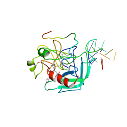
4NIY
 | | Crystal structure of trypsiligase (K60E/N143H/Y151H/D189K trypsin) complexed to YRH-ecotin (M84Y/M85R/A86H ecotin) | | 分子名称: | CALCIUM ION, Cationic trypsin, Ecotin, ... | | 著者 | Schoepfel, M, Parthier, C, Stubbs, M.T. | | 登録日 | 2013-11-08 | | 公開日 | 2014-02-19 | | 最終更新日 | 2014-03-19 | | 実験手法 | X-RAY DIFFRACTION (2.84 Å) | | 主引用文献 | N-terminal protein modification by substrate-activated reverse proteolysis.
Angew.Chem.Int.Ed.Engl., 53, 2014
|
|
3CEN
 | |
3L5F
 | | Structure of BACE Bound to SCH736201 | | 分子名称: | (2E,5R)-5-(2-cyclohexylethyl)-5-(cyclohexylmethyl)-2-imino-3-methylimidazolidin-4-one, Beta-secretase 1, D(-)-TARTARIC ACID | | 著者 | Strickland, C, Zhu, Z. | | 登録日 | 2009-12-21 | | 公開日 | 2010-02-16 | | 最終更新日 | 2017-11-01 | | 実験手法 | X-RAY DIFFRACTION (1.7 Å) | | 主引用文献 | Discovery of Cyclic Acylguanidines as Highly Potent and Selective beta-Site Amyloid Cleaving Enzyme (BACE) Inhibitors: Part I-Inhibitor Design and Validation
J.Med.Chem., 53, 2010
|
|
3Q5Y
 | | V beta/V beta homodimerization-based pre-TCR model suggested by TCR beta crystal structures | | 分子名称: | 4-(2-HYDROXYETHYL)-1-PIPERAZINE ETHANESULFONIC ACID, DI(HYDROXYETHYL)ETHER, GLYCEROL, ... | | 著者 | Chen, Q, Zhang, H, Wang, J.-H. | | 登録日 | 2010-12-30 | | 公開日 | 2011-03-09 | | 最終更新日 | 2014-10-15 | | 実験手法 | X-RAY DIFFRACTION (1.9 Å) | | 主引用文献 | A conserved hydrophobic patch on Vbeta domains revealed by TCRbeta chain crystal structures: implications for pre-TCR dimerization
Front Immunol, 2, 2011
|
|
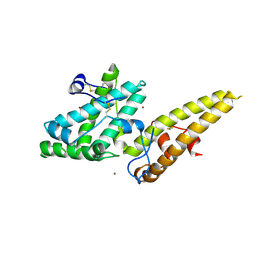
4NXI
 | | Rv2466c Mediates the Activation of TP053 To Kill Replicating and Non-replicating Mycobacterium tuberculosis | | 分子名称: | 1,2-ETHANEDIOL, MAGNESIUM ION, Uncharacterized protein | | 著者 | Albesa-Jove, D, Urresti, S, Comino, N, Guerin, M.E. | | 登録日 | 2013-12-09 | | 公開日 | 2014-10-22 | | 最終更新日 | 2023-11-08 | | 実験手法 | X-RAY DIFFRACTION (1.698 Å) | | 主引用文献 | Rv2466c mediates the activation of TP053 to kill replicating and non-replicating Mycobacterium tuberculosis.
Acs Chem.Biol., 9, 2014
|
|
4HX1
 | | Structure of HLA-A68 complexed with a tumor antigen derived peptide | | 分子名称: | 9-mer peptide from Tyrosinase-related protein-2, Beta-2-microglobulin, GLYCEROL, ... | | 著者 | Niu, L, Cheng, H, Zhang, S, Tan, S, Zhang, Y, Qi, J, Liu, J, Gao, G.F. | | 登録日 | 2012-11-09 | | 公開日 | 2013-10-02 | | 実験手法 | X-RAY DIFFRACTION (1.802 Å) | | 主引用文献 | Structural basis for the differential classification of HLA-A*6802 and HLA-A*6801 into the A2 and A3 supertypes
Mol.Immunol., 55, 2013
|
|
3QO4
 | | The Crystal Structure of Death Receptor 6 | | 分子名称: | ACETATE ION, SULFATE ION, Tumor necrosis factor receptor superfamily member 21 | | 著者 | Kuester, M, Kemmerzehl, S, Dahms, S.O, Roeser, D, Than, M.E. | | 登録日 | 2011-02-09 | | 公開日 | 2011-05-18 | | 最終更新日 | 2012-02-29 | | 実験手法 | X-RAY DIFFRACTION (2.2 Å) | | 主引用文献 | The crystal structure of death receptor 6 (DR6): a potential receptor of the amyloid precursor protein (APP).
J.Mol.Biol., 409, 2011
|
|
4KZW
 | | Structure of the carbohydrate-recognition domain of the C-type lectin mincle | | 分子名称: | C-TYPE LECTIN MINCLE, CALCIUM ION, CITRATE ANION, ... | | 著者 | Feinberg, H, Jegouzo, S.A.F, Rowntree, T.J.W, Guan, Y, Brash, M.A, Taylor, M.E, Weis, W.I, Drickamer, K. | | 登録日 | 2013-05-30 | | 公開日 | 2013-08-28 | | 最終更新日 | 2017-11-15 | | 実験手法 | X-RAY DIFFRACTION (1.85 Å) | | 主引用文献 | Mechanism for Recognition of an Unusual Mycobacterial Glycolipid by the Macrophage Receptor Mincle.
J.Biol.Chem., 288, 2013
|
|
3CS7
 | | FACTOR XA IN COMPLEX WITH THE INHIBITOR 1-(4-methoxyphenyl)-6-(4-(1-(pyrrolidin-1-ylmethyl)cyclopropyl)phenyl)-3-(trifluoromethyl)-5,6-dihydro-1H-pyrazolo[3,4-c]pyridin-7(4H)-one | | 分子名称: | 1-(4-methoxyphenyl)-6-(4-(1-(pyrrolidin-1-ylmethyl)cyclopropyl)phenyl)-3-(trifluoromethyl)-5,6-dihydro-1H-pyrazolo[3,4-c]pyridin-7(4H)-one, CALCIUM ION, Coagulation factor X | | 著者 | Alexander, R.S. | | 登録日 | 2008-04-09 | | 公開日 | 2008-07-08 | | 最終更新日 | 2011-07-13 | | 実験手法 | X-RAY DIFFRACTION (2.2 Å) | | 主引用文献 | Achieving structural diversity using the perpendicular conformation of alpha-substituted phenylcyclopropanes to mimic the bioactive conformation of ortho-substituted biphenyl P4 moieties: discovery of novel, highly potent inhibitors of Factor Xa.
Bioorg.Med.Chem.Lett., 18, 2008
|
|
1HUT
 | | THE STRUCTURE OF ALPHA-THROMBIN INHIBITED BY A 15-MER SINGLE-STRANDED DNA APTAMER | | 分子名称: | ALPHA-Thrombin heavy chain, ALPHA-Thrombin light chain, D-phenylalanyl-N-[(3S)-6-carbamimidamido-1-chloro-2-oxohexan-3-yl]-L-prolinamide, ... | | 著者 | Padmanabhan, K, Padmanabhan, K.P, Ferrara, J.D, Sadler, J.E, Tulinsky, A. | | 登録日 | 1993-05-27 | | 公開日 | 1994-06-22 | | 最終更新日 | 2013-02-27 | | 実験手法 | X-RAY DIFFRACTION (2.9 Å) | | 主引用文献 | The structure of alpha-thrombin inhibited by a 15-mer single-stranded DNA aptamer.
J.Biol.Chem., 268, 1993
|
|
6DLP
 | | Crystal structure of LRRK2 WD40 domain dimer | | 分子名称: | Leucine-rich repeat serine/threonine-protein kinase 2, PLATINUM (II) ION | | 著者 | Zhang, P, Ru, H, Wang, L, Wu, H. | | 登録日 | 2018-06-02 | | 公開日 | 2019-01-09 | | 最終更新日 | 2024-03-13 | | 実験手法 | X-RAY DIFFRACTION (4 Å) | | 主引用文献 | Crystal structure of the WD40 domain dimer of LRRK2.
Proc. Natl. Acad. Sci. U.S.A., 116, 2019
|
|
3VUV
 | | Crystal structure of the merozoite surface protein MSPDBL2 from P. falciparum bound to zinc | | 分子名称: | Erythrocyte membrane protein, putative, ZINC ION | | 著者 | Czabotar, P.E, Hodder, A.N, Clarke, O.B, Lin, C.S, Smith, B.J, Cowman, A.F. | | 登録日 | 2012-07-09 | | 公開日 | 2012-08-08 | | 最終更新日 | 2023-11-08 | | 実験手法 | X-RAY DIFFRACTION (2.114 Å) | | 主引用文献 | Insights into Duffy binding-like domains through the crystal structure and function of the merozoite surface protein MSPDBL2 from Plasmodium falciparum
J.Biol.Chem., 287, 2012
|
|
6R6T
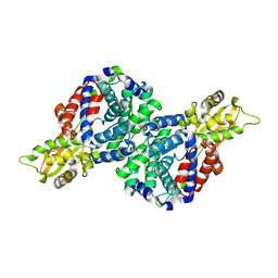 | | Crystal structure of mouse cis-aconitate decarboxylase | | 分子名称: | Cis-aconitate decarboxylase | | 著者 | Lukat, P, Chen, F, Saile, K, Buessow, K, Pessler, F, Blankenfeldt, W. | | 登録日 | 2019-03-28 | | 公開日 | 2019-09-25 | | 最終更新日 | 2024-01-24 | | 実験手法 | X-RAY DIFFRACTION (2.535 Å) | | 主引用文献 | Crystal structure ofcis-aconitate decarboxylase reveals the impact of naturally occurring human mutations on itaconate synthesis.
Proc.Natl.Acad.Sci.USA, 116, 2019
|
|
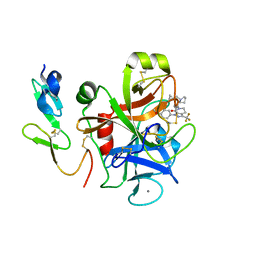
1IA3
 | | Candida albicans dihydrofolate reductase complex in which the dihydronicotinamide moiety of dihydro-nicotinamide-adenine-dinucleotide phosphate (NADPH) is displaced by 5-[(4-TERT-BUTYLPHENYL)SULFANYL]-2,4-QUINAZOLINEDIAMINE (GW995) | | 分子名称: | 2-(N-MORPHOLINO)-ETHANESULFONIC ACID, 5-[4-TERT-BUTYLPHENYLSULFANYL]-2,4-QUINAZOLINEDIAMINE, DIHYDROFOLATE REDUCTASE, ... | | 著者 | Whitlow, M, Howard, A.J, Kuyper, L.F. | | 登録日 | 2001-03-22 | | 公開日 | 2001-04-11 | | 最終更新日 | 2023-08-09 | | 実験手法 | X-RAY DIFFRACTION (1.78 Å) | | 主引用文献 | X-ray Crystal Structures of Candida albicans Dihydrofolate Reductase: High Resolution Ternary Complexes in Which the Dihydronicotinamide Moiety of NADPH is Displaced by an inhibitor
J.Med.Chem., 44, 2001
|
|
3VUU
 | | Crystal structure of the merozoite surface protein MSPDBL2 from P. falciparum | | 分子名称: | CHLORIDE ION, Erythrocyte membrane protein, putative | | 著者 | Czabotar, P.E, Hodder, A.N, Clarke, O.B, Lin, C.S, Smith, B.J, Cowman, A.F. | | 登録日 | 2012-07-09 | | 公開日 | 2012-08-08 | | 最終更新日 | 2023-11-08 | | 実験手法 | X-RAY DIFFRACTION (2.093 Å) | | 主引用文献 | Insights into Duffy binding-like domains through the crystal structure and function of the merozoite surface protein MSPDBL2 from Plasmodium falciparum
J.Biol.Chem., 287, 2012
|
|
6E08
 | |
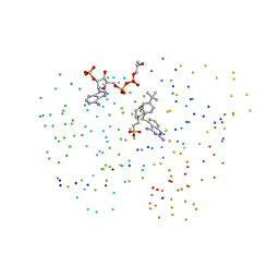
6E07
 | | Crystal structure of Canton G6PD in complex with structural NADP | | 分子名称: | GLYCEROL, Glucose-6-phosphate 1-dehydrogenase, NADP NICOTINAMIDE-ADENINE-DINUCLEOTIDE PHOSPHATE, ... | | 著者 | Rahighi, S, Mochly-Rosen, D, Wakatsuki, S. | | 登録日 | 2018-07-06 | | 公開日 | 2018-07-25 | | 最終更新日 | 2023-10-11 | | 実験手法 | X-RAY DIFFRACTION (2.6 Å) | | 主引用文献 | Correcting glucose-6-phosphate dehydrogenase deficiency with a small-molecule activator.
Nat Commun, 9, 2018
|
|


