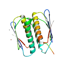
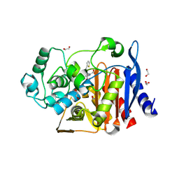
6WE8
 
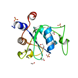 | | YTH domain of human YTHDC1 | | 分子名称: | 1,2-ETHANEDIOL, GLYCEROL, SULFATE ION, ... | | 著者 | Horton, J.R, Cheng, X. | | 登録日 | 2020-04-01 | | 公開日 | 2020-07-15 | | 最終更新日 | 2023-10-18 | | 実験手法 | X-RAY DIFFRACTION (1.18 Å) | | 主引用文献 | Biochemical and structural basis for YTH domain of human YTHDC1 binding to methylated adenine in DNA.
Nucleic Acids Res., 48, 2020
|
|
7JRE
 | |
6E5Z
 | |
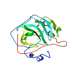
4KIF
 | | Crystal structure of methyltransferase from Streptomyces hygroscopicus complexed with phenylpyruvic acid | | 分子名称: | (2R)-2-hydroxy-3-phenylpropanoic acid, 3-PHENYLPYRUVIC ACID, CALCIUM ION, ... | | 著者 | Liu, Y.C, Zou, X.W, Chan, H.C, Huang, C.J, Li, T.L. | | 登録日 | 2013-05-02 | | 公開日 | 2014-05-07 | | 最終更新日 | 2023-11-15 | | 実験手法 | X-RAY DIFFRACTION (2.5 Å) | | 主引用文献 | Structure and mechanism of a nonhaem-iron SAM-dependent C-methyltransferase and its engineering to a hydratase and an O-methyltransferase
Acta Crystallogr.,Sect.D, 70, 2014
|
|
7JVS
 | |
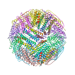
6WIV
 | | Structure of human GABA(B) receptor in an inactive state | | 分子名称: | (2R)-3-{[(S)-(2-aminoethoxy)(hydroxy)phosphoryl]oxy}-2-{[(9Z)-octadec-9-enoyl]oxy}propyl (5Z,8Z,11Z,14Z)-icosa-5,8,11,14-tetraenoate, 2-acetamido-2-deoxy-beta-D-glucopyranose, CALCIUM ION, ... | | 著者 | Park, J, Fu, Z, Frangaj, A, Liu, J, Mosyak, L, Shen, T, Slavkovich, V.N, Ray, K.M, Taura, J, Cao, B, Geng, Y, Zuo, H, Kou, Y, Grassucci, R, Chen, S, Liu, Z, Lin, X, Williams, J.P, Rice, W.J, Eng, E.T, Huang, R.K, Soni, R.K, Kloss, B, Yu, Z, Javitch, J.A, Hendrickson, W.A, Slesinger, P.A, Quick, M, Graziano, J, Yu, H, Fiehn, O, Clarke, O.B, Frank, J, Fan, Q.R. | | 登録日 | 2020-04-10 | | 公開日 | 2020-07-01 | | 最終更新日 | 2024-10-09 | | 実験手法 | ELECTRON MICROSCOPY (3.3 Å) | | 主引用文献 | Structure of human GABABreceptor in an inactive state.
Nature, 584, 2020
|
|
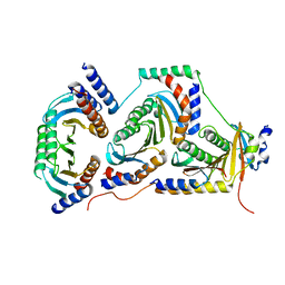
7JT8
 | | Apo structure of a pseudomurein peptide ligase type E from Methanothermus fervidus | | 分子名称: | MAGNESIUM ION, Mur ligase middle domain protein, SULFATE ION | | 著者 | Carbone, V, Schofield, L.R, Sutherland-Smith, A.J, Ronimus, R.S, Subedi, B.P. | | 登録日 | 2020-08-17 | | 公開日 | 2021-09-01 | | 最終更新日 | 2024-10-30 | | 実験手法 | X-RAY DIFFRACTION (1.84 Å) | | 主引用文献 | Structural characterisation of methanogen pseudomurein cell wall peptide ligases homologous to bacterial MurE/F murein peptide ligases.
Microbiology (Reading, Engl.), 168, 2022
|
|
7JQI
 | |
6E99
 | |
7JQH
 | |
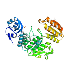
6WHF
 | | class C beta-lactamase from Escherichia coli in complex with cephalothin | | 分子名称: | (2R)-5-[(acetyloxy)methyl]-2-{(1R)-2-oxo-1-[(thiophen-2-ylacetyl)amino]ethyl}-3,6-dihydro-2H-1,3-thiazine-4-carboxylic acid, 1,2-ETHANEDIOL, Beta-lactamase | | 著者 | Chang, C, Maltseva, N, Endres, M, Joachimiak, A, Center for Structural Genomics of Infectious Diseases (CSGID) | | 登録日 | 2020-04-08 | | 公開日 | 2020-04-22 | | 最終更新日 | 2024-10-16 | | 実験手法 | X-RAY DIFFRACTION (1.4 Å) | | 主引用文献 | class C beta-lactamase from Escherichia coli in complex with Cephalothin
To Be Published
|
|
4PLD
 | |
1ZAH
 | | Fructose-1,6-bisphosphate aldolase from rabbit muscle | | 分子名称: | Fructose-bisphosphate aldolase A | | 著者 | St-Jean, M, Lafrance-Vanasse, J, Liotard, B, Sygusch, J. | | 登録日 | 2005-04-06 | | 公開日 | 2005-05-10 | | 最終更新日 | 2023-08-23 | | 実験手法 | X-RAY DIFFRACTION (1.8 Å) | | 主引用文献 | High Resolution Reaction Intermediates of Rabbit Muscle Fructose-1,6-bisphosphate Aldolase: substrate cleavage and induced fit.
J.Biol.Chem., 280, 2005
|
|
7JQJ
 | |
5C6F
 | | Crystal structures of ferritin mutants reveal side-on binding to diiron and end-on cleavage of oxygen | | 分子名称: | Bacterial non-heme ferritin, FE (III) ION, IMIDAZOLE | | 著者 | Kim, S, Kim, K.H, Seok, J.H, Park, Y.H, Jung, S.W, Chung, Y.B, Lee, D.B, Lee, J.H, Han, K.R, Cho, A.E, Lee, C, Chung, M.S. | | 登録日 | 2015-06-23 | | 公開日 | 2016-07-27 | | 最終更新日 | 2024-03-20 | | 実験手法 | X-RAY DIFFRACTION (2 Å) | | 主引用文献 | Structural Basis of Novel Iron-Uptake Route and Reaction Intermediates in Ferritins from Gram-Negative Bacteria.
J. Mol. Biol., 428, 2016
|
|
6ECX
 | |
6EEA
 | | Bioreductive 4-hydroxy-3-nitro-5-ureido-benzenesulfonamides selectively target the tumor-associated carbonic anhydrase isoforms IX and XII and show hypoxia-enhanced cytotoxicity against human cancer cell lines. | | 分子名称: | 4-hydroxy-3-nitro-5-({[4-(trifluoromethyl)phenyl]carbamoyl}amino)benzene-1-sulfonamide, Carbonic anhydrase 2, ZINC ION | | 著者 | Singh, S, McKenna, R, Supuran, C.T, Nocentini, A, Lomelino, C, Lucarini, E, Bartolucci, G, Mannelli, L.D.C, Ghelardini, C, Gratteri, P. | | 登録日 | 2018-08-13 | | 公開日 | 2018-11-28 | | 最終更新日 | 2023-10-11 | | 実験手法 | X-RAY DIFFRACTION (1.63 Å) | | 主引用文献 | 4-Hydroxy-3-nitro-5-ureido-benzenesulfonamides Selectively Target the Tumor-Associated Carbonic Anhydrase Isoforms IX and XII Showing Hypoxia-Enhanced Antiproliferative Profiles.
J. Med. Chem., 61, 2018
|
|
4PNX
 | | Crystal structure of the complex of lactoperoxidase with bromo methane at 2.41 angstrom resolution | | 分子名称: | 2-acetamido-2-deoxy-beta-D-glucopyranose, BROMOMETHANE, CALCIUM ION, ... | | 著者 | Sirohi, H.V, Tyagi, T.K, Singh, A.K, Sinha, M, Bhushan, A, Kaur, P, Sharma, S, Singh, T.P. | | 登録日 | 2014-02-22 | | 公開日 | 2014-03-12 | | 最終更新日 | 2024-10-16 | | 実験手法 | X-RAY DIFFRACTION (2.41 Å) | | 主引用文献 | Structure of bovine lactoperoxidase with a partially linked heme moiety at 1.98 angstrom resolution.
Biochim.Biophys.Acta, 1865, 2017
|
|
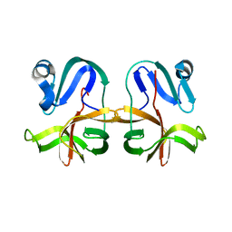
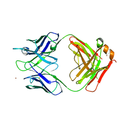
6X4T
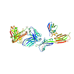 | | Crystal structure of ICOS-L in complex with Prezalumab and VNAR domain | | 分子名称: | 2-acetamido-2-deoxy-beta-D-glucopyranose, 2-acetamido-2-deoxy-beta-D-glucopyranose-(1-4)-2-acetamido-2-deoxy-beta-D-glucopyranose, GLYCEROL, ... | | 著者 | Rujas, E, Sicard, T, Julien, J.P. | | 登録日 | 2020-05-23 | | 公開日 | 2020-10-14 | | 最終更新日 | 2024-10-09 | | 実験手法 | X-RAY DIFFRACTION (3.15 Å) | | 主引用文献 | Structural characterization of the ICOS/ICOS-L immune complex reveals high molecular mimicry by therapeutic antibodies.
Nat Commun, 11, 2020
|
|
6X5E
 | |
6EHR
 | |
5CA2
 | |
1ZKY
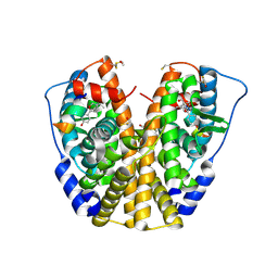 | | Human Estrogen Receptor Alpha Ligand-Binding Domain In Complex With OBCP-3M and A Glucocorticoid Receptor Interacting Protein 1 Nr Box II Peptide | | 分子名称: | 4-[(1S,2S,5S)-5-(HYDROXYMETHYL)-6,8,9-TRIMETHYL-3-OXABICYCLO[3.3.1]NON-7-EN-2-YL]PHENOL, Estrogen receptor, Nuclear receptor coactivator 2 | | 著者 | Rajan, S.S, Hsieh, R.W, Sharma, S.K, Hahm, J.B, Nettles, K.W, Greene, G.L. | | 登録日 | 2005-05-04 | | 公開日 | 2006-05-09 | | 最終更新日 | 2024-10-30 | | 実験手法 | X-RAY DIFFRACTION (2.25 Å) | | 主引用文献 | Identification of ligands with bicyclic scaffolds provides insights into mechanisms of estrogen receptor subtype selectivity.
J.Biol.Chem., 281, 2006
|
|
6WER
 | |
5CAC
 | | REFINED STRUCTURE OF HUMAN CARBONIC ANHYDRASE II AT 2.0 ANGSTROMS RESOLUTION | | 分子名称: | CARBONIC ANHYDRASE FORM C, SULFITE ION, ZINC ION | | 著者 | Lindahl, M, Habash, D, Harrop, S, Helliwell, D.R, Liljas, A. | | 登録日 | 1991-09-05 | | 公開日 | 1994-01-31 | | 最終更新日 | 2024-03-06 | | 実験手法 | X-RAY DIFFRACTION (2.2 Å) | | 主引用文献 | Refined structure of human carbonic anhydrase II at 2.0 A resolution.
Proteins, 4, 1988
|
|


