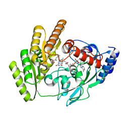
8JDF
 
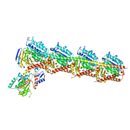
 | | Crystal structure of H405A mLDHD in complex with D-lactate | | 分子名称: | FLAVIN-ADENINE DINUCLEOTIDE, LACTIC ACID, Probable D-lactate dehydrogenase, ... | | 著者 | Jin, S, Chen, X, Yang, J, Ding, J. | | 登録日 | 2023-05-13 | | 公開日 | 2023-10-18 | | 最終更新日 | 2024-02-21 | | 実験手法 | X-RAY DIFFRACTION (1.69 Å) | | 主引用文献 | Lactate dehydrogenase D is a general dehydrogenase for D-2-hydroxyacids and is associated with D-lactic acidosis.
Nat Commun, 14, 2023
|
|
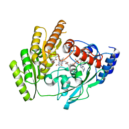
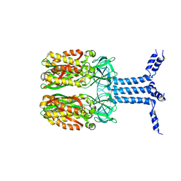
8JDS
 | | Crystal structure of mLDHD in complex with Pyruvate | | 分子名称: | FLAVIN-ADENINE DINUCLEOTIDE, MANGANESE (II) ION, PYRUVIC ACID, ... | | 著者 | Jin, S, Chen, X, Yang, J, Ding, J. | | 登録日 | 2023-05-15 | | 公開日 | 2023-10-18 | | 最終更新日 | 2024-02-21 | | 実験手法 | X-RAY DIFFRACTION (1.636 Å) | | 主引用文献 | Lactate dehydrogenase D is a general dehydrogenase for D-2-hydroxyacids and is associated with D-lactic acidosis.
Nat Commun, 14, 2023
|
|
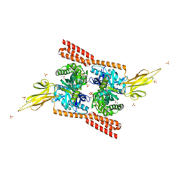
8JDE
 | | Crystal structure of mLDHD in complex with D-lactate | | 分子名称: | FLAVIN-ADENINE DINUCLEOTIDE, LACTIC ACID, MANGANESE (II) ION, ... | | 著者 | Jin, S, Chen, X, Yang, J, Ding, J. | | 登録日 | 2023-05-13 | | 公開日 | 2023-10-18 | | 最終更新日 | 2024-02-21 | | 実験手法 | X-RAY DIFFRACTION (1.729 Å) | | 主引用文献 | Lactate dehydrogenase D is a general dehydrogenase for D-2-hydroxyacids and is associated with D-lactic acidosis.
Nat Commun, 14, 2023
|
|
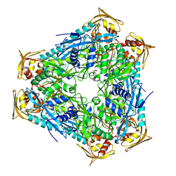
8JDP
 | | Crystal structure of H405A mLDHD in complex with D-2-hydroxyisovaleric acid | | 分子名称: | DEAMINOHYDROXYVALINE, FLAVIN-ADENINE DINUCLEOTIDE, Probable D-lactate dehydrogenase, ... | | 著者 | Jin, S, Chen, X, Yang, J, Ding, J. | | 登録日 | 2023-05-15 | | 公開日 | 2023-10-18 | | 最終更新日 | 2024-02-21 | | 実験手法 | X-RAY DIFFRACTION (1.6 Å) | | 主引用文献 | Lactate dehydrogenase D is a general dehydrogenase for D-2-hydroxyacids and is associated with D-lactic acidosis.
Nat Commun, 14, 2023
|
|
8JDY
 | | Crystal structure of mLDHD in complex with 2-ketoisocaproic acid | | 分子名称: | 2-OXO-4-METHYLPENTANOIC ACID, FLAVIN-ADENINE DINUCLEOTIDE, MANGANESE (II) ION, ... | | 著者 | Jin, S, Chen, X, Yang, J, Ding, J. | | 登録日 | 2023-05-15 | | 公開日 | 2023-10-18 | | 最終更新日 | 2024-02-21 | | 実験手法 | X-RAY DIFFRACTION (1.59 Å) | | 主引用文献 | Lactate dehydrogenase D is a general dehydrogenase for D-2-hydroxyacids and is associated with D-lactic acidosis.
Nat Commun, 14, 2023
|
|
8JDV
 | | Crystal structure of mLDHD in complex with 2-ketohexanoic acid | | 分子名称: | 2-Ketohexanoic acid, FLAVIN-ADENINE DINUCLEOTIDE, MANGANESE (II) ION, ... | | 著者 | Jin, S, Chen, X, Yang, J, Ding, J. | | 登録日 | 2023-05-15 | | 公開日 | 2023-10-18 | | 最終更新日 | 2024-02-21 | | 実験手法 | X-RAY DIFFRACTION (1.96 Å) | | 主引用文献 | Lactate dehydrogenase D is a general dehydrogenase for D-2-hydroxyacids and is associated with D-lactic acidosis.
Nat Commun, 14, 2023
|
|
8JDZ
 | | Crystal structure of mLDHD in complex with 2-keto-3-methylvaleric acid | | 分子名称: | (3S)-3-methyl-2-oxopentanoic acid, FLAVIN-ADENINE DINUCLEOTIDE, MANGANESE (II) ION, ... | | 著者 | Jin, S, Chen, X, Yang, J, Ding, J. | | 登録日 | 2023-05-15 | | 公開日 | 2023-10-18 | | 最終更新日 | 2024-02-21 | | 実験手法 | X-RAY DIFFRACTION (1.56 Å) | | 主引用文献 | Lactate dehydrogenase D is a general dehydrogenase for D-2-hydroxyacids and is associated with D-lactic acidosis.
Nat Commun, 14, 2023
|
|
4MRV
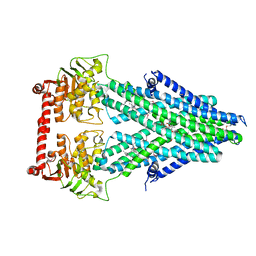 | | Structure of a bacterial Atm1-family ABC transporter | | 分子名称: | ABC transporter related protein, LAURYL DIMETHYLAMINE-N-OXIDE, PHOSPHATE ION, ... | | 著者 | Lee, J.Y, Yang, J.G, Zhitnitsky, D, Lewinson, O, Rees, D.C. | | 登録日 | 2013-09-17 | | 公開日 | 2014-03-19 | | 最終更新日 | 2019-07-17 | | 実験手法 | X-RAY DIFFRACTION (2.5 Å) | | 主引用文献 | Structural basis for heavy metal detoxification by an Atm1-type ABC exporter.
Science, 343, 2014
|
|
4MRP
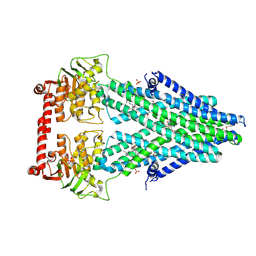 | | Structure of a bacterial Atm1-family ABC transporter | | 分子名称: | ABC transporter related protein, GLUTATHIONE, LAURYL DIMETHYLAMINE-N-OXIDE, ... | | 著者 | Lee, J.Y, Yang, J.G, Zhitnitsky, D, Lewinson, O, Rees, D.C. | | 登録日 | 2013-09-17 | | 公開日 | 2014-03-19 | | 最終更新日 | 2019-07-17 | | 実験手法 | X-RAY DIFFRACTION (2.5 Å) | | 主引用文献 | Structural basis for heavy metal detoxification by an Atm1-type ABC exporter.
Science, 343, 2014
|
|
7CE8
 | | Crystal structure of T2R-TTL-Compound11 complex | | 分子名称: | 2-(N-MORPHOLINO)-ETHANESULFONIC ACID, CALCIUM ION, GLYCEROL, ... | | 著者 | Chen, L.J, Chen, Q, Yu, Y, Yang, J.H. | | 登録日 | 2020-06-22 | | 公開日 | 2021-06-23 | | 最終更新日 | 2023-11-29 | | 実験手法 | X-RAY DIFFRACTION (2.725 Å) | | 主引用文献 | Small Molecules Promote Selective Denaturation and Degradation of Tubulin Heterodimers through a Low-Barrier Hydrogen Bond.
J.Med.Chem., 65, 2022
|
|
8J4I
 | | Unveiling the Role of Human Prohibitin 2 as a Mitochondrial Calcium Channel in Parkinson's Disease | | 分子名称: | Prohibitin-2 | | 著者 | Li, Y.Y, Fang, Y, Gao, Y.X, Ding, W, Yang, J, Liu, Y, Shen, B, Wang, J.F, Zhou, S. | | 登録日 | 2023-04-20 | | 公開日 | 2024-06-05 | | 実験手法 | SOLUTION NMR | | 主引用文献 | Human Prohibitin 2 is a Mitochondrial Ca2+-Selective Channel
To Be Published
|
|
4JNE
 | | Allosteric opening of the polypeptide-binding site when an Hsp70 binds ATP | | 分子名称: | ADENOSINE-5'-TRIPHOSPHATE, GLYCEROL, Hsp70 CHAPERONE DnaK, ... | | 著者 | Qi, R, Sarbeng, E.B, Liu, Q, Le, K.Q, Xu, X, Xu, H, Yang, J, Wong, J.L, Vorvis, C, Hendrickson, W.A, Zhou, L, Liu, Q. | | 登録日 | 2013-03-15 | | 公開日 | 2013-05-29 | | 最終更新日 | 2024-02-28 | | 実験手法 | X-RAY DIFFRACTION (1.96 Å) | | 主引用文献 | Allosteric opening of the polypeptide-binding site when an Hsp70 binds ATP.
Nat.Struct.Mol.Biol., 20, 2013
|
|
4JN4
 | | Allosteric opening of the polypeptide-binding site when an Hsp70 binds ATP | | 分子名称: | ADENOSINE-5'-TRIPHOSPHATE, Chaperone protein DnaK, GLYCEROL, ... | | 著者 | Qi, R, Sarbeng, E.B, Liu, Q, Le, K.Q, Xu, X, Xu, H, Yang, J, Wong, J.L, Vorvis, C, Hendrickson, W.A, Zhou, L, Liu, Q. | | 登録日 | 2013-03-14 | | 公開日 | 2013-05-29 | | 最終更新日 | 2024-02-28 | | 実験手法 | X-RAY DIFFRACTION (2.3 Å) | | 主引用文献 | Allosteric opening of the polypeptide-binding site when an Hsp70 binds ATP.
Nat.Struct.Mol.Biol., 20, 2013
|
|
2L31
 | | Human PARP-1 zinc finger 2 | | 分子名称: | Poly [ADP-ribose] polymerase 1, ZINC ION | | 著者 | Neuhaus, D, Eustermann, S, Yang, J, Videler, H. | | 登録日 | 2010-08-30 | | 公開日 | 2011-02-02 | | 最終更新日 | 2024-05-01 | | 実験手法 | SOLUTION NMR | | 主引用文献 | The DNA-binding domain of human PARP-1 interacts with DNA single-strand breaks as a monomer through its second zinc finger.
J.Mol.Biol., 407, 2011
|
|
2L30
 | | Human PARP-1 zinc finger 1 | | 分子名称: | Poly [ADP-ribose] polymerase 1, ZINC ION | | 著者 | Neuhaus, D, Eustermann, S, Yang, J, Videler, H. | | 登録日 | 2010-08-30 | | 公開日 | 2011-02-02 | | 最終更新日 | 2024-05-01 | | 実験手法 | SOLUTION NMR | | 主引用文献 | The DNA-binding domain of human PARP-1 interacts with DNA single-strand breaks as a monomer through its second zinc finger.
J.Mol.Biol., 407, 2011
|
|
4QB9
 | | Crystal structure of Mycobacterium smegmatis Eis in complex with paromomycin | | 分子名称: | Enhanced intracellular survival protein, PAROMOMYCIN, SULFATE ION | | 著者 | Kim, K.H, Ahn, D.R, Yoon, H.J, Yang, J.K, Suh, S.W. | | 登録日 | 2014-05-06 | | 公開日 | 2015-04-01 | | 最終更新日 | 2024-03-20 | | 実験手法 | X-RAY DIFFRACTION (3.293 Å) | | 主引用文献 | Structure of Mycobacterium smegmatis Eis in complex with paromomycin.
Acta Crystallogr.,Sect.F, 70, 2014
|
|
3G6M
 | | crystal structure of a chitinase CrChi1 from the nematophagous fungus Clonostachys rosea in complex with a potent inhibitor caffeine | | 分子名称: | CAFFEINE, Chitinase | | 著者 | Gan, Z, Yang, J, Lou, Z, Rao, Z, Zhang, K.-Q. | | 登録日 | 2009-02-06 | | 公開日 | 2010-02-16 | | 最終更新日 | 2023-11-01 | | 実験手法 | X-RAY DIFFRACTION (1.65 Å) | | 主引用文献 | Crystal structure and mutagenesis analysis of chitinase CrChi1 from the nematophagous fungus Clonostachys rosea in complex with the inhibitor caffeine
Microbiology, 156, 2010
|
|
2N8A
 | | 1H, 13C and 15N chemical shift assignments and solution structure for PARP-1 F1F2 domains in complex with a DNA single-strand break | | 分子名称: | DNA (45-MER), Poly [ADP-ribose] polymerase 1, ZINC ION | | 著者 | Neuhaus, D, Eustermann, S, Yang, J, Wu, W. | | 登録日 | 2015-10-08 | | 公開日 | 2015-12-02 | | 最終更新日 | 2024-05-01 | | 実験手法 | SOLUTION NMR | | 主引用文献 | Structural Basis of Detection and Signaling of DNA Single-Strand Breaks by Human PARP-1.
Mol.Cell, 60, 2015
|
|
6AYF
 | | TRPML3/ML-SA1 complex at pH 7.4 | | 分子名称: | 2-acetamido-2-deoxy-beta-D-glucopyranose, Mucolipin-3 | | 著者 | Zhou, X, Li, M, Su, D, Jia, Q, Li, H, Li, X, Yang, J. | | 登録日 | 2017-09-08 | | 公開日 | 2017-11-08 | | 最終更新日 | 2024-03-13 | | 実験手法 | ELECTRON MICROSCOPY (3.62 Å) | | 主引用文献 | Cryo-EM structures of the human endolysosomal TRPML3 channel in three distinct states.
Nat. Struct. Mol. Biol., 24, 2017
|
|
5E85
 | | isolated SBD of BiP | | 分子名称: | 78 kDa glucose-regulated protein, SULFATE ION | | 著者 | Liu, Q, Yang, J, Nune, M, Zong, Y, Zhou, L. | | 登録日 | 2015-10-13 | | 公開日 | 2015-12-30 | | 最終更新日 | 2024-03-06 | | 実験手法 | X-RAY DIFFRACTION (2.57 Å) | | 主引用文献 | Close and Allosteric Opening of the Polypeptide-Binding Site in a Human Hsp70 Chaperone BiP.
Structure, 23, 2015
|
|
5E86
 | | isolated SBD of BiP with loop34 modification | | 分子名称: | 78 kDa glucose-regulated protein | | 著者 | Liu, Q, Yang, J, Nune, M, Zong, Y, Zhou, L. | | 登録日 | 2015-10-13 | | 公開日 | 2015-12-30 | | 最終更新日 | 2024-03-06 | | 実験手法 | X-RAY DIFFRACTION (2.681 Å) | | 主引用文献 | Close and Allosteric Opening of the Polypeptide-Binding Site in a Human Hsp70 Chaperone BiP.
Structure, 23, 2015
|
|
2K0A
 | | 1H, 15N and 13C chemical shift assignments for Rds3 protein | | 分子名称: | Pre-mRNA-splicing factor RDS3, ZINC ION | | 著者 | Loening, N, van Roon, A, Yang, J, Nagai, K, Neuhaus, D. | | 登録日 | 2008-01-31 | | 公開日 | 2008-07-22 | | 最終更新日 | 2024-05-29 | | 実験手法 | SOLUTION NMR | | 主引用文献 | Solution structure of the U2 snRNP protein Rds3p reveals a knotted zinc-finger motif.
Proc.Natl.Acad.Sci.Usa, 105, 2008
|
|
4JNF
 | | Allosteric opening of the polypeptide-binding site when an Hsp70 binds ATP | | 分子名称: | Hsp70 CHAPERONE DnaK | | 著者 | Qi, R, Sarbeng, E.B, Liu, Q, Le, K.Q, Xu, X, Xu, H, Yang, J, Wong, J.L, Vorvis, C, Hendrickson, W.A, Zhou, L, Liu, Q. | | 登録日 | 2013-03-15 | | 公開日 | 2013-05-29 | | 最終更新日 | 2023-09-20 | | 実験手法 | X-RAY DIFFRACTION (1.621 Å) | | 主引用文献 | Allosteric opening of the polypeptide-binding site when an Hsp70 binds ATP.
Nat.Struct.Mol.Biol., 20, 2013
|
|
2LD1
 | |
2IGH
 | | DETERMINATION OF THE SOLUTION STRUCTURES OF DOMAINS II AND III OF PROTEIN G FROM STREPTOCOCCUS BY 1H NMR | | 分子名称: | PROTEIN G | | 著者 | Lian, L.-Y, Derrick, J.P, Sutcliffe, M.J, Yang, J.C, Roberts, G.C.K. | | 登録日 | 1992-08-26 | | 公開日 | 1994-01-31 | | 最終更新日 | 2024-05-29 | | 実験手法 | SOLUTION NMR | | 主引用文献 | Determination of the solution structures of domains II and III of protein G from Streptococcus by 1H nuclear magnetic resonance.
J.Mol.Biol., 228, 1992
|
|


