8E6Q
 
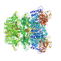 | | Human TRPM2 ion channel in 1 mM ADPR | | 分子名称: | ADENOSINE-5-DIPHOSPHORIBOSE, Transient receptor potential cation channel subfamily M member 2 | | 著者 | Wang, L, Fu, T.M, Xia, S, Wu, H. | | 登録日 | 2022-08-23 | | 公開日 | 2024-08-14 | | 実験手法 | ELECTRON MICROSCOPY (3.4 Å) | | 主引用文献 | A unified mechanism for human TRPM2 activation, desensitization and inhibition
To Be Published
|
|
8E6R
 
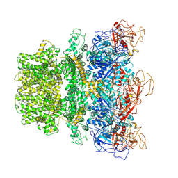 | | Human TRPM2 ion channel in 1 mM dADPR | | 分子名称: | ADENOSINE-5-DIPHOSPHORIBOSE, Transient receptor potential cation channel subfamily M member 2, [(2R,3S,4R,5R)-5-(6-AMINOPURIN-9-YL)-3,4-DIHYDROXY-OXOLAN-2-YL]METHYL [HYDROXY-[[(2R,3S,4R,5S)-3,4,5-TRIHYDROXYOXOLAN-2-YL]METHOXY]PHOSPHORYL] HYDROGEN PHOSPHATE | | 著者 | Wang, L, Fu, T.M, Xia, S, Wu, H. | | 登録日 | 2022-08-23 | | 公開日 | 2024-08-14 | | 実験手法 | ELECTRON MICROSCOPY (5.6 Å) | | 主引用文献 | A unified mechanism for human TRPM2 activation, desensitization and inhibition
To Be Published
|
|
8E6T
 
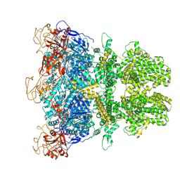 | | Human TRPM2 ion channel in 1 mM BR-ADPR | | 分子名称: | (3R,4R,5R)-5-(6-amino-8-bromo-9H-purin-9-yl)-3,4,5-trihydroxypentyl [(2S,3S,4S,5R)-3,4,5-trihydroxyoxolan-2-yl]methyl dihydrogen diphosphate (non-preferred name), Transient receptor potential cation channel subfamily M member 2 | | 著者 | Wang, L, Fu, T.M, Xia, S, Wu, H. | | 登録日 | 2022-08-23 | | 公開日 | 2024-08-14 | | 実験手法 | ELECTRON MICROSCOPY (3.7 Å) | | 主引用文献 | A unified mechanism for human TRPM2 activation, desensitization and inhibition
To Be Published
|
|
4YPH
 
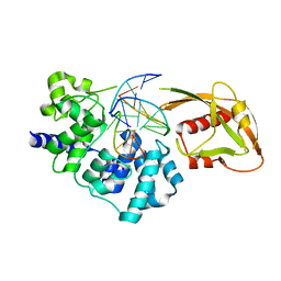 | | Crystal Structure of MutY bound to its anti-substrate with the disulfide cross-linker reduced | | 分子名称: | A/G-specific adenine glycosylase, DNA (5'-D(*AP*AP*GP*AP*CP*(8OG)P*TP*GP*GP*AP*C)-3'), DNA (5'-D(TP*GP*TP*CP*CP*AP*CP*GP*TP*CP*T)-3'), ... | | 著者 | Wang, L, Lee, S, Verdine, G.L. | | 登録日 | 2015-03-12 | | 公開日 | 2015-05-27 | | 最終更新日 | 2023-09-27 | | 実験手法 | X-RAY DIFFRACTION (2.32 Å) | | 主引用文献 | Structural Basis for Avoidance of Promutagenic DNA Repair by MutY Adenine DNA Glycosylase.
J.Biol.Chem., 290, 2015
|
|
8ERP
 
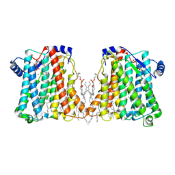 | |
8ERO
 
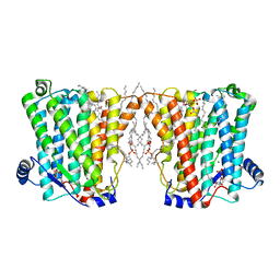 | |
4YOQ
 
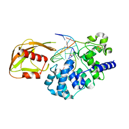 | | Crystal Structure of MutY bound to its anti-substrate | | 分子名称: | A/G-specific adenine glycosylase, DNA (5'-D(*AP*AP*GP*AP*CP*(8OG)P*TP*GP*GP*AP*C)-3'), DNA (5'-D(*T*GP*TP*CP*CP*AP*CP*GP*TP*CP*T)-3'), ... | | 著者 | Wang, L, Lee, S, Verdine, G.L. | | 登録日 | 2015-03-11 | | 公開日 | 2015-05-27 | | 最終更新日 | 2023-09-27 | | 実験手法 | X-RAY DIFFRACTION (2.21 Å) | | 主引用文献 | Structural Basis for Avoidance of Promutagenic DNA Repair by MutY Adenine DNA Glycosylase.
J.Biol.Chem., 290, 2015
|
|
6ND0
 
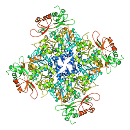 | |
4JR6
 
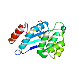 | | Crystal structure of DsbA from Mycobacterium tuberculosis (reduced) | | 分子名称: | Possible conserved membrane or secreted protein, SULFATE ION | | 著者 | Wang, L. | | 登録日 | 2013-03-21 | | 公開日 | 2013-07-17 | | 最終更新日 | 2017-11-15 | | 実験手法 | X-RAY DIFFRACTION (1.902 Å) | | 主引用文献 | Structure analysis of the extracellular domain reveals disulfide bond forming-protein properties of Mycobacterium tuberculosis Rv2969c.
Protein Cell, 4, 2013
|
|
4JR4
 
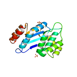 | | Crystal structure of Mtb DsbA (Oxidized) | | 分子名称: | Possible conserved membrane or secreted protein, SULFATE ION | | 著者 | Wang, L. | | 登録日 | 2013-03-21 | | 公開日 | 2013-07-17 | | 最終更新日 | 2024-10-16 | | 実験手法 | X-RAY DIFFRACTION (2.498 Å) | | 主引用文献 | Structure analysis of the extracellular domain reveals disulfide bond forming-protein properties of Mycobacterium tuberculosis Rv2969c.
Protein Cell, 4, 2013
|
|
4NSC
 
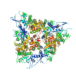 | | Crystal Structure of CBARA1 in the Apo-form | | 分子名称: | Calcium uptake protein 1, mitochondrial | | 著者 | Wang, L, Yang, X, Li, S, Shen, Y. | | 登録日 | 2013-11-28 | | 公開日 | 2014-02-26 | | 最終更新日 | 2024-02-28 | | 実験手法 | X-RAY DIFFRACTION (3.2 Å) | | 主引用文献 | Structural and mechanistic insights into MICU1 regulation of mitochondrial calcium uptake.
Embo J., 33, 2014
|
|
8E6V
 
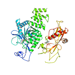 | | MHR1/2 and NUDT9H of human TRPM2 in 1 mM dADPR (local refinement) | | 分子名称: | ADENOSINE-5-DIPHOSPHORIBOSE, Transient receptor potential cation channel subfamily M member 2, ZINC ION | | 著者 | Wang, L, Fu, T.M, Xia, S, Wu, H. | | 登録日 | 2022-08-23 | | 公開日 | 2024-08-14 | | 実験手法 | ELECTRON MICROSCOPY (3.3 Å) | | 主引用文献 | A unified mechanism for human TRPM2 activation, desensitization and inhibition
To Be Published
|
|
7TRJ
 
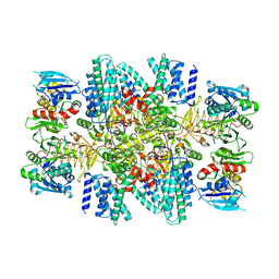 | | The eukaryotic translation initiation factor 2B from Homo sapiens with a H160D mutation in the beta subunit | | 分子名称: | Translation initiation factor eIF-2B subunit alpha, Translation initiation factor eIF-2B subunit beta, Translation initiation factor eIF-2B subunit delta, ... | | 著者 | Wang, L, Schoof, M, Lawrence, R, Boone, M, Frost, A, Walter, P. | | 登録日 | 2022-01-29 | | 公開日 | 2022-04-27 | | 最終更新日 | 2024-02-21 | | 実験手法 | ELECTRON MICROSCOPY (2.8 Å) | | 主引用文献 | A point mutation in the nucleotide exchange factor eIF2B constitutively activates the integrated stress response by allosteric modulation.
Elife, 11, 2022
|
|
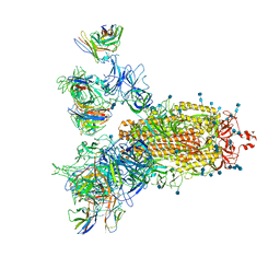
7CWS
 
 | |
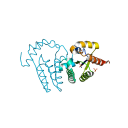
1LY1
 
 | |
7Y0N
 
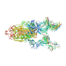 | | SARS-CoV-2 WT Spike in complex with R15 Fab and P14 Nanobody | | 分子名称: | 2-acetamido-2-deoxy-beta-D-glucopyranose, 2-acetamido-2-deoxy-beta-D-glucopyranose-(1-4)-2-acetamido-2-deoxy-beta-D-glucopyranose, Heavy chain of R15-F7, ... | | 著者 | Wang, L. | | 登録日 | 2022-06-05 | | 公開日 | 2023-09-13 | | 実験手法 | ELECTRON MICROSCOPY (3.8 Å) | | 主引用文献 | SARS-CoV-2 WT Spike in complex with R15 Fab and P14 Nanobody
To Be Published
|
|
1BLR
 
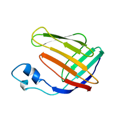 | | NMR SOLUTION STRUCTURE OF HUMAN CELLULAR RETINOIC ACID BINDING PROTEIN-TYPE II, 22 STRUCTURES | | 分子名称: | CELLULAR RETINOIC ACID BINDING PROTEIN-TYPE II | | 著者 | Wang, L, Li, Y, Abilddard, F, Yan, H, Markely, J. | | 登録日 | 1998-07-20 | | 公開日 | 1999-01-13 | | 最終更新日 | 2024-05-22 | | 実験手法 | SOLUTION NMR | | 主引用文献 | NMR solution structure of type II human cellular retinoic acid binding protein: implications for ligand binding.
Biochemistry, 37, 1998
|
|
7FAP
 
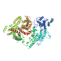 | | Structure of VAR2CSA-CSA 3D7 | | 分子名称: | 2-acetamido-2-deoxy-4-O-sulfo-beta-D-galactopyranose-(1-3)-beta-D-glucopyranuronic acid-(1-3)-2-acetamido-2-deoxy-4-O-sulfo-beta-D-galactopyranose-(1-4)-beta-D-glucopyranuronic acid-(1-3)-2-acetamido-2-deoxy-4-O-sulfo-beta-D-galactopyranose-(1-4)-beta-D-glucopyranuronic acid-(1-3)-2-acetamido-2-deoxy-4-O-sulfo-beta-D-galactopyranose-(1-4)-beta-D-glucopyranuronic acid-(1-3)-2-acetamido-2-deoxy-4-O-sulfo-beta-D-galactopyranose-(1-4)-beta-D-glucopyranuronic acid-(1-3)-2-acetamido-2-deoxy-4-O-sulfo-beta-D-galactopyranose-(1-4)-beta-D-glucopyranuronic acid, Erythrocyte membrane protein 1, PfEMP1 | | 著者 | Wang, L, Wang, Z. | | 登録日 | 2021-07-07 | | 公開日 | 2022-05-04 | | 最終更新日 | 2024-10-09 | | 実験手法 | ELECTRON MICROSCOPY (3.4 Å) | | 主引用文献 | The molecular mechanism of cytoadherence to placental or tumor cells through VAR2CSA from Plasmodium falciparum.
Cell Discov, 7, 2021
|
|
4J6O
 
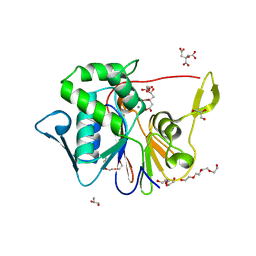 | | Crystal Structure of the Phosphatase Domain of C. thermocellum (Bacterial) PnkP | | 分子名称: | CITRIC ACID, GLYCEROL, MANGANESE (II) ION, ... | | 著者 | Wang, L, Smith, P, Shuman, S. | | 登録日 | 2013-02-11 | | 公開日 | 2013-04-10 | | 最終更新日 | 2024-02-28 | | 実験手法 | X-RAY DIFFRACTION (1.6 Å) | | 主引用文献 | Structure and mechanism of the 2',3' phosphatase component of the bacterial Pnkp-Hen1 RNA repair system.
Nucleic Acids Res., 41, 2013
|
|
8WWC
 
 | |
7Y0O
 
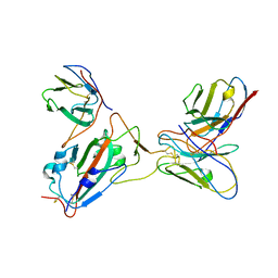 | |
6MJ2
 
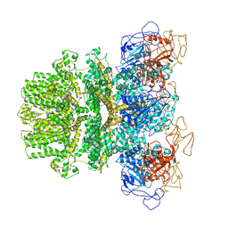 | | Human TRPM2 ion channel in a calcium- and ADPR-bound state | | 分子名称: | CALCIUM ION, Transient receptor potential cation channel subfamily M member 2 | | 著者 | Wang, L, Fu, T.M, Xia, S, Wu, H. | | 登録日 | 2018-09-20 | | 公開日 | 2018-12-12 | | 最終更新日 | 2024-10-09 | | 実験手法 | ELECTRON MICROSCOPY (6.36 Å) | | 主引用文献 | Structures and gating mechanism of human TRPM2.
Science, 362, 2018
|
|
6MIX
 
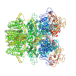 | |
6MIZ
 
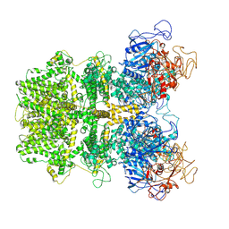 | |
7DWV
 
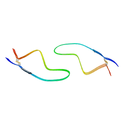 | | Cryo-EM structure of amyloid fibril formed by familial prion disease-related mutation E196K | | 分子名称: | Major prion protein | | 著者 | Wang, L.Q, Zhao, K, Yuan, H.Y, Li, X.N, Dang, H.B, Ma, Y.Y, Wang, Q, Wang, C, Sun, Y.P, Chen, J, Li, D, Zhang, D.L, Yin, P, Liu, C, Liang, Y. | | 登録日 | 2021-01-18 | | 公開日 | 2021-10-13 | | 実験手法 | ELECTRON MICROSCOPY (3.07 Å) | | 主引用文献 | Genetic prion disease-related mutation E196K displays a novel amyloid fibril structure revealed by cryo-EM.
Sci Adv, 7, 2021
|
|
