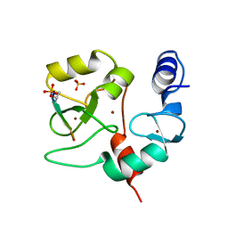
5THR
 
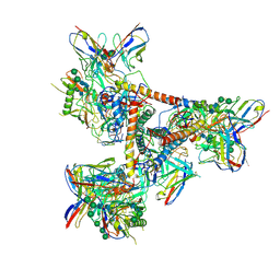 | |
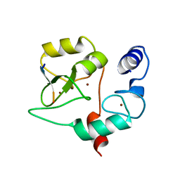
6A9S
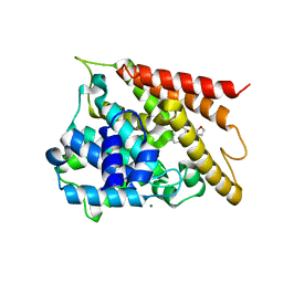
 | | The crystal structure of vaccinia virus A26 (residues 1-397) | | 分子名称: | 1,2-ETHANEDIOL, Protein A26 | | 著者 | Wang, H.C, Ko, T.Z, Luo, Y.C, Liao, Y.T, Chang, W. | | 登録日 | 2018-07-16 | | 公開日 | 2019-06-12 | | 最終更新日 | 2019-07-10 | | 実験手法 | X-RAY DIFFRACTION (1.18 Å) | | 主引用文献 | Vaccinia viral A26 protein is a fusion suppressor of mature virus and triggers membrane fusion through conformational change at low pH.
Plos Pathog., 15, 2019
|
|
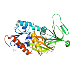
5BYB
 | | Crystal structure of the catalytic domain of human diphosphoinositol pentakisphosphate kinase 2 (PPIP5K2) in complex with ADP and 1,5-(PA)2-IP4 | | 分子名称: | 1,2-ETHANEDIOL, ACETATE ION, ADENOSINE-5'-DIPHOSPHATE, ... | | 著者 | Wang, H, Shears, S.B. | | 登録日 | 2015-06-10 | | 公開日 | 2015-07-22 | | 最終更新日 | 2023-09-27 | | 実験手法 | X-RAY DIFFRACTION (2.3 Å) | | 主引用文献 | Synthetic tools for studying the chemical biology of InsP8.
Chem.Commun.(Camb.), 51, 2015
|
|
5BYA
 | | Crystal structure of the catalytic domain of human diphosphoinositol pentakisphosphate kinase 2 (PPIP5K2) in complex with ADP and 1,5-(PCP)2-IP4 | | 分子名称: | 1,2-ETHANEDIOL, ACETATE ION, ADENOSINE-5'-DIPHOSPHATE, ... | | 著者 | Wang, H, Shears, S.B. | | 登録日 | 2015-06-10 | | 公開日 | 2015-07-22 | | 最終更新日 | 2023-09-27 | | 実験手法 | X-RAY DIFFRACTION (1.9 Å) | | 主引用文献 | Synthetic tools for studying the chemical biology of InsP8.
Chem.Commun.(Camb.), 51, 2015
|
|
4QBS
 | |
4QBR
 | |
5DGH
 | |
1BM5
 | |
5VIY
 | |
5WH5
 | |
8J8Y
 | |
3USJ
 | |
6KA0
 | |
3USO
 | |
6KA1
 | |
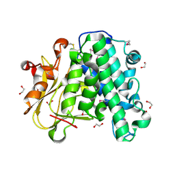
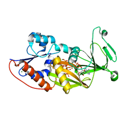
3T9D
 | | Crystal structure of the catalytic domain of human diphosphoinositol pentakisphosphate kinase 2 (PPIP5K2) in complex with AMPPNP and 5-(PP)-IP5 (5-IP7) | | 分子名称: | (1r,2R,3S,4s,5R,6S)-2,3,4,5,6-pentakis(phosphonooxy)cyclohexyl trihydrogen diphosphate, Inositol Pyrophosphate Kinase, MAGNESIUM ION, ... | | 著者 | Wang, H, Falck, J, Hall, T.M.T, Shears, S.B. | | 登録日 | 2011-08-02 | | 公開日 | 2011-12-07 | | 最終更新日 | 2024-02-28 | | 実験手法 | X-RAY DIFFRACTION (1.85 Å) | | 主引用文献 | Structural basis for an inositol pyrophosphate kinase surmounting phosphate crowding.
Nat.Chem.Biol., 8, 2011
|
|
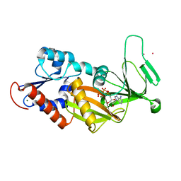
3T9E
 | | Crystal structure of the catalytic domain of human diphosphoinositol pentakisphosphate kinase 2 (PPIP5K2) in complex with ADP, 5-(PP)-IP5 (5-IP7) and MgF3 (transition state mimic) | | 分子名称: | (1r,2R,3S,4s,5R,6S)-2,3,4,5,6-pentakis(phosphonooxy)cyclohexyl trihydrogen diphosphate, ADENOSINE-5'-DIPHOSPHATE, Inositol Pyrophosphate Kinase, ... | | 著者 | Wang, H, Falck, J, Hall, T.M.T, Shears, S.B. | | 登録日 | 2011-08-02 | | 公開日 | 2011-12-07 | | 最終更新日 | 2024-02-28 | | 実験手法 | X-RAY DIFFRACTION (1.9 Å) | | 主引用文献 | Structural basis for an inositol pyrophosphate kinase surmounting phosphate crowding.
Nat.Chem.Biol., 8, 2011
|
|
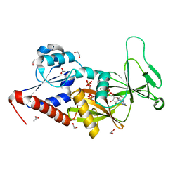
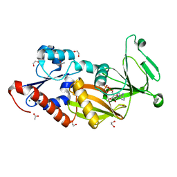
3T9A
 | | Crystal structure of the catalytic domain of human diphosphoinositol pentakisphosphate kinase 2 (PPIP5K2) in complex with AMPPNP at pH 7.0 | | 分子名称: | CADMIUM ION, Inositol Pyrophosphate Kinase, MAGNESIUM ION, ... | | 著者 | Wang, H, Falck, J, Hall, T.M.T, Shears, S.B. | | 登録日 | 2011-08-02 | | 公開日 | 2011-12-07 | | 最終更新日 | 2024-02-28 | | 実験手法 | X-RAY DIFFRACTION (1.8 Å) | | 主引用文献 | Structural basis for an inositol pyrophosphate kinase surmounting phosphate crowding.
Nat.Chem.Biol., 8, 2011
|
|
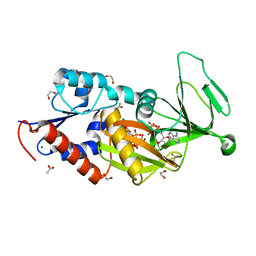
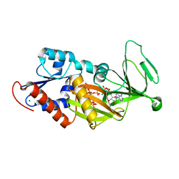
3T7A
 | | Crystal structure of the catalytic domain of human diphosphoinositol pentakisphosphate kinase 2 (PPIP5K2) in complex with ADP at pH 5.2 | | 分子名称: | 1,2-ETHANEDIOL, ACETATE ION, ADENOSINE-5'-DIPHOSPHATE, ... | | 著者 | Wang, H, Falck, J, Hall, T.M.T, Shears, S.B. | | 登録日 | 2011-07-29 | | 公開日 | 2011-12-07 | | 最終更新日 | 2023-09-13 | | 実験手法 | X-RAY DIFFRACTION (1.7 Å) | | 主引用文献 | Structural basis for an inositol pyrophosphate kinase surmounting phosphate crowding.
Nat.Chem.Biol., 8, 2011
|
|
3T9F
 | | Crystal structure of the catalytic domain of human diphosphoinositol pentakisphosphate kinase 2 (PPIP5K2) in complex with ADP and 1,5-(PP)2-IP4 (1,5-IP8) | | 分子名称: | (1R,3S,4R,5S,6R)-2,4,5,6-tetrakis(phosphonooxy)cyclohexane-1,3-diyl bis[trihydrogen (diphosphate)], ADENOSINE-5'-DIPHOSPHATE, CADMIUM ION, ... | | 著者 | Wang, H, Falck, J, Hall, T.M.T, Shears, S.B. | | 登録日 | 2011-08-02 | | 公開日 | 2011-12-07 | | 最終更新日 | 2024-02-28 | | 実験手法 | X-RAY DIFFRACTION (2 Å) | | 主引用文献 | Structural basis for an inositol pyrophosphate kinase surmounting phosphate crowding.
Nat.Chem.Biol., 8, 2011
|
|
3T54
 | | Crystal structure of the catalytic domain of human diphosphoinositol pentakisphosphate kinase 2 (PPIP5K2) in complex with ATP and Cadmium | | 分子名称: | ADENOSINE-5'-TRIPHOSPHATE, CADMIUM ION, Inositol Pyrophosphate Kinase | | 著者 | Wang, H, Falck, J, Hall, T.M.T, Shears, S.B. | | 登録日 | 2011-07-26 | | 公開日 | 2011-12-07 | | 最終更新日 | 2024-02-28 | | 実験手法 | X-RAY DIFFRACTION (1.9 Å) | | 主引用文献 | Structural basis for an inositol pyrophosphate kinase surmounting phosphate crowding.
Nat.Chem.Biol., 8, 2011
|
|
3T9C
 | | Crystal structure of the catalytic domain of human diphosphoinositol pentakisphosphate kinase 2 (PPIP5K2) in complex with AMPPNP and inositol hexakisphosphate (IP6) | | 分子名称: | INOSITOL HEXAKISPHOSPHATE, Inositol Pyrophosphate Kinase, MAGNESIUM ION, ... | | 著者 | Wang, H, Falck, J, Hall, T.M.T, Shears, S.B. | | 登録日 | 2011-08-02 | | 公開日 | 2011-12-07 | | 最終更新日 | 2023-09-13 | | 実験手法 | X-RAY DIFFRACTION (1.9 Å) | | 主引用文献 | Structural basis for an inositol pyrophosphate kinase surmounting phosphate crowding.
Nat.Chem.Biol., 8, 2011
|
|
3USG
 | | Crystal structure of LeuT bound to L-leucine in space group C2 from lipid bicelles | | 分子名称: | ACETATE ION, DI(HYDROXYETHYL)ETHER, LEUCINE, ... | | 著者 | Wang, H, Elferich, J, Gouaux, E. | | 登録日 | 2011-11-23 | | 公開日 | 2012-01-11 | | 最終更新日 | 2023-09-13 | | 実験手法 | X-RAY DIFFRACTION (2.502 Å) | | 主引用文献 | Structures of LeuT in bicelles define conformation and substrate binding in a membrane-like context.
Nat.Struct.Mol.Biol., 19, 2012
|
|
3USK
 | |
3USL
 | | Crystal Structure of LeuT bound to L-selenomethionine in space group C2 from lipid bicelles | | 分子名称: | ACETATE ION, IODIDE ION, PHOSPHOCHOLINE, ... | | 著者 | Wang, H, Elferich, J, Gouaux, E. | | 登録日 | 2011-11-23 | | 公開日 | 2012-01-11 | | 最終更新日 | 2023-12-06 | | 実験手法 | X-RAY DIFFRACTION (2.71 Å) | | 主引用文献 | Structures of LeuT in bicelles define conformation and substrate binding in a membrane-like context.
Nat.Struct.Mol.Biol., 19, 2012
|
|


