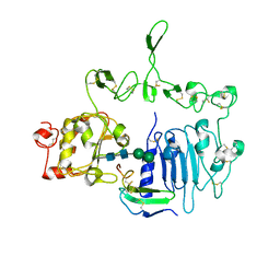
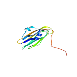
7LIR
 
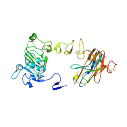
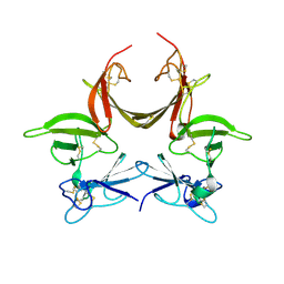
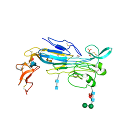 | | Structure of the invertebrate ALK GRD | | 分子名称: | 2-acetamido-2-deoxy-beta-D-glucopyranose, 2-acetamido-2-deoxy-beta-D-glucopyranose-(1-4)-2-acetamido-2-deoxy-beta-D-glucopyranose, ALK tyrosine kinase receptor homolog scd-2, ... | | 著者 | Stayrook, S, Li, T, Klein, D.E. | | 登録日 | 2021-01-27 | | 公開日 | 2021-11-24 | | 最終更新日 | 2021-12-15 | | 実験手法 | X-RAY DIFFRACTION (2.6 Å) | | 主引用文献 | Structural basis for ligand reception by anaplastic lymphoma kinase.
Nature, 600, 2021
|
|
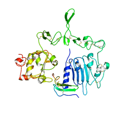
7LS0
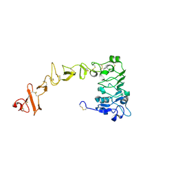
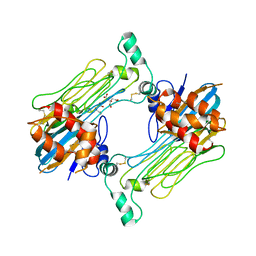 | | Structure of the Human ALK GRD bound to AUG | | 分子名称: | 2-acetamido-2-deoxy-beta-D-glucopyranose, ALK tyrosine kinase receptor fused with ALK and LTK ligand 2, CITRIC ACID | | 著者 | Stayrook, S, Li, T, Klein, D.E. | | 登録日 | 2021-02-17 | | 公開日 | 2021-11-24 | | 最終更新日 | 2023-10-18 | | 実験手法 | X-RAY DIFFRACTION (3.05 Å) | | 主引用文献 | Structural basis for ligand reception by anaplastic lymphoma kinase.
Nature, 600, 2021
|
|
7LRZ
 | |
8UKX
 | |
8UKV
 | |
8UKW
 | |
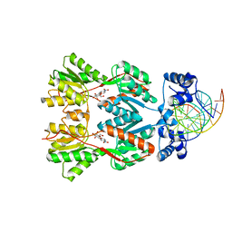
3BDN
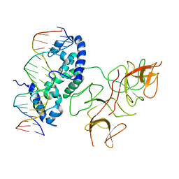 | | Crystal Structure of the Lambda Repressor | | 分子名称: | DNA (5'-D(*DAP*DAP*DTP*DAP*DCP*DCP*DAP*DCP*DTP*DGP*DGP*DCP*DGP*DGP*DTP*DGP*DAP*DTP*DAP*DT)-3'), DNA (5'-D(*DTP*DAP*DTP*DAP*DTP*DCP*DAP*DCP*DCP*DGP*DCP*DCP*DAP*DGP*DTP*DGP*DGP*DTP*DAP*DT)-3'), Lambda Repressor | | 著者 | Stayrook, S.E, Jaru-Ampornpan, P, Hochschild, A, Lewis, M. | | 登録日 | 2007-11-15 | | 公開日 | 2008-04-15 | | 最終更新日 | 2023-08-30 | | 実験手法 | X-RAY DIFFRACTION (3.909 Å) | | 主引用文献 | Crystal structure of the lambda repressor and a model for pairwise cooperative operator binding
Nature, 452, 2008
|
|
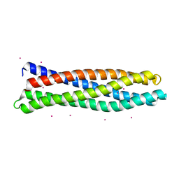
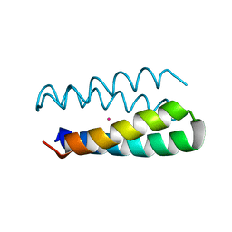
1MFT
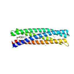
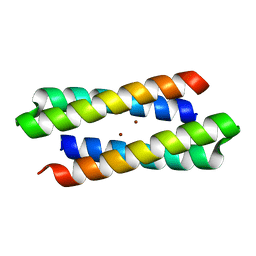 | | Crystal Structure Of Four-Helix Bundle Model | | 分子名称: | Four-helix bundle model, ZINC ION | | 著者 | Lahr, S.J, Stayrook, S.E, North, B, Kaplan, J, Geremia, S, DeGrado, W. | | 登録日 | 2002-08-13 | | 公開日 | 2004-01-20 | | 最終更新日 | 2024-02-14 | | 実験手法 | X-RAY DIFFRACTION (2.5 Å) | | 主引用文献 | Analysis and Design of Turns in alpha-Helical Hairpins
J.Mol.Biol., 346, 2005
|
|
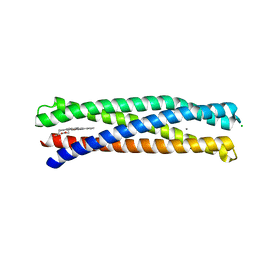
5VJS
 | | De Novo Photosynthetic Reaction Center Protein Equipped with Heme B, a synthetic Zn porphyrin, and Zn(II) cations | | 分子名称: | CHLORIDE ION, PROTOPORPHYRIN IX CONTAINING FE, Reaction Center Maquette, ... | | 著者 | Ennist, N.M, Dutton, P.L, Stayrook, S.E, Moser, C.C. | | 登録日 | 2017-04-19 | | 公開日 | 2018-04-25 | | 最終更新日 | 2024-05-22 | | 実験手法 | X-RAY DIFFRACTION (2 Å) | | 主引用文献 | De novo protein design of photochemical reaction centers.
Nat Commun, 13, 2022
|
|
5VJU
 | | De Novo Photosynthetic Reaction Center Protein Variant Equipped with His-Tyr H-bond, Heme B, and Cd(II) ions | | 分子名称: | CADMIUM ION, PROTOPORPHYRIN IX CONTAINING FE, Reaction Center Maquette Leu71His variant | | 著者 | Ennist, N.M, Stayrook, S.E, Dutton, P.L, Moser, C.C. | | 登録日 | 2017-04-19 | | 公開日 | 2018-04-25 | | 最終更新日 | 2024-05-22 | | 実験手法 | X-RAY DIFFRACTION (2.08 Å) | | 主引用文献 | De novo protein design of photochemical reaction centers.
Nat Commun, 13, 2022
|
|
7TVD
 | |
1PI1
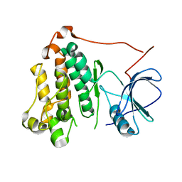
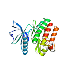
 | | Crystal structure of a human Mob1 protein; toward understanding Mob-regulated cell cycle pathways. | | 分子名称: | Mob1A, ZINC ION | | 著者 | Stavridi, E.S, Harris, K.G, Huyen, Y, Bothos, J, Voewerd, P.M, Stayrook, S.E, Jeffrey, P.D, Pavletich, N.P, Luca, F.C. | | 登録日 | 2003-05-29 | | 公開日 | 2003-09-30 | | 最終更新日 | 2024-05-22 | | 実験手法 | X-RAY DIFFRACTION (2 Å) | | 主引用文献 | Crystal structure of a human mob1 protein. Toward understanding mob-regulated cell cycle pathways.
Structure, 11, 2003
|
|
7LFS
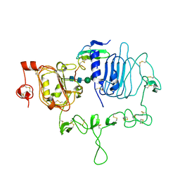 | | Crystal structure of the epidermal growth factor receptor extracellular region with A265V mutation in complex with epiregulin | | 分子名称: | 2-acetamido-2-deoxy-beta-D-glucopyranose, 2-acetamido-2-deoxy-beta-D-glucopyranose-(1-4)-2-acetamido-2-deoxy-beta-D-glucopyranose, Isoform 4 of Epidermal growth factor receptor, ... | | 著者 | Hu, C, Leche II, C.A, Stayrook, S.E, Ferguson, K.M, Lemmon, M.A. | | 登録日 | 2021-01-18 | | 公開日 | 2021-11-17 | | 最終更新日 | 2023-10-18 | | 実験手法 | X-RAY DIFFRACTION (3.5 Å) | | 主引用文献 | Glioblastoma mutations alter EGFR dimer structure to prevent ligand bias.
Nature, 602, 2022
|
|
7LEN
 | | Crystal structure of the epidermal growth factor receptor extracellular region with R84K mutation in complex with epiregulin crystallized with trehalose | | 分子名称: | 2-acetamido-2-deoxy-beta-D-glucopyranose, 2-acetamido-2-deoxy-beta-D-glucopyranose-(1-4)-2-acetamido-2-deoxy-beta-D-glucopyranose, 2-acetamido-2-deoxy-beta-D-glucopyranose-(1-6)-2-acetamido-2-deoxy-beta-D-glucopyranose, ... | | 著者 | Hu, C, Leche II, C.A, Stayrook, S.E, Ferguson, K.M, Lemmon, M.A. | | 登録日 | 2021-01-14 | | 公開日 | 2021-11-17 | | 最終更新日 | 2023-10-18 | | 実験手法 | X-RAY DIFFRACTION (2.9 Å) | | 主引用文献 | Glioblastoma mutations alter EGFR dimer structure to prevent ligand bias.
Nature, 602, 2022
|
|
7LFR
 | | Crystal structure of the epidermal growth factor receptor extracellular region with R84K mutation in complex with epiregulin crystallized with spermine | | 分子名称: | 2-acetamido-2-deoxy-beta-D-glucopyranose, Epidermal growth factor receptor, Proepiregulin, ... | | 著者 | Hu, C, Leche II, C.A, Stayrook, S.E, Ferguson, K.M, Lemmon, M.A. | | 登録日 | 2021-01-18 | | 公開日 | 2021-11-17 | | 最終更新日 | 2023-10-18 | | 実験手法 | X-RAY DIFFRACTION (3.2 Å) | | 主引用文献 | Glioblastoma mutations alter EGFR dimer structure to prevent ligand bias.
Nature, 602, 2022
|
|
8D9O
 | | De Novo Photosynthetic Reaction Center Protein in Apo-State | | 分子名称: | CADMIUM ION, Reaction Center Maquette | | 著者 | Ennist, N.M, Stayrook, S.E, Dutton, P.L, Moser, C.C. | | 登録日 | 2022-06-10 | | 公開日 | 2022-09-28 | | 最終更新日 | 2023-10-18 | | 実験手法 | X-RAY DIFFRACTION (1.78 Å) | | 主引用文献 | Rational design of photosynthetic reaction center protein maquettes.
Front Mol Biosci, 9, 2022
|
|
8D9P
 | | De Novo Photosynthetic Reaction Center Protein Equipped with Heme B and Mn(II) cations | | 分子名称: | CHLORIDE ION, MANGANESE (II) ION, PROTOPORPHYRIN IX CONTAINING FE, ... | | 著者 | Ennist, N.M, Stayrook, S.E, Dutton, P.L, Moser, C.C. | | 登録日 | 2022-06-10 | | 公開日 | 2022-09-28 | | 最終更新日 | 2023-10-18 | | 実験手法 | X-RAY DIFFRACTION (1.9 Å) | | 主引用文献 | Rational design of photosynthetic reaction center protein maquettes.
Front Mol Biosci, 9, 2022
|
|
3CGU
 | |
6VG3
 | |
5VOE
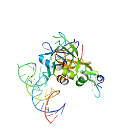 | | DesGla-XaS195A Bound to Aptamer 11F7t | | 分子名称: | Aptamer 11F7t (36-MER), CALCIUM ION, Coagulation factor X, ... | | 著者 | Gunaratne, R, Kumar, S, Frederiksen, J.W, Stayrook, S, Lohrmann, J.L, Perry, K, Chabata, C.V, Thalji, N.K, Ho, M.D, Arepally, G, Camire, R.M, Krishnaswamy, S.K, Sullenger, B.A. | | 登録日 | 2017-05-02 | | 公開日 | 2018-06-20 | | 最終更新日 | 2023-10-04 | | 実験手法 | X-RAY DIFFRACTION (2 Å) | | 主引用文献 | Combination of aptamer and drug for reversible anticoagulation in cardiopulmonary bypass.
Nat. Biotechnol., 36, 2018
|
|
7ME4
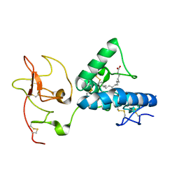 | | Structure of the extracellular WNT-binding module in Drosophila Ror2/Nrk | | 分子名称: | 2-acetamido-2-deoxy-beta-D-glucopyranose-(1-4)-2-acetamido-2-deoxy-beta-D-glucopyranose, PALMITOLEIC ACID, Tyrosine-protein kinase transmembrane receptor Ror2 | | 著者 | Mendrola, J.M, Shi, F, Perry, K, Stayrook, S.E, Lemmon, M.A. | | 登録日 | 2021-04-06 | | 公開日 | 2021-10-13 | | 最終更新日 | 2023-10-18 | | 実験手法 | X-RAY DIFFRACTION (1.75 Å) | | 主引用文献 | ROR and RYK extracellular region structures suggest that receptor tyrosine kinases have distinct WNT-recognition modes.
Cell Rep, 37, 2021
|
|
7ME5
 | | Structure of the extracellular WNT-binding module in Drl-2 | | 分子名称: | 2-acetamido-2-deoxy-beta-D-glucopyranose, Tyrosine-protein kinase transmembrane receptor DRL-2 | | 著者 | Shi, F, Mendrola, J.M, Perry, K, Stayrook, S.E, Lemmon, M.A. | | 登録日 | 2021-04-06 | | 公開日 | 2021-10-13 | | 最終更新日 | 2023-10-18 | | 実験手法 | X-RAY DIFFRACTION (2 Å) | | 主引用文献 | ROR and RYK extracellular region structures suggest that receptor tyrosine kinases have distinct WNT-recognition modes.
Cell Rep, 37, 2021
|
|
2PE5
 | | Crystal Structure of the Lac Repressor bound to ONPG in repressed state | | 分子名称: | 2-nitrophenyl beta-D-galactopyranoside, DNA (5'-D(*DAP*DAP*DTP*DTP*DGP*DTP*DGP*DAP*DGP*DCP*DGP*DCP*DTP*DCP*DAP*DCP*DAP*DAP*DTP*DT)-3'), Lactose operon repressor | | 著者 | Daber, R, Stayrook, S.E, Rosenberg, A, Lewis, M. | | 登録日 | 2007-04-02 | | 公開日 | 2008-03-18 | | 最終更新日 | 2023-08-30 | | 実験手法 | X-RAY DIFFRACTION (3.5 Å) | | 主引用文献 | Structural analysis of lac repressor bound to allosteric effectors
J.Mol.Biol., 370, 2007
|
|
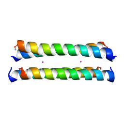
1Y47
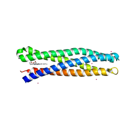
 | | Structural studies of designed alpha-helical hairpins | | 分子名称: | CADMIUM ION, dueferri (DF2) | | 著者 | Lahr, S.J, Engel, D.E, Stayrook, S.E, Maglio, O, North, B, Geremia, S, Lombardi, A, DeGrado, W.F. | | 登録日 | 2004-11-30 | | 公開日 | 2005-03-22 | | 最終更新日 | 2023-08-23 | | 実験手法 | X-RAY DIFFRACTION (2.7 Å) | | 主引用文献 | Analysis and design of turns in alpha-helical hairpins
J.Mol.Biol., 346, 2005
|
|
1M3W
 | | Crystal Structure of a Molecular Maquette Scaffold | | 分子名称: | H10H24, MERCURY (II) ION | | 著者 | Huang, S.S, Gibney, B.R, Stayrook, S.E, Dutton, P.L, Lewis, M. | | 登録日 | 2002-07-01 | | 公開日 | 2003-02-18 | | 最終更新日 | 2011-07-13 | | 実験手法 | X-RAY DIFFRACTION (2.8 Å) | | 主引用文献 | X-ray Structure of a Maquette Scaffold
J.Mol.Biol., 326, 2003
|
|


