Loading
PDBj
MenuPDBj@FacebookPDBj@TwitterPDBj@YouTubewwPDB FoundationwwPDB
RCSB PDBPDBeBMRBAdv. SearchSearch help
PDB: 787 results

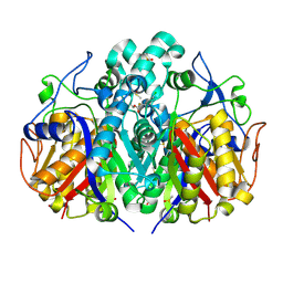
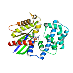
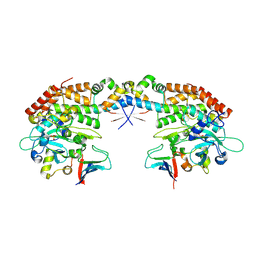
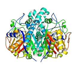
5SN6
DownloadVisualize
BU of 5sn6 by Molmil
PanDDA analysis group deposition -- Crystal Structure of Pseudomonas Aeruginosa FabF-C164Q mutant protein in complex with Z2856434942
Descriptor: 3-oxoacyl-[acyl-carrier-protein] synthase 2, DIMETHYL SULFOXIDE, PHOSPHATE ION, ...
Authors:Brenk, R, Georgiou, C.
Deposit date:2022-05-30
Release date:2023-12-20
Last modified:2024-01-10
Method:X-RAY DIFFRACTION (1.75 Å)
Cite:PanDDA analysis group deposition
Chemrxiv, 2023
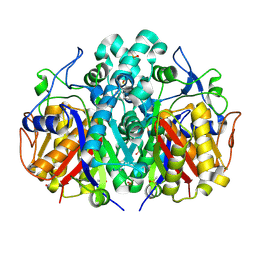
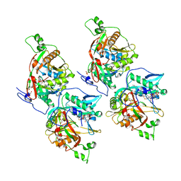
5SNN
DownloadVisualize
BU of 5snn by Molmil
PanDDA analysis group deposition -- Crystal Structure of Pseudomonas Aeruginosa FabF-C164Q mutant protein in complex with Z1954800564
Descriptor: 3-oxoacyl-[acyl-carrier-protein] synthase 2, 5-methoxy-1,3-benzothiazol-2-amine, DIMETHYL SULFOXIDE, ...
Authors:Brenk, R, Georgiou, C.
Deposit date:2022-05-30
Release date:2023-12-20
Last modified:2024-01-10
Method:X-RAY DIFFRACTION (2.08 Å)
Cite:PanDDA analysis group deposition
Chemrxiv, 2023
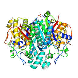
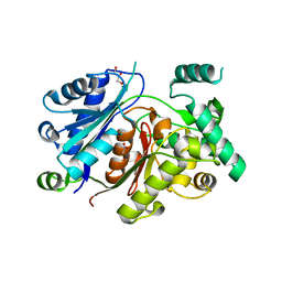
5SO5
DownloadVisualize
BU of 5so5 by Molmil
PanDDA analysis group deposition -- Crystal Structure of Pseudomonas Aeruginosa FabF-C164Q mutant protein in complex with JKH93B
Descriptor: 3-oxoacyl-[acyl-carrier-protein] synthase 2, DIMETHYL SULFOXIDE, PHOSPHATE ION, ...
Authors:Brenk, R, Georgiou, C.
Deposit date:2022-05-30
Release date:2023-12-20
Last modified:2024-01-10
Method:X-RAY DIFFRACTION (1.68 Å)
Cite:PanDDA analysis group deposition
Chemrxiv, 2023
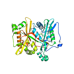
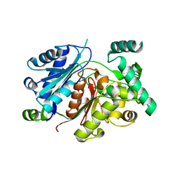
5SO1
DownloadVisualize
BU of 5so1 by Molmil
PanDDA analysis group deposition -- Crystal Structure of Pseudomonas Aeruginosa FabF-C164Q mutant protein in complex with Z30620520
Descriptor: 3-oxoacyl-[acyl-carrier-protein] synthase 2, DIMETHYL SULFOXIDE, PHOSPHATE ION, ...
Authors:Brenk, R, Georgiou, C.
Deposit date:2022-05-30
Release date:2023-12-20
Last modified:2024-01-10
Method:X-RAY DIFFRACTION (2.07 Å)
Cite:PanDDA analysis group deposition
Chemrxiv, 2023
5SOH
DownloadVisualize
BU of 5soh by Molmil
PanDDA analysis group deposition -- Crystal Structure of Pseudomonas Aeruginosa FabF-C164Q mutant protein in complex with Z31432226
Descriptor: (azepan-1-yl)(2H-1,3-benzodioxol-5-yl)methanone, 3-oxoacyl-[acyl-carrier-protein] synthase 2, DIMETHYL SULFOXIDE, ...
Authors:Brenk, R, Georgiou, C.
Deposit date:2022-05-30
Release date:2023-12-20
Last modified:2024-01-10
Method:X-RAY DIFFRACTION (1.61 Å)
Cite:PanDDA analysis group deposition
Chemrxiv, 2023
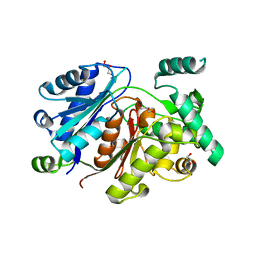
6EU5
DownloadVisualize
BU of 6eu5 by Molmil
Leishmania major N-myristoyltransferase with bound myristoyl-CoA and inhibitor
Descriptor: 4-[3-[(8~{a}~{R})-3,4,6,7,8,8~{a}-hexahydro-1~{H}-pyrrolo[1,2-a]pyrazin-2-yl]propyl]-2,6-bis(chloranyl)-~{N}-methyl-~{N}-(1,3,5-trimethylpyrazol-4-yl)benzenesulfonamide, Glycylpeptide N-tetradecanoyltransferase, TETRADECANOYL-COA
Authors:Brenk, R, Kehrein, J, Kersten, C.
Deposit date:2017-10-27
Release date:2019-02-06
Last modified:2024-01-17
Method:X-RAY DIFFRACTION (1.496083 Å)
Cite:How To Design Selective Ligands for Highly Conserved Binding Sites: A Case Study UsingN-Myristoyltransferases as a Model System.
J.Med.Chem., 2019
6M8H
DownloadVisualize
BU of 6m8h by Molmil
Crystal Structure of the R208Q mutant of G(i) subunit alpha-1
Descriptor: 5'-GUANOSINE-DIPHOSPHATE-MONOTHIOPHOSPHATE, Guanine nucleotide-binding protein G(i) subunit alpha-1, MAGNESIUM ION
Authors:Mascarenhas, R, Goossens, J, Leverson, B, Kothawala, S, Ballicora, M, Olsen, K, de freitas, D, Liu, D.
Deposit date:2018-08-21
Release date:2019-08-21
Last modified:2024-03-13
Method:X-RAY DIFFRACTION (2.07 Å)
Cite:FUNCTIONAL CONSEQUENCES OF ONCOGENIC MUTATIONS IN THE SWITCH II REGION OF Galphai1 and Galphas PROTEINS
To Be Published
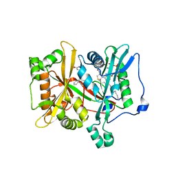
6HSP
DownloadVisualize
BU of 6hsp by Molmil
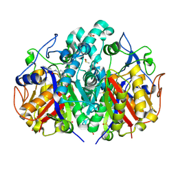
Crystal structure of the zebrafish peroxisomal SCP2-thiolase (type-1) in complex with CoA and octanoyl-CoA
Descriptor: COENZYME A, GLYCEROL, OCTANOYL-COENZYME A, ...
Authors:Wierenga, R.K, Kiema, T.R, Thapa, C.J.
Deposit date:2018-10-01
Release date:2019-01-02
Last modified:2024-01-24
Method:X-RAY DIFFRACTION (1.73 Å)
Cite:The peroxisomal zebrafish SCP2-thiolase (type-1) is a weak transient dimer as revealed by crystal structures and native mass spectrometry.
Biochem. J., 476, 2019
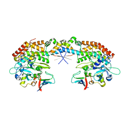
7AB5
DownloadVisualize
BU of 7ab5 by Molmil
Crystal structure of the Escherichia coli toxin-antitoxin system HipBST (HipT D233Q)
Descriptor: Couple_hipA domain-containing protein, HipA_C domain-containing protein, Predicted transcriptional regulator, ...
Authors:Baerentsen, R.L, Brodersen, D.E.
Deposit date:2020-09-06
Release date:2022-01-12
Last modified:2024-05-01
Method:X-RAY DIFFRACTION (2.9 Å)
Cite:Structural basis for regulation of a tripartite toxin-antitoxin system by dual phosphorylation
Biorxiv, 2022
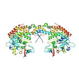
7AB4
DownloadVisualize
BU of 7ab4 by Molmil
Crystal structure of the Escherichia coli toxin-antitoxin system HipBST (HipT S59A)
Descriptor: Couple_hipA domain-containing protein, HipA_C domain-containing protein, Predicted transcriptional regulator, ...
Authors:Baerentsen, R.L, Brodersen, D.E.
Deposit date:2020-09-06
Release date:2022-01-12
Last modified:2024-05-01
Method:X-RAY DIFFRACTION (3.34 Å)
Cite:Structural basis for regulation of a tripartite toxin-antitoxin system by dual phosphorylation
Biorxiv, 2022
7AB3
DownloadVisualize
BU of 7ab3 by Molmil
Crystal structure of the Escherichia coli toxin-antitoxin system HipBST (HipT S57A)
Descriptor: Couple_hipA domain-containing protein, HipA_C domain-containing protein, Predicted transcriptional regulator, ...
Authors:Baerentsen, R.L, Brodersen, D.E.
Deposit date:2020-09-06
Release date:2022-01-12
Last modified:2024-01-31
Method:X-RAY DIFFRACTION (2.4 Å)
Cite:Structural basis for regulation of a tripartite toxin-antitoxin system by dual phosphorylation
Biorxiv, 2022
6F56
DownloadVisualize
BU of 6f56 by Molmil
Mutant of Human N-myristoyltransferase with bound myristoyl-CoA
Descriptor: GLYCEROL, Glycylpeptide N-tetradecanoyltransferase 1, MAGNESIUM ION, ...
Authors:Brenk, R, Kehrein, J, Kersten, C.
Deposit date:2017-11-30
Release date:2019-03-20
Last modified:2024-01-17
Method:X-RAY DIFFRACTION (1.94019878 Å)
Cite:How To Design Selective Ligands for Highly Conserved Binding Sites: A Case Study UsingN-Myristoyltransferases as a Model System.
J.Med.Chem., 2019
6HRV
DownloadVisualize
BU of 6hrv by Molmil
Crystal structure of the zebrafish peroxisomal SCP2-thiolase (type-1)
Descriptor: ACETATE ION, GLYCEROL, SCP2-thiolase (type-1)
Authors:Wierenga, R.K, Kiema, T.R, Thapa, C.J.
Deposit date:2018-09-28
Release date:2019-01-02
Last modified:2024-01-24
Method:X-RAY DIFFRACTION (1.95 Å)
Cite:The peroxisomal zebrafish SCP2-thiolase (type-1) is a weak transient dimer as revealed by crystal structures and native mass spectrometry.
Biochem. J., 476, 2019
6HSJ
DownloadVisualize
BU of 6hsj by Molmil
Crystal structure of the zebrafish peroxisomal SCP2-thiolase (type-1) in complex with CoA
Descriptor: ACETATE ION, COENZYME A, GLYCEROL, ...
Authors:Wierenga, R.K, Kiema, T.R, Thapa, C.J.
Deposit date:2018-10-01
Release date:2019-01-02
Last modified:2024-01-24
Method:X-RAY DIFFRACTION (1.46 Å)
Cite:The peroxisomal zebrafish SCP2-thiolase (type-1) is a weak transient dimer as revealed by crystal structures and native mass spectrometry.
Biochem. J., 476, 2019
5KJY
DownloadVisualize
BU of 5kjy by Molmil
Co-crystal structure of PKA RI alpha CNB-B mutant (G316R/A336T) with cAMP
Descriptor: ADENOSINE-3',5'-CYCLIC-MONOPHOSPHATE, cAMP-dependent protein kinase type I-alpha regulatory subunit
Authors:Lorenz, R, Moon, E, Kim, J.J, Huang, G.Y, Kim, C, Herberg, F.W.
Deposit date:2016-06-20
Release date:2017-06-28
Last modified:2023-09-27
Method:X-RAY DIFFRACTION (2 Å)
Cite:Mutations of PKA cyclic nucleotide-binding domains reveal novel aspects of cyclic nucleotide selectivity.
Biochem. J., 474, 2017
5KJX
DownloadVisualize
BU of 5kjx by Molmil
Co-crystal Structure of PKA RI alpha CNB-B domain with cAMP
Descriptor: ADENOSINE-3',5'-CYCLIC-MONOPHOSPHATE, cAMP-dependent protein kinase type I-alpha regulatory subunit
Authors:Lorenz, R, Moon, E, Kim, J.J, Huang, G.Y, Kim, C, Herberg, F.W.
Deposit date:2016-06-20
Release date:2017-06-28
Last modified:2023-09-27
Method:X-RAY DIFFRACTION (1.9 Å)
Cite:Mutations of PKA cyclic nucleotide-binding domains reveal novel aspects of cyclic nucleotide selectivity.
Biochem. J., 474, 2017
6EWF
DownloadVisualize
BU of 6ewf by Molmil
Leishmania major N-myristoyltransferase with bound myristoyl-CoA and inhibitor
Descriptor: Glycylpeptide N-tetradecanoyltransferase, N-[2-(3-methoxyphenyl)ethanimidoyl]-2-piperidin-4-yloxy-benzamide, TETRADECANOYL-COA
Authors:Brenk, R, Kehrein, J, Kersten, C.
Deposit date:2017-11-03
Release date:2019-02-06
Last modified:2024-01-17
Method:X-RAY DIFFRACTION (1.5351733 Å)
Cite:How To Design Selective Ligands for Highly Conserved Binding Sites: A Case Study UsingN-Myristoyltransferases as a Model System.
J.Med.Chem., 2019
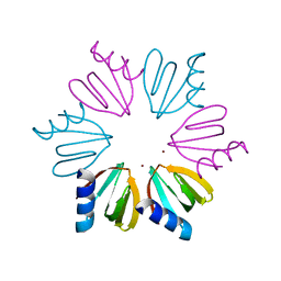
3QSU
DownloadVisualize
BU of 3qsu by Molmil
Structure of Staphylococcus aureus Hfq in complex with A7 RNA
Descriptor: RNA (5'-R(P*AP*AP*AP*AP*AP*AP*A)-3'), RNA chaperone Hfq, ZINC ION
Authors:Brennan, R, Horstmann, N, Link, T.M.
Deposit date:2011-02-21
Release date:2012-09-12
Last modified:2024-02-21
Method:X-RAY DIFFRACTION (2.2 Å)
Cite:Structural mechanism of Staphylococcus aureus Hfq binding to an RNA A-tract.
Nucleic Acids Res., 40, 2012
4ZDP
DownloadVisualize
BU of 4zdp by Molmil
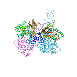
The crystal structure of Y334C mutant of human SepSecS in complex with selenocysteine tRNA (tRNASec)
Descriptor: (5-HYDROXY-4,6-DIMETHYLPYRIDIN-3-YL)METHYL DIHYDROGEN PHOSPHATE, O-phosphoseryl-tRNA(Sec) selenium transferase, PHOSPHATE ION, ...
Authors:French, R.L, Simonovic, M.
Deposit date:2015-04-17
Release date:2016-04-20
Last modified:2023-09-27
Method:X-RAY DIFFRACTION (2.703 Å)
Cite:Structural basis for early-onset neurological disorders caused by mutations in human selenocysteine synthase.
Sci Rep, 6, 2016
4ZDO
DownloadVisualize
BU of 4zdo by Molmil
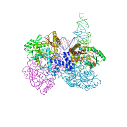
The crystal structure of T325S mutant of human SepSecS in complex with selenocysteine tRNA (tRNASec)
Descriptor: (5-HYDROXY-4,6-DIMETHYLPYRIDIN-3-YL)METHYL DIHYDROGEN PHOSPHATE, O-phosphoseryl-tRNA(Sec) selenium transferase, PHOSPHATE ION, ...
Authors:French, R.L, Simonovic, M.
Deposit date:2015-04-17
Release date:2016-04-20
Last modified:2023-09-27
Method:X-RAY DIFFRACTION (2.65 Å)
Cite:Structural basis for early-onset neurological disorders caused by mutations in human selenocysteine synthase.
Sci Rep, 6, 2016
8QAI
DownloadVisualize
BU of 8qai by Molmil
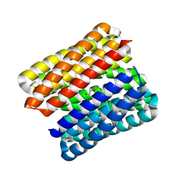
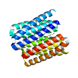
X-ray crystal structure of a de novo designed single-chain parallel coiled-coil alpha-helical barrel with 7 inner helices, sc-CC-7-LI
Descriptor: DI(HYDROXYETHYL)ETHER, sc-CC-7-LI
Authors:Petrenas, R, Albanese, K.I, Woolfson, D.N.
Deposit date:2023-08-22
Release date:2024-07-03
Last modified:2024-08-07
Method:X-RAY DIFFRACTION (2.5 Å)
Cite:Rationally seeded computational protein design of ɑ-helical barrels.
Nat.Chem.Biol., 20, 2024
8QAH
DownloadVisualize
BU of 8qah by Molmil
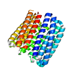
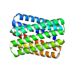
X-ray crystal structure of a de novo designed single-chain parallel coiled-coil alpha-helical barrel with 8 inner helices, sc-CC-8-58
Descriptor: sc-CC-8-58
Authors:Petrenas, R, Albanese, K.I, Woolfson, D.N.
Deposit date:2023-08-22
Release date:2024-07-03
Last modified:2024-08-07
Method:X-RAY DIFFRACTION (2.35 Å)
Cite:Rationally seeded computational protein design of ɑ-helical barrels.
Nat.Chem.Biol., 20, 2024
8QAG
DownloadVisualize
BU of 8qag by Molmil
X-ray crystal structure of a de novo designed single-chain parallel coiled-coil alpha-helical barrel with 6 inner helices, sc-CC-6-95
Descriptor: DI(HYDROXYETHYL)ETHER, TETRAETHYLENE GLYCOL, sc-CC-6-95
Authors:Petrenas, R, Albanese, K.I, Woolfson, D.N.
Deposit date:2023-08-22
Release date:2024-07-03
Last modified:2024-08-07
Method:X-RAY DIFFRACTION (2.8 Å)
Cite:Rationally seeded computational protein design of ɑ-helical barrels.
Nat.Chem.Biol., 20, 2024
8QKD
DownloadVisualize
BU of 8qkd by Molmil
X-ray crystal structure of a de novo designed single-chain parallel coiled-coil alpha-helical barrel with 5 inner helices, sc-CC-5-24
Descriptor: DI(HYDROXYETHYL)ETHER, sc-CC-5-24
Authors:Petrenas, R, Albanese, K.I, Woolfson, D.N.
Deposit date:2023-09-15
Release date:2024-07-03
Last modified:2024-08-07
Method:X-RAY DIFFRACTION (1.9 Å)
Cite:Rationally seeded computational protein design of ɑ-helical barrels.
Nat.Chem.Biol., 20, 2024
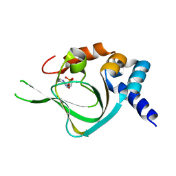
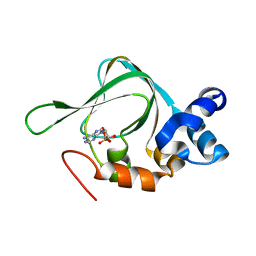
4WYZ
DownloadVisualize
BU of 4wyz by Molmil
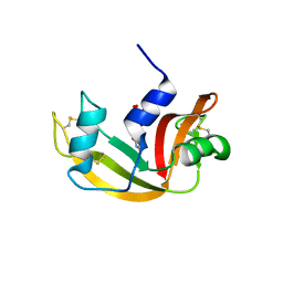
The crystal structure of the A109G mutant of RNase A in complex with 3'UMP
Descriptor: 3'-URIDINEMONOPHOSPHATE, Ribonuclease pancreatic
Authors:French, R.L, Gagne, D, Doucet, N, Simonovic, M.
Deposit date:2014-11-18
Release date:2015-11-18
Last modified:2019-12-25
Method:X-RAY DIFFRACTION (1.449 Å)
Cite:Perturbation of the Conformational Dynamics of an Active-Site Loop Alters Enzyme Activity.
Structure, 23, 2015

223790

數據於2024-08-14公開中

PDB statisticsPDBj update infoContact PDBjnumon