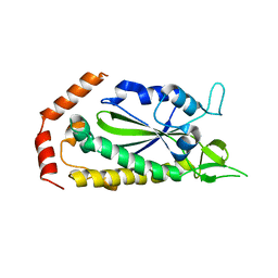
2Q7T
 
 | |
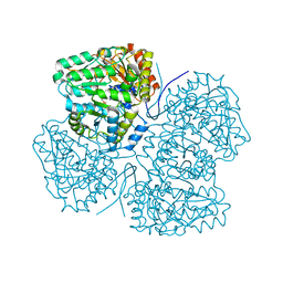
8SBG
 
 | |
8UGT
 
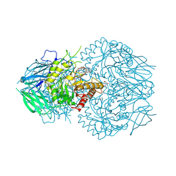 | | E. eligens beta-glucuronidase bound to UNC10206581-G | | 分子名称: | 8-(4-beta-D-glucopyranuronosylpiperazin-1-yl)-5-(methylamino)-1,2,3,4-tetrahydro[1,2,3]triazino[4',5':4,5]thieno[2,3-c]isoquinoline, Beta-glucuronidase, GLYCEROL, ... | | 著者 | Simpson, J.B, Redinbo, M.R. | | 登録日 | 2023-10-06 | | 公開日 | 2024-07-24 | | 最終更新日 | 2024-09-11 | | 実験手法 | X-RAY DIFFRACTION (2.65 Å) | | 主引用文献 | Advanced piperazine-containing inhibitors target microbial beta-glucuronidases linked to gut toxicity.
Rsc Chem Biol, 5, 2024
|
|
8UMG
 
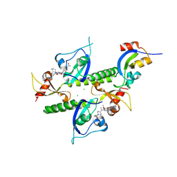 | | Chromodomains of human CHD1 complexed with UNC10142 | | 分子名称: | 1-{4-[{2-(azonan-1-yl)-6-methoxy-7-[3-(piperidin-1-yl)propoxy]quinazolin-4-yl}(methyl)amino]piperidin-1-yl}ethan-1-one, CHLORIDE ION, Chromodomain-helicase-DNA-binding protein 1 | | 著者 | Graboski, A.L, Redinbo, M.R. | | 登録日 | 2023-10-17 | | 公開日 | 2024-10-23 | | 実験手法 | X-RAY DIFFRACTION (3.1 Å) | | 主引用文献 | Optimization of a quinazoline-based scaffold against human CHD1
To Be Published
|
|
8W1Q
 
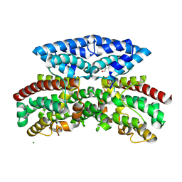 | | Aerobic crystal structure of iron-bound FlcD from Pseudomonas aeruginosa | | 分子名称: | FE (II) ION, GLYCEROL, MAGNESIUM ION, ... | | 著者 | Walker, M.E, Grove, T.L, Li, B, Redinbo, M.R. | | 登録日 | 2024-02-17 | | 公開日 | 2024-07-31 | | 最終更新日 | 2024-09-18 | | 実験手法 | X-RAY DIFFRACTION (1.56 Å) | | 主引用文献 | Structural Basis for Methine Excision by a Heme Oxygenase-like Enzyme.
Acs Cent.Sci., 10, 2024
|
|
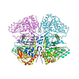
8SIJ
 
 | |
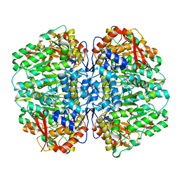
8SL7
 
 | |
5CZK
 
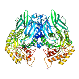 | | Structure of E. coli beta-glucuronidase bound with a novel, potent inhibitor 1-((6,8-dimethyl-2-oxo-1,2-dihydroquinolin-3-yl)methyl)-1-(2-hydroxyethyl)-3-(4-hydroxyphenyl)thiourea | | 分子名称: | 1-[(6,8-dimethyl-2-oxo-1,2-dihydroquinolin-3-yl)methyl]-1-(2-hydroxyethyl)-3-(4-hydroxyphenyl)thiourea, Beta-glucuronidase | | 著者 | Roberts, A.R, Wallace, B.R, Redinbo, M.R. | | 登録日 | 2015-07-31 | | 公開日 | 2015-10-14 | | 最終更新日 | 2019-12-04 | | 実験手法 | X-RAY DIFFRACTION (2.39 Å) | | 主引用文献 | Structure and Inhibition of Microbiome beta-Glucuronidases Essential to the Alleviation of Cancer Drug Toxicity.
Chem.Biol., 22, 2015
|
|
9B9M
 
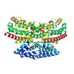 | | Crystal structure of iron-bound FlcD from Pseudomonas aeruginosa | | 分子名称: | FE (II) ION, Pyrroloquinoline quinone (Coenzyme PQQ) biosynthesis protein C | | 著者 | Walker, M.E, Grove, T.L, Li, B, Redinbo, M.R. | | 登録日 | 2024-04-02 | | 公開日 | 2024-07-31 | | 最終更新日 | 2024-09-18 | | 実験手法 | X-RAY DIFFRACTION (2.07 Å) | | 主引用文献 | Structural Basis for Methine Excision by a Heme Oxygenase-like Enzyme.
Acs Cent.Sci., 10, 2024
|
|
6ED2
 
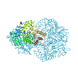 | | Faecalibacterium prausnitzii beta-glucuronidase | | 分子名称: | FORMIC ACID, GLYCEROL, Glycosyl hydrolase family 2, ... | | 著者 | Pellock, S.J, Biernat, K.A, Redinbo, M.R. | | 登録日 | 2018-08-08 | | 公開日 | 2019-02-13 | | 最終更新日 | 2024-03-13 | | 実験手法 | X-RAY DIFFRACTION (2.3 Å) | | 主引用文献 | Structure, function, and inhibition of drug reactivating human gut microbial beta-glucuronidases.
Sci Rep, 9, 2019
|
|
9B9O
 
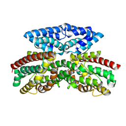 | | Crystal structure of FlcD from Pseudomonas aeruginosa bond to iron(II) and substrate | | 分子名称: | (2R)-2-{[(2Z)-2-(hydroxyimino)ethyl]sulfanyl}butanedioic acid, FE (II) ION, Pyrroloquinoline quinone (Coenzyme PQQ) biosynthesis protein C | | 著者 | Walker, M.E, Grove, T.L, Li, B, Redinbo, M.R. | | 登録日 | 2024-04-02 | | 公開日 | 2024-07-31 | | 最終更新日 | 2024-09-18 | | 実験手法 | X-RAY DIFFRACTION (2.16 Å) | | 主引用文献 | Structural Basis for Methine Excision by a Heme Oxygenase-like Enzyme.
Acs Cent.Sci., 10, 2024
|
|
9B9N
 
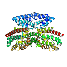 | | Crystal structure of FlcD from Pseudomonas aeruginosa bound to iron (II) and substrate | | 分子名称: | (2R)-2-{[(2Z)-2-(hydroxyimino)ethyl]sulfanyl}butanedioic acid, FE (III) ION, Pyrroloquinoline quinone (Coenzyme PQQ) biosynthesis protein C | | 著者 | Walker, M.E, Grove, T.L, Li, B, Redinbo, M.R. | | 登録日 | 2024-04-02 | | 公開日 | 2024-07-31 | | 最終更新日 | 2024-09-18 | | 実験手法 | X-RAY DIFFRACTION (2.28 Å) | | 主引用文献 | Structural Basis for Methine Excision by a Heme Oxygenase-like Enzyme.
Acs Cent.Sci., 10, 2024
|
|
2QNV
 
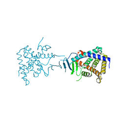 | | Crystal Structure of the Pregnane X Receptor bound to Colupulone | | 分子名称: | 3,5-dihydroxy-4,6,6-tris(3-methylbut-2-en-1-yl)-2-(2-methylpropanoyl)cyclohexa-2,4-dien-1-one, Orphan nuclear receptor PXR | | 著者 | Teotico, D.G, Bischof, J, Redinbo, M.R. | | 登録日 | 2007-07-19 | | 公開日 | 2008-07-29 | | 最終更新日 | 2023-08-30 | | 実験手法 | X-RAY DIFFRACTION (2.8 Å) | | 主引用文献 | Crystal Structure of the Pregnane X Receptor bound to Colupulone
TO BE PUBLISHED
|
|
6ED1
 
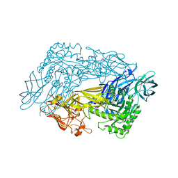 | | Bacteroides dorei Beta-glucuronidase | | 分子名称: | Glycosyl hydrolase family 2, sugar binding domain protein, SODIUM ION | | 著者 | Biernat, K.A, Redinbo, M.R. | | 登録日 | 2018-08-08 | | 公開日 | 2019-02-13 | | 最終更新日 | 2023-10-11 | | 実験手法 | X-RAY DIFFRACTION (2.9 Å) | | 主引用文献 | Structure, function, and inhibition of drug reactivating human gut microbial beta-glucuronidases.
Sci Rep, 9, 2019
|
|
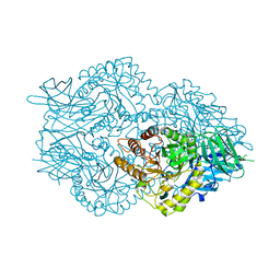
6EC6
 
 | |
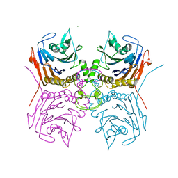
7SVH
 
 | |
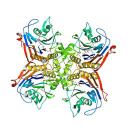
7SVG
 
 | |
7SVJ
 
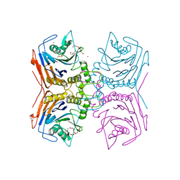 | | Bile Salt Hydrolase from Lactobacillus ingluviei | | 分子名称: | CALCIUM ION, Choloylglycine hydrolase, DI(HYDROXYETHYL)ETHER, ... | | 著者 | Walker, M.E, Patel, S, Redinbo, M.R. | | 登録日 | 2021-11-19 | | 公開日 | 2023-01-25 | | 最終更新日 | 2023-10-25 | | 実験手法 | X-RAY DIFFRACTION (1.43 Å) | | 主引用文献 | Bile salt hydrolases shape the bile acid landscape and restrict Clostridioides difficile growth in the murine gut.
Nat Microbiol, 8, 2023
|
|
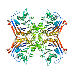
7SVI
 
 | |
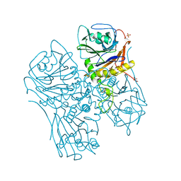
7SVK
 
 | |
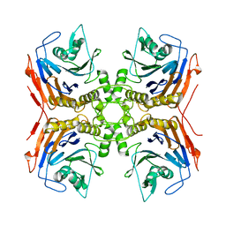
7SVE
 
 | |
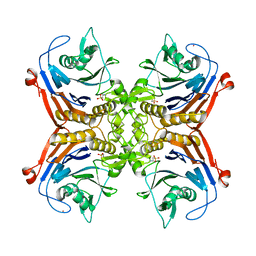
7SVF
 
 | |
3B7Q
 
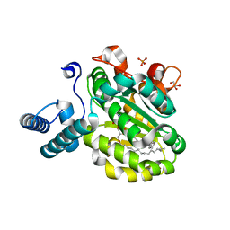 | | Crystal Structure of Yeast Sec14 Homolog Sfh1 in Complex with Phosphatidylcholine | | 分子名称: | (4S,7R)-4-HYDROXY-N,N,N-TRIMETHYL-9-OXO-7-[(PALMITOYLOXY)METHYL]-3,5,8-TRIOXA-4-PHOSPHAHEXACOSAN-1-AMINIUM 4-OXIDE, GLYCEROL, PHOSPHATE ION, ... | | 著者 | Ortlund, E.A, Schaaf, G, Redinbo, M.R, Bankaitis, V. | | 登録日 | 2007-10-31 | | 公開日 | 2008-02-19 | | 最終更新日 | 2023-08-30 | | 実験手法 | X-RAY DIFFRACTION (2.03 Å) | | 主引用文献 | Functional anatomy of phospholipid binding and regulation of phosphoinositide homeostasis by proteins of the sec14 superfamily
Mol.Cell, 29, 2008
|
|
3B74
 
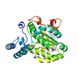 | | Crystal Structure of Yeast Sec14 Homolog Sfh1 in Complex with Phosphatidylethanolamine | | 分子名称: | 1,2-dioleoyl-sn-glycero-3-phosphoethanolamine, Uncharacterized protein YKL091C | | 著者 | Ortlund, E.A, Schaaf, G, Redinbo, M.R, Bankaitis, V. | | 登録日 | 2007-10-30 | | 公開日 | 2008-02-19 | | 最終更新日 | 2023-08-30 | | 実験手法 | X-RAY DIFFRACTION (1.9 Å) | | 主引用文献 | Functional anatomy of phospholipid binding and regulation of phosphoinositide homeostasis by proteins of the sec14 superfamily
Mol.Cell, 29, 2008
|
|
3B7N
 
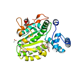 | | Crystal Structure of Yeast Sec14 Homolog Sfh1 in Complex with Phosphatidylinositol | | 分子名称: | (1R)-2-{[(S)-hydroxy{[(1S,2R,3R,4S,5S,6R)-2,3,4,5,6-pentahydroxycyclohexyl]oxy}phosphoryl]oxy}-1-[(octadecanoyloxy)methyl]ethyl (9Z)-octadec-9-enoate, ACETATE ION, PHOSPHATE ION, ... | | 著者 | Ortlund, E.A, Schaaf, G, Redinbo, M.R, Bankaitis, V. | | 登録日 | 2007-10-31 | | 公開日 | 2008-02-19 | | 最終更新日 | 2024-02-21 | | 実験手法 | X-RAY DIFFRACTION (1.86 Å) | | 主引用文献 | Functional anatomy of phospholipid binding and regulation of phosphoinositide homeostasis by proteins of the sec14 superfamily
Mol.Cell, 29, 2008
|
|
