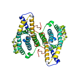
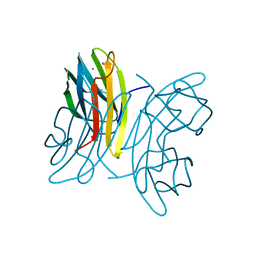
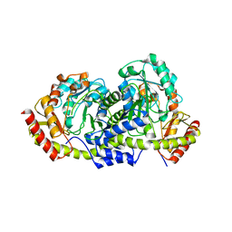
1WT5
 
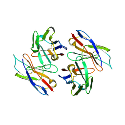 | | The Crystal Structure Of A Humanized Antibody Fv 528 | | 分子名称: | ANTI EGFR ANTIBODY FV REGION | | 著者 | Makabe, K, Tsumoto, K, Asano, R, Kondo, H, Kumagai, I. | | 登録日 | 2004-11-16 | | 公開日 | 2005-05-16 | | 最終更新日 | 2011-07-13 | | 実験手法 | X-RAY DIFFRACTION (2.1 Å) | | 主引用文献 | Thermodynamic consequences of mutations in vernier zone residues of a humanized anti-human epidermal growth factor receptor murine antibody, 528
J.Biol.Chem., 283, 2008
|
|
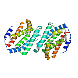
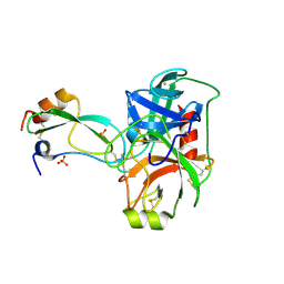
1P0N
 | | IPP:DMAPP isomerase type II, FMN complex | | 分子名称: | FLAVIN MONONUCLEOTIDE, Isopentenyl-diphosphate delta-isomerase | | 著者 | Steinbacher, S, Kaiser, J, Gerhardt, S, Eisenreich, W, Huber, R, Bacher, A, Rohdich, F. | | 登録日 | 2003-04-10 | | 公開日 | 2003-06-17 | | 最終更新日 | 2023-10-25 | | 実験手法 | X-RAY DIFFRACTION (2.8 Å) | | 主引用文献 | Crystal Structure of the Type II Isopentenyl Diphosphate:Dimethylallyl Diphosphate Isomerase from Bacillus subtilis
J.Mol.Biol., 329, 2003
|
|
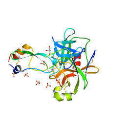
6F23
 | | Complex between MTH1 and compound 16 (a 4-amino-7-azaindole derivative) | | 分子名称: | 4-[(2~{R})-2-phenylpyrrolidin-1-yl]-1~{H}-pyrrolo[2,3-b]pyridine, 7,8-dihydro-8-oxoguanine triphosphatase, GLYCEROL, ... | | 著者 | Viklund, J, Tresaugues, L, Talagas, A, Andersson, M, Ericsson, U, Forsblom, R, Ginman, T, Hallberg, K, Lindstrom, J, Persson, L, Silvander, C, Rahm, F. | | 登録日 | 2017-11-23 | | 公開日 | 2018-03-07 | | 最終更新日 | 2024-01-17 | | 実験手法 | X-RAY DIFFRACTION (1.84 Å) | | 主引用文献 | Creation of a Novel Class of Potent and Selective MutT Homologue 1 (MTH1) Inhibitors Using Fragment-Based Screening and Structure-Based Drug Design.
J. Med. Chem., 61, 2018
|
|
1WLY
 | |
1OTV
 | | PqqC, Pyrroloquinolinquinone Synthase C | | 分子名称: | Coenzyme PQQ synthesis protein C | | 著者 | Magnusson, O.T, Toyama, H, Saeki, M, Rojas, A, Reed, J.C, Adachi, O, Klinman, J.P, SChwarzenbacher, R. | | 登録日 | 2003-03-23 | | 公開日 | 2004-05-11 | | 最終更新日 | 2024-02-14 | | 実験手法 | X-RAY DIFFRACTION (2.1 Å) | | 主引用文献 | Quinone Biogenesis: Structure and Mechanism of PqqC, the Final Catalyst in the Production of Pyrroloquinoline Quinone.
Proc.Natl.Acad.Sci.USA, 101, 2004
|
|
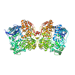
6FA9
 | | CRYSTAL STRUCTURE OF THE DEAH-BOX HELICASE PRP2 | | 分子名称: | Putative mRNA splicing factor, SULFATE ION | | 著者 | Schmitt, A, Hamann, F, Neumann, P, Ficner, R. | | 登録日 | 2017-12-15 | | 公開日 | 2018-07-11 | | 最終更新日 | 2024-01-17 | | 実験手法 | X-RAY DIFFRACTION (2.6 Å) | | 主引用文献 | Crystal structure of the spliceosomal DEAH-box ATPase Prp2.
Acta Crystallogr D Struct Biol, 74, 2018
|
|
3NWN
 | | Crystal structure of the human KIF9 motor domain in complex with ADP | | 分子名称: | ADENOSINE-5'-DIPHOSPHATE, CHLORIDE ION, Kinesin-like protein KIF9, ... | | 著者 | Zhu, H, Tempel, W, He, H, Shen, Y, Wang, J, Brothers, G, Landry, R, Arrowsmith, C.H, Edwards, A.M, Sundstrom, M, Weigelt, J, Bochkarev, A, Park, H, Structural Genomics Consortium (SGC) | | 登録日 | 2010-07-09 | | 公開日 | 2010-07-21 | | 最終更新日 | 2023-09-06 | | 実験手法 | X-RAY DIFFRACTION (2 Å) | | 主引用文献 | Crystal structure of the human KIF9 motor domain in complex with ADP
To be Published
|
|
6FB9
 | |
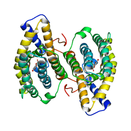
1WUR
 | | Structure of GTP cyclohydrolase I Complexed with 8-oxo-dGTP | | 分子名称: | 8-OXO-2'-DEOXYGUANOSINE-5'-TRIPHOSPHATE, GTP cyclohydrolase I, ZINC ION | | 著者 | Tanaka, Y, Nakagawa, N, Masui, R, Yokoyama, S, Kuramitsu, S, RIKEN Structural Genomics/Proteomics Initiative (RSGI) | | 登録日 | 2004-12-08 | | 公開日 | 2005-07-19 | | 最終更新日 | 2024-03-13 | | 実験手法 | X-RAY DIFFRACTION (1.82 Å) | | 主引用文献 | Novel reaction mechanism of GTP cyclohydrolase I. High-resolution X-ray crystallography of Thermus thermophilus HB8 enzyme complexed with a transition state analogue, the 8-oxoguanine derivative
J.Biochem.(Tokyo), 138, 2005
|
|
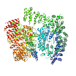
4IFH
 | | Crystal structure of human insulin degrading enzyme (IDE) in complex with compound BDM44619 | | 分子名称: | Insulin-degrading enzyme, N-({1-[(2R)-4-(hydroxyamino)-1-(naphthalen-2-yl)-4-oxobutan-2-yl]-1H-1,2,3-triazol-4-yl}methyl)-4-methylbenzamide, ZINC ION | | 著者 | Liang, W.G, Guo, Q, Deprez, R, Deprez, B, Tang, W. | | 登録日 | 2012-12-14 | | 公開日 | 2013-12-18 | | 最終更新日 | 2024-02-28 | | 実験手法 | X-RAY DIFFRACTION (3.286 Å) | | 主引用文献 | Catalytic site inhibition of insulin-degrading enzyme by a small molecule induces glucose intolerance in mice.
Nat Commun, 6, 2015
|
|
1WWM
 | | Crystal Structure of Conserved Hypothetical Protein TT2028 from an Extremely Thermophilic Bacterium Thermus thermophilus HB8 | | 分子名称: | hypothetical protein TT2028 | | 著者 | Mizohata, E, Ushikoshi-Nakayama, R, Terada, T, Murayama, K, Sakai, H, Kuramitsu, S, Shirouzu, M, Yokoyama, S, RIKEN Structural Genomics/Proteomics Initiative (RSGI) | | 登録日 | 2005-01-07 | | 公開日 | 2005-07-07 | | 最終更新日 | 2011-07-13 | | 実験手法 | X-RAY DIFFRACTION (2.61 Å) | | 主引用文献 | Crystal Structure of TT2028 from an Extremely Thermophilic Bacterium Thermus thermophilus HB8
To be Published
|
|
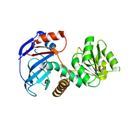
3OC0
 | | Structure of human DPP-IV with HTS hit (2S,3S,11bS)-3-butyl-9,10-dimethoxy-1,3,4,6,7,11b-hexahydro-2H-pyrido[2,1-a]isoquinolin-2-ylamine | | 分子名称: | (2S,3R,11bR)-3-butyl-9,10-dimethoxy-1,3,4,6,7,11b-hexahydro-2H-pyrido[2,1-a]isoquinolin-2-amine, 2-acetamido-2-deoxy-beta-D-glucopyranose, Dipeptidyl peptidase 4 | | 著者 | Hennig, M, Stihle, M, Thoma, R. | | 登録日 | 2010-08-09 | | 公開日 | 2010-08-25 | | 最終更新日 | 2020-07-29 | | 実験手法 | X-RAY DIFFRACTION (2.7 Å) | | 主引用文献 | Discovery of carmegliptin: A potent and long-acting dipeptidyl peptidase IV inhibitor for the treatment of type 2 diabetes.
Bioorg.Med.Chem.Lett., 20, 2010
|
|
1OTW
 | | Crystal structure of PqqC in complex with PQQ and a putative H2O2 | | 分子名称: | Coenzyme PQQ synthesis protein C, HYDROGEN PEROXIDE, PYRROLOQUINOLINE QUINONE | | 著者 | Magnusson, O.T, Toyama, H, Saeki, M, Rojas, A, Reed, J.C, Liddington, R.C, Klinman, J.P, Schwarzenbacher, R. | | 登録日 | 2003-03-23 | | 公開日 | 2004-05-11 | | 最終更新日 | 2023-08-16 | | 実験手法 | X-RAY DIFFRACTION (2.3 Å) | | 主引用文献 | Quinone biogenesis: Structure and mechanism of PqqC, the final catalyst in the production of pyrroloquinoline quinone.
Proc.Natl.Acad.Sci.USA, 101, 2004
|
|
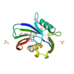
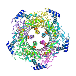
1OUR
 | | LecB (PA-LII) in complex with mannose | | 分子名称: | CALCIUM ION, alpha-D-mannopyranose, hypothetical protein LecB | | 著者 | Loris, R, Tielker, D, Jaeger, K.-E, Wyns, L. | | 登録日 | 2003-03-25 | | 公開日 | 2003-09-09 | | 最終更新日 | 2024-03-13 | | 実験手法 | X-RAY DIFFRACTION (1.42 Å) | | 主引用文献 | Structural Basis of Carbohydrate Recognition by the Lectin LecB from Pseudomonas aeruginosa
J.MOL.BIOL., 331, 2003
|
|
6EWJ
 | | Putative sugar aminotransferase Spr1654 from Streptococcus pneumoniae, apo-form | | 分子名称: | Putative capsular polysaccharide biosynthesis protein | | 著者 | Achour, A, Sun, R, Sandalova, T, Han, X. | | 登録日 | 2017-11-04 | | 公開日 | 2018-05-02 | | 最終更新日 | 2024-01-17 | | 実験手法 | X-RAY DIFFRACTION (1.9 Å) | | 主引用文献 | Structural and functional studies of Spr1654: an essential aminotransferase in teichoic acid biosynthesis inStreptococcus pneumoniae.
Open Biol, 8, 2018
|
|
1P1M
 | | Structure of Thermotoga maritima amidohydrolase TM0936 bound to Ni and methionine | | 分子名称: | Hypothetical protein TM0936, METHIONINE, NICKEL (II) ION | | 著者 | Kniewel, R, Buglino, J.A, Lima, C.D, Burley, S.K, New York SGX Research Center for Structural Genomics (NYSGXRC) | | 登録日 | 2003-04-12 | | 公開日 | 2003-04-29 | | 最終更新日 | 2024-02-14 | | 実験手法 | X-RAY DIFFRACTION (1.5 Å) | | 主引用文献 | Structure of the hypothetical protein TM0936 from Thermotoga maritima at
1.5A bound to Ni and methionine
To be Published, 2003
|
|
1P2K
 | | Structural consequences of accommodation of four non-cognate amino-acid residues in the S1 pocket of bovine trypsin and chymotrypsin | | 分子名称: | CALCIUM ION, Pancreatic trypsin inhibitor, SULFATE ION, ... | | 著者 | Helland, R, Czapinska, H, Leiros, I, Olufsen, M, Otlewski, J, Smalaas, A.O. | | 登録日 | 2003-04-15 | | 公開日 | 2004-04-20 | | 最終更新日 | 2023-08-16 | | 実験手法 | X-RAY DIFFRACTION (1.6 Å) | | 主引用文献 | Structural consequences of accommodation of four non-cognate amino acid residues in the S1 pocket of bovine trypsin and chymotrypsin.
J.Mol.Biol., 333, 2003
|
|
1P2Q
 | | Structural consequences of accommodation of four non-cognate amino-acid residues in the S1 pocket of bovine trypsin and chymotrypsin | | 分子名称: | 2-AMINO-2-HYDROXYMETHYL-PROPANE-1,3-DIOL, Chymotrypsinogen A, Pancreatic trypsin inhibitor, ... | | 著者 | Helland, R, Czapinska, H, Leiros, I, Olufsen, M, Otlewski, J, Smalaas, A.O. | | 登録日 | 2003-04-15 | | 公開日 | 2004-04-20 | | 最終更新日 | 2023-08-16 | | 実験手法 | X-RAY DIFFRACTION (1.8 Å) | | 主引用文献 | Structural consequences of accommodation of four non-cognate amino acid residues in the S1 pocket of bovine trypsin and chymotrypsin.
J.Mol.Biol., 333, 2003
|
|
1P3E
 | | Structure of Glu endopeptidase in complex with MPD | | 分子名称: | (4S)-2-METHYL-2,4-PENTANEDIOL, glutamyl-endopeptidase | | 著者 | Meijers, R, Blagova, E.V, Levdikov, V.M, Rudenskaya, G.N, Chestukhina, G.G, Akimkina, T.V, Kostrov, S.V, Lamzin, V.S, Kuranova, I.P. | | 登録日 | 2003-04-17 | | 公開日 | 2004-04-27 | | 最終更新日 | 2023-08-16 | | 実験手法 | X-RAY DIFFRACTION (1.72 Å) | | 主引用文献 | The crystal structure of glutamyl endopeptidase from Bacillus intermedius reveals a structural link between zymogen activation and charge compensation.
Biochemistry, 43, 2004
|
|
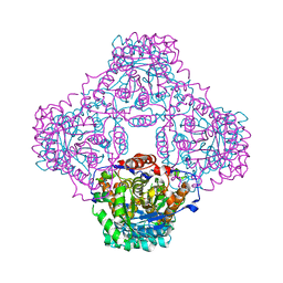
6EZ8
 | | Human Huntingtin-HAP40 complex structure | | 分子名称: | Factor VIII intron 22 protein, Huntingtin | | 著者 | Guo, Q, Bin, H, Cheng, J, Pfeifer, G, Baumeister, W, Fernandez-Busnadiego, R, Kochanek, S. | | 登録日 | 2017-11-14 | | 公開日 | 2018-02-21 | | 最終更新日 | 2019-12-11 | | 実験手法 | ELECTRON MICROSCOPY (4 Å) | | 主引用文献 | The cryo-electron microscopy structure of huntingtin.
Nature, 555, 2018
|
|
4IOR
 | |
6EVM
 | | Crystal structure of a Pro-9 complexed peptide-substrate-binding domain of human type II collagen prolyl 4-hydroxylase | | 分子名称: | DIMETHYL SULFOXIDE, Pro-9, Prolyl 4-hydroxylase subunit alpha-2, ... | | 著者 | Murthy, A.V, Sulu, R, Koski, M.K, Wierenga, R.K. | | 登録日 | 2017-11-02 | | 公開日 | 2018-09-12 | | 最終更新日 | 2024-01-17 | | 実験手法 | X-RAY DIFFRACTION (2 Å) | | 主引用文献 | Structural enzymology binding studies of the peptide-substrate-binding domain of human collagen prolyl 4-hydroxylase (type-II): High affinity peptides have a PxGP sequence motif.
Protein Sci., 27, 2018
|
|
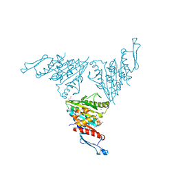
1P8T
 | | Crystal structure of Nogo-66 Receptor | | 分子名称: | 2-acetamido-2-deoxy-alpha-D-glucopyranose, 2-acetamido-2-deoxy-beta-D-glucopyranose, Reticulon 4 receptor | | 著者 | Barton, W.A, Liu, B.P, Tzvetkova, D, Jeffrey, P.D, Fournier, A.E, Sah, D, Cate, R, Strittmatter, S.M, Nikolov, D.B. | | 登録日 | 2003-05-07 | | 公開日 | 2003-05-20 | | 最終更新日 | 2020-07-29 | | 実験手法 | X-RAY DIFFRACTION (3.2 Å) | | 主引用文献 | Structure and axon outgrowth inhibitor binding of the Nogo-66 receptor and related proteins
Embo J., 22, 2003
|
|
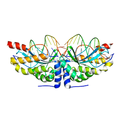
1P9U
 | | Coronavirus Main Proteinase (3CLpro) Structure: Basis for Design of anti-SARS Drugs | | 分子名称: | (4R)-2-METHYLPENTANE-2,4-DIOL, PHQ-VNSTLQ-CHLOROMETHYLKETONE INHIBITOR, SULFATE ION, ... | | 著者 | Anand, K, Ziebuhr, J, Wadhwani, P, Mesters, J.R, Hilgenfeld, R. | | 登録日 | 2003-05-12 | | 公開日 | 2003-05-20 | | 最終更新日 | 2017-10-11 | | 実験手法 | X-RAY DIFFRACTION (2.37 Å) | | 主引用文献 | Coronavirus Main Proteinase (3CLpro) Structure: Basis for Design of anti-SARS Drugs
Science, 300, 2003
|
|
6F29
 | | Crystal structure of the kainate receptor GluK3 ligand binding domain in complex with (S)-1-[2-Amino-2-carboxyethyl]-5,7-dihydrothieno[3,4-d]pyrimidin-2,4(1H,3H)-dione at resolution 2.6A | | 分子名称: | (2~{S})-2-azanyl-3-[2,4-bis(oxidanylidene)-5,7-dihydrothieno[3,4-d]pyrimidin-1-yl]propanoic acid, CHLORIDE ION, Glutamate receptor ionotropic, ... | | 著者 | Venskutonyte, R, Frydenvang, K, Kastrup, J.S. | | 登録日 | 2017-11-23 | | 公開日 | 2018-02-28 | | 最終更新日 | 2024-01-17 | | 実験手法 | X-RAY DIFFRACTION (2.6 Å) | | 主引用文献 | ( S)-2-Amino-3-(5-methyl-3-hydroxyisoxazol-4-yl)propanoic Acid (AMPA) and Kainate Receptor Ligands: Further Exploration of Bioisosteric Replacements and Structural and Biological Investigation.
J. Med. Chem., 61, 2018
|
|


