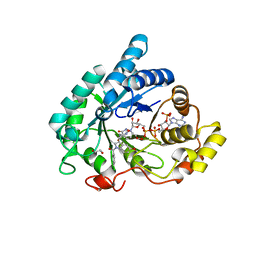
4DPO
 
 | | Crystal structure of a conserved protein MM_1583 from Methanosarcina mazei Go1 | | 分子名称: | Conserved protein | | 著者 | Agarwal, R, Chamala, S, Evans, R, Gizzi, A, Hillerich, B, Kar, A, LaFleur, J, Foti, R, Siedel, R, Zencheck, W, Villigas, G, Almo, S.C, Swaminathan, S, New York Structural Genomics Research Consortium (NYSGRC) | | 登録日 | 2012-02-13 | | 公開日 | 2012-02-29 | | 最終更新日 | 2017-11-15 | | 実験手法 | X-RAY DIFFRACTION (2.73 Å) | | 主引用文献 | Crystal structure of a conserved protein MM_1583 from Methanosarcina mazei Go1
To be Published
|
|
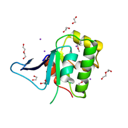
6IU8
 | | Crystal structure of cytoplasmic metal binding domain with cobalt ions | | 分子名称: | COBALT (II) ION, VIT1, ZINC ION | | 著者 | Kato, T, Nishizawa, T, Yamashita, K, Kumazaki, K, Ishitani, R, Nureki, O. | | 登録日 | 2018-11-27 | | 公開日 | 2019-02-06 | | 最終更新日 | 2023-11-22 | | 実験手法 | X-RAY DIFFRACTION (2.7 Å) | | 主引用文献 | Crystal structure of plant vacuolar iron transporter VIT1.
Nat Plants, 5, 2019
|
|
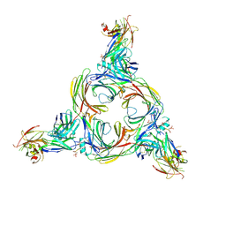
8OQM
 | | Structure of Mycobacterium tuberculosis beta-oxidation trifunctional enzyme in complex with Fragment-M-10 | | 分子名称: | 3-hydroxyacyl-CoA dehydrogenase, 6-[(6-azanyl-4-oxidanyl-naphthalen-2-yl)sulfonylamino]-4-oxidanyl-naphthalene-2-sulfonic acid, 6-azanyl-4-oxidanyl-naphthalene-2-sulfonic acid, ... | | 著者 | Dalwani, S, Wierenga, R.K, Venkatesan, R. | | 登録日 | 2023-04-12 | | 公開日 | 2024-01-24 | | 最終更新日 | 2024-08-14 | | 実験手法 | X-RAY DIFFRACTION (3.2 Å) | | 主引用文献 | Crystallographic fragment-binding studies of the Mycobacterium tuberculosis trifunctional enzyme suggest binding pockets for the tails of the acyl-CoA substrates at its active sites and a potential substrate-channeling path between them.
Acta Crystallogr D Struct Biol, 80, 2024
|
|
8GO8
 | | Structure of beta-arrestin1 in complex with a phosphopeptide corresponding to the human C5a anaphylatoxin chemotactic receptor 1, C5aR1 | | 分子名称: | Beta-arrestin-1, C5a anaphylatoxin chemotactic receptor 1, Fab30 heavy chain, ... | | 著者 | Maharana, J, Sarma, P, Yadav, M.K, Banerjee, R, Shukla, A.K. | | 登録日 | 2022-08-24 | | 公開日 | 2023-05-17 | | 最終更新日 | 2024-07-17 | | 実験手法 | ELECTRON MICROSCOPY (3.41 Å) | | 主引用文献 | Structural snapshots uncover a key phosphorylation motif in GPCRs driving beta-arrestin activation.
Mol.Cell, 83, 2023
|
|
8GP3
 | | Structure of beta-arrestin1 in complex with a phosphopeptide corresponding to the human C-X-C chemokine receptor type 4, CXCR4 | | 分子名称: | Beta-arrestin-1, C-X-C chemokine receptor type 4, Fab30 Heavy Chain, ... | | 著者 | Maharana, J, Sarma, P, Yadav, M.K, Banerjee, R, Shukla, A.K. | | 登録日 | 2022-08-25 | | 公開日 | 2023-05-17 | | 最終更新日 | 2024-07-17 | | 実験手法 | ELECTRON MICROSCOPY (4.8 Å) | | 主引用文献 | Structural snapshots uncover a key phosphorylation motif in GPCRs driving beta-arrestin activation.
Mol.Cell, 83, 2023
|
|
8GOO
 | | Structure of beta-arrestin2 in complex with a phosphopeptide corresponding to the human C5a anaphylatoxin chemotactic receptor 1, C5aR1 | | 分子名称: | Beta-arrestin-2, C5a anaphylatoxin chemotactic receptor 1, Fab30 Heavy Chain, ... | | 著者 | Maharana, J, Sarma, P, Yadav, M.K, Banerjee, R, Shukla, A.K. | | 登録日 | 2022-08-25 | | 公開日 | 2023-05-17 | | 最終更新日 | 2024-10-16 | | 実験手法 | ELECTRON MICROSCOPY (4.4 Å) | | 主引用文献 | Structural snapshots uncover a key phosphorylation motif in GPCRs driving beta-arrestin activation.
Mol.Cell, 83, 2023
|
|
1M4Y
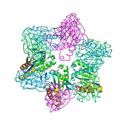 | | Crystal structure of HslV from Thermotoga maritima | | 分子名称: | ATP-dependent protease hslV, SODIUM ION | | 著者 | Song, H.K, Ramachandran, R, Bochtler, M.B, Hartmann, C, Azim, M.K, Huber, R. | | 登録日 | 2002-07-05 | | 公開日 | 2003-05-06 | | 最終更新日 | 2024-02-14 | | 実験手法 | X-RAY DIFFRACTION (2.1 Å) | | 主引用文献 | Isolation and characterization of the prokaryotic proteasome homolog HslVU (ClpQY) from Thermotoga maritima and the crystal structure of HslV.
BIOPHYS.CHEM., 100, 2003
|
|
1MKJ
 | | Human Kinesin Motor Domain With Docked Neck Linker | | 分子名称: | ADENOSINE-5'-DIPHOSPHATE, KINESIN HEAVY CHAIN, MAGNESIUM ION, ... | | 著者 | Sindelar, C.V, Budny, M.J, Rice, S, Naber, N, Fletterick, R, Cooke, R. | | 登録日 | 2002-08-29 | | 公開日 | 2002-10-30 | | 最終更新日 | 2024-02-14 | | 実験手法 | X-RAY DIFFRACTION (2.7 Å) | | 主引用文献 | Two conformations in the human kinesin power stroke defined by X-ray crystallography and EPR spectroscopy.
Nat.Struct.Biol., 9, 2002
|
|
8GLB
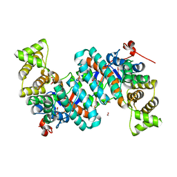 | | Selenomethionine Derivatized Citrate Synthase (CitA) in Mycobacterium tuberculosis with Pyruvate | | 分子名称: | 1,2-ETHANEDIOL, PYRUVIC ACID, citrate synthase | | 著者 | Pathirage, R, Ronning, D, Favrot, L. | | 登録日 | 2023-03-21 | | 公開日 | 2023-06-07 | | 最終更新日 | 2023-11-15 | | 実験手法 | X-RAY DIFFRACTION (2.1 Å) | | 主引用文献 | Mycobacterium tuberculosis CitA activity is modulated by cysteine oxidation and pyruvate binding.
Rsc Med Chem, 14, 2023
|
|
8GMI
 | |
3SNL
 | | Highly Potent, Selective, and Orally Active Phosphodiestarase 10A Inhibitors | | 分子名称: | 6-chloro-3,4-dimethyl-1-(3-methylpyridin-4-yl)-8-(trifluoromethyl)imidazo[1,5-a]quinoxaline, MAGNESIUM ION, ZINC ION, ... | | 著者 | Malamas, M.S, Ni, Y, Erdei, J, Stange, H, Schindler, R, Lankau, H.-J, Grunwald, C, Fan, K.Y, Parris, K.D, Langen, B, Egerland, U, Hage, T, Marquis, K.L, Grauer, S, Brennan, J, Navarra, R, Graf, R, Harrison, B.L, Robichaud, A, Kronbach, T, Pangalos, M, Hofgen, N, Brandon, N.J. | | 登録日 | 2011-06-29 | | 公開日 | 2011-10-26 | | 最終更新日 | 2024-02-28 | | 実験手法 | X-RAY DIFFRACTION (2.4 Å) | | 主引用文献 | Highly Potent, Selective, and Orally Active Phosphodiesterase 10A Inhibitors.
J.Med.Chem., 54, 2011
|
|
8GMK
 | | Pyruvate bound structure of Citrate Synthase (CitA) in Mycobacterium Tuberculosis | | 分子名称: | 1,2-ETHANEDIOL, DI(HYDROXYETHYL)ETHER, PYRUVIC ACID, ... | | 著者 | Pathirage, R, Ronning, D, Yamsek, M. | | 登録日 | 2023-03-26 | | 公開日 | 2023-06-07 | | 最終更新日 | 2023-11-15 | | 実験手法 | X-RAY DIFFRACTION (1.81 Å) | | 主引用文献 | Mycobacterium tuberculosis CitA activity is modulated by cysteine oxidation and pyruvate binding.
Rsc Med Chem, 14, 2023
|
|
8GI7
 | |
8GM9
 | | Structure of Citrate Synthase(CitA) in Mycobacterium Tuberculosis | | 分子名称: | 1,2-ETHANEDIOL, ACRYLIC ACID, DI(HYDROXYETHYL)ETHER, ... | | 著者 | Pathirage, R, Ronning, D, Favrot, L. | | 登録日 | 2023-03-24 | | 公開日 | 2023-06-07 | | 最終更新日 | 2023-11-08 | | 実験手法 | X-RAY DIFFRACTION (2.4 Å) | | 主引用文献 | Mycobacterium tuberculosis CitA activity is modulated by cysteine oxidation and pyruvate binding.
Rsc Med Chem, 14, 2023
|
|
8GLL
 | | R149E variant of Citrate Synthase (CitA) in Mycobacterium tuberculosis | | 分子名称: | 1,2-ETHANEDIOL, DI(HYDROXYETHYL)ETHER, SULFATE ION, ... | | 著者 | Pathirage, R, Ronning, D, Petit, C. | | 登録日 | 2023-03-22 | | 公開日 | 2023-06-07 | | 最終更新日 | 2023-11-08 | | 実験手法 | X-RAY DIFFRACTION (2.65 Å) | | 主引用文献 | Mycobacterium tuberculosis CitA activity is modulated by cysteine oxidation and pyruvate binding.
Rsc Med Chem, 14, 2023
|
|
8GMF
 | |
8GIW
 | |
5EUU
 | | Rat prestin STAS domain in complex with chloride | | 分子名称: | 1,2-ETHANEDIOL, CHLORIDE ION, Prestin,Prestin, ... | | 著者 | Lolli, G, Pasqualetto, E, Costanzi, E, Bonetto, G, Battistutta, R. | | 登録日 | 2015-11-19 | | 公開日 | 2015-12-16 | | 最終更新日 | 2024-01-10 | | 実験手法 | X-RAY DIFFRACTION (1.87 Å) | | 主引用文献 | The STAS domain of mammalian SLC26A5 prestin harbours an anion-binding site.
Biochem.J., 473, 2016
|
|
5EUS
 | | Rat prestin STAS domain in complex with bromide | | 分子名称: | 1,2-ETHANEDIOL, BROMIDE ION, Prestin,Rat prestin STAS domain, ... | | 著者 | Lolli, G, Pasqualetto, E, Costanzi, E, Bonetto, G, Battistutta, R. | | 登録日 | 2015-11-19 | | 公開日 | 2015-12-16 | | 最終更新日 | 2024-01-10 | | 実験手法 | X-RAY DIFFRACTION (1.833 Å) | | 主引用文献 | The STAS domain of mammalian SLC26A5 prestin harbours an anion-binding site.
Biochem.J., 473, 2016
|
|
5EUW
 | | Rat prestin STAS domain in complex with nitrate | | 分子名称: | 1,2-ETHANEDIOL, DI(HYDROXYETHYL)ETHER, NITRATE ION, ... | | 著者 | Lolli, G, Pasqualetto, E, Costanzi, E, Bonetto, G, Battistutta, R. | | 登録日 | 2015-11-19 | | 公開日 | 2015-12-16 | | 最終更新日 | 2024-01-10 | | 実験手法 | X-RAY DIFFRACTION (1.81 Å) | | 主引用文献 | The STAS domain of mammalian SLC26A5 prestin harbours an anion-binding site.
Biochem.J., 473, 2016
|
|
5EUX
 | | Rat prestin STAS domain in complex with thiocyanate | | 分子名称: | 1,2-ETHANEDIOL, DI(HYDROXYETHYL)ETHER, Prestin,Prestin, ... | | 著者 | Lolli, G, Pasqualetto, E, Costanzi, E, Bonetto, G, Battistutta, R. | | 登録日 | 2015-11-19 | | 公開日 | 2015-12-16 | | 最終更新日 | 2024-01-10 | | 実験手法 | X-RAY DIFFRACTION (2.038 Å) | | 主引用文献 | The STAS domain of mammalian SLC26A5 prestin harbours an anion-binding site.
Biochem.J., 473, 2016
|
|
6GXK
 | | Crystal structure of Aldo-Keto Reductase 1C3 (AKR1C3) complexed with inhibitor. | | 分子名称: | 1,2-ETHANEDIOL, 4-[[1-(4-chlorophenyl)carbonyl-5-methoxy-2-methyl-indol-3-yl]methyl]-1,2,5-oxadiazol-3-one, Aldo-keto reductase family 1 member C3, ... | | 著者 | Goyal, P, Wahlgren, W.Y, Friemann, R. | | 登録日 | 2018-06-27 | | 公開日 | 2019-05-08 | | 最終更新日 | 2024-05-15 | | 実験手法 | X-RAY DIFFRACTION (1.7 Å) | | 主引用文献 | Bioisosteres of Indomethacin as Inhibitors of Aldo-Keto Reductase 1C3.
Acs Med.Chem.Lett., 10, 2019
|
|
5EUZ
 | | Rat prestin STAS domain in complex with iodide | | 分子名称: | 1,2-ETHANEDIOL, DI(HYDROXYETHYL)ETHER, IODIDE ION, ... | | 著者 | Lolli, G, Pasqualetto, E, Costanzi, E, Bonetto, G, Battistutta, R. | | 登録日 | 2015-11-19 | | 公開日 | 2015-12-16 | | 最終更新日 | 2024-10-16 | | 実験手法 | X-RAY DIFFRACTION (2.403 Å) | | 主引用文献 | The STAS domain of mammalian SLC26A5 prestin harbours an anion-binding site.
Biochem.J., 473, 2016
|
|
6IAA
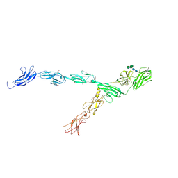 | | hRobo2 ectodomain | | 分子名称: | 2-acetamido-2-deoxy-beta-D-glucopyranose-(1-4)-2-acetamido-2-deoxy-beta-D-glucopyranose, Roundabout homolog 2, alpha-D-mannopyranose-(1-3)-[alpha-D-mannopyranose-(1-6)]beta-D-mannopyranose-(1-4)-2-acetamido-2-deoxy-beta-D-glucopyranose-(1-4)-2-acetamido-2-deoxy-beta-D-glucopyranose | | 著者 | Barak, R, Isupov, N.M, Opatowsky, Y. | | 登録日 | 2018-11-26 | | 公開日 | 2019-03-13 | | 最終更新日 | 2024-10-16 | | 実験手法 | X-RAY DIFFRACTION (3.6 Å) | | 主引用文献 | Structural Principles in Robo Activation and Auto-inhibition.
Cell, 177, 2019
|
|
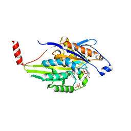
3ZHO
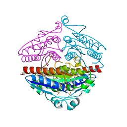 | | X-ray structure of E.coli Wrba in complex with FMN at 1.2 A resolution | | 分子名称: | FLAVIN MONONUCLEOTIDE, FLAVOPROTEIN WRBA | | 著者 | Kishko, I, Lapkouski, M, Brynda, J, Kuty, M, Carey, J, Smatanova, I.K, Ettrich, R. | | 登録日 | 2012-12-23 | | 公開日 | 2013-08-28 | | 最終更新日 | 2023-12-20 | | 実験手法 | X-RAY DIFFRACTION (1.2 Å) | | 主引用文献 | 1.2 A Resolution Crystal Structure of Escherichia Coli Wrba Holoprotein
Acta Crystallogr.,Sect.D, 69, 2013
|
|


