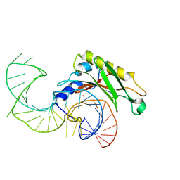
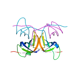
5ML7
 
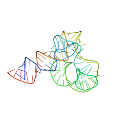 | | CRYSTAL STRUCTURE OF L1-STALK FRAGMENT OF 23S rRNA FROM HALOARCULA MARISMORTUI | | 分子名称: | 23S ribosomal RNA, CALCIUM ION, MAGNESIUM ION | | 著者 | Gabdulkhakov, A.G, Tishchenko, T.V, Nevskaya, N.A, Nikonov, S.V. | | 登録日 | 2016-12-06 | | 公開日 | 2017-12-20 | | 最終更新日 | 2024-01-17 | | 実験手法 | X-RAY DIFFRACTION (3.3 Å) | | 主引用文献 | Crystal Structure of the 23S rRNA Fragment Specific to r-Protein L1 and Designed Model of the Ribosomal L1 Stalk from Haloarcula marismortui
Crystals, 2017
|
|
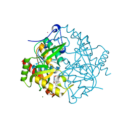
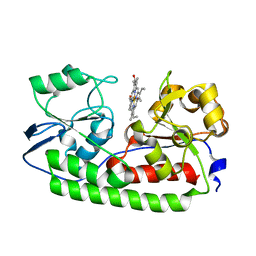
6T8N
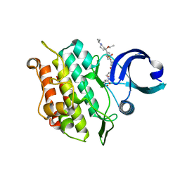 | | Crystal structure of the ACVR1 (ALK2) kinase in complex with the compound M4K3007 | | 分子名称: | 1,2-ETHANEDIOL, Activin receptor type I, DIMETHYL SULFOXIDE, ... | | 著者 | Adamson, R.J, Williams, E.P, Bonomo, S, Rankin, S, Bacos, D, Rae, A, Cramp, S, Burgess-Brown, N, von Delft, F, Arrowsmith, C.H, Edwards, A.M, Bountra, C, Bullock, A.N. | | 登録日 | 2019-10-24 | | 公開日 | 2019-11-27 | | 最終更新日 | 2024-01-24 | | 実験手法 | X-RAY DIFFRACTION (1.77 Å) | | 主引用文献 | Crystal structure of the ACVR1 (ALK2) kinase in complex with the compound M4K3007
To Be Published
|
|
2HW8
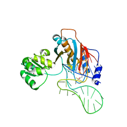 | | Structure of ribosomal protein L1-mRNA complex at 2.1 resolution. | | 分子名称: | 1,4-BUTANEDIOL, 36-MER, 50S ribosomal protein L1, ... | | 著者 | Tishchenko, S, Nikonova, E, Nikulin, A, Nevskaya, N, Volchkov, S, Piendl, W, Garber, M, Nikonov, S. | | 登録日 | 2006-08-01 | | 公開日 | 2006-12-19 | | 最終更新日 | 2024-10-09 | | 実験手法 | X-RAY DIFFRACTION (2.1 Å) | | 主引用文献 | Structure of the ribosomal protein L1-mRNA complex at 2.1 A resolution: common features of crystal packing of L1-RNA complexes.
ACTA CRYSTALLOGR.,SECT.D, 62, 2006
|
|
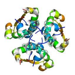
4AIY
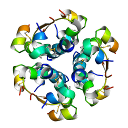 | | R6 HUMAN INSULIN HEXAMER (SYMMETRIC), NMR, 'GREEN' SUBSTATE, AVERAGE STRUCTURE | | 分子名称: | PHENOL, PROTEIN (INSULIN) | | 著者 | O'Donoghue, S.I, Chang, X, Abseher, R, Nilges, M, Led, J.J. | | 登録日 | 1998-12-29 | | 公開日 | 2000-02-28 | | 最終更新日 | 2023-12-27 | | 実験手法 | SOLUTION NMR | | 主引用文献 | Unraveling the symmetry ambiguity in a hexamer: calculation of the R6 human insulin structure.
J.Biomol.NMR, 16, 2000
|
|
4F9T
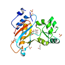 | | Ribosomal protein L1 from Thermus thermophilus with substitution Thr217Ala | | 分子名称: | (4R)-2-METHYLPENTANE-2,4-DIOL, (4S)-2-METHYL-2,4-PENTANEDIOL, 50S ribosomal protein L1, ... | | 著者 | Kljashtorny, V.G, Volchkov, S.A, Nikonova, E.Y, Kostareva, O.S, Tishchenko, S.V, Nevskaya, N.A, Nikonov, S.V. | | 登録日 | 2012-05-21 | | 公開日 | 2012-08-08 | | 最終更新日 | 2024-02-28 | | 実験手法 | X-RAY DIFFRACTION (1.46 Å) | | 主引用文献 | [Crystal structures of mutant ribosomal proteins L1].
MOL.BIOL.(MOSCOW), 41, 2007
|
|
4PJ3
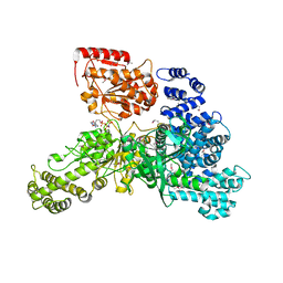 | | Structural insight into the function and evolution of the spliceosomal helicase Aquarius, Structure of Aquarius in complex with AMPPNP | | 分子名称: | Intron-binding protein aquarius, MAGNESIUM ION, PHOSPHOAMINOPHOSPHONIC ACID-ADENYLATE ESTER | | 著者 | De, I, Bessonov, S, Hofele, R, dos Santos, K.F, Will, C.L, Urlaub, H, Luhrmann, R, Pena, V. | | 登録日 | 2014-05-11 | | 公開日 | 2015-01-21 | | 最終更新日 | 2024-10-09 | | 実験手法 | X-RAY DIFFRACTION (2.3 Å) | | 主引用文献 | The RNA helicase Aquarius exhibits structural adaptations mediating its recruitment to spliceosomes.
Nat.Struct.Mol.Biol., 22, 2015
|
|
5DSZ
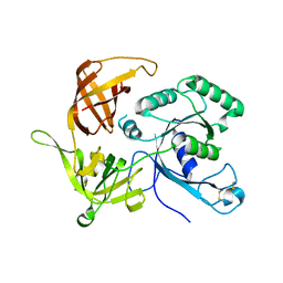 | | Gamma-subunit of the translation initiation factor 2 from S. solfataricus | | 分子名称: | Translation initiation factor 2 subunit gamma | | 著者 | Kravchenko, O.V, Nikonov, O.S, Stolboushkina, E.A, Nevskaya, N.A, Nikulin, A.D, Garber, M.B, Nikonov, S.V. | | 登録日 | 2015-09-17 | | 公開日 | 2016-08-24 | | 最終更新日 | 2024-01-10 | | 実験手法 | X-RAY DIFFRACTION (2.5 Å) | | 主引用文献 | Gamma-subunit of the translation initiation factor 2 from S. solfataricus
To Be Published
|
|
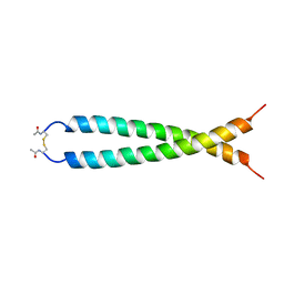
1AXH
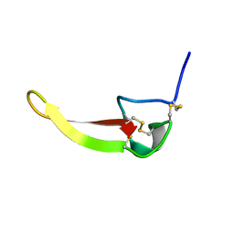 | | ATRACOTOXIN-HVI FROM HADRONYCHE VERSUTA (AUSTRALIAN FUNNEL-WEB SPIDER, NMR, 20 STRUCTURES | | 分子名称: | ATRACOTOXIN-HVI | | 著者 | Fletcher, J.I, O'Donoghue, S.I, Nilges, M, King, G.F. | | 登録日 | 1996-11-04 | | 公開日 | 1997-11-12 | | 最終更新日 | 2021-02-03 | | 実験手法 | SOLUTION NMR | | 主引用文献 | The structure of a novel insecticidal neurotoxin, omega-atracotoxin-HV1, from the venom of an Australian funnel web spider.
Nat.Struct.Biol., 4, 1997
|
|
8ILL
 | | Crystal structure of a highly photostable and bright green fluorescent protein at pH5.6 | | 分子名称: | CHLORIDE ION, alpha-D-glucopyranose-(1-1)-alpha-D-glucopyranose, green fluorescent protein | | 著者 | Ago, H, Ando, R, Hirano, M, Shimozono, S, Miyawaki, A, Yamamoto, M. | | 登録日 | 2023-03-03 | | 公開日 | 2023-10-04 | | 最終更新日 | 2024-04-24 | | 実験手法 | X-RAY DIFFRACTION (2.2 Å) | | 主引用文献 | StayGold variants for molecular fusion and membrane-targeting applications.
Nat.Methods, 21, 2024
|
|
8ILK
 | | Crystal structure of a highly photostable and bright green fluorescent protein at pH8.5 | | 分子名称: | CHLORIDE ION, Green FLUORESCENT PROTEIN | | 著者 | Ago, H, Ando, R, Hirano, M, Shimozono, S, Miyawaki, A, Yamamoto, M. | | 登録日 | 2023-03-03 | | 公開日 | 2023-10-04 | | 最終更新日 | 2024-04-24 | | 実験手法 | X-RAY DIFFRACTION (1.56 Å) | | 主引用文献 | StayGold variants for molecular fusion and membrane-targeting applications.
Nat.Methods, 21, 2024
|
|
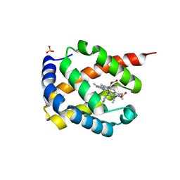
7F30
 | | Crystal structure of OxdB E85A in complex with Z-2- (3-bromophenyl) propanal oxime | | 分子名称: | PROTOPORPHYRIN IX CONTAINING FE, Phenylacetaldoxime dehydratase, Z-2-(3-bromophenyl) propanal oxime | | 著者 | Muraki, N, Matsui, D, Asano, Y, Aono, S. | | 登録日 | 2021-06-15 | | 公開日 | 2022-04-27 | | 最終更新日 | 2023-11-29 | | 実験手法 | X-RAY DIFFRACTION (2 Å) | | 主引用文献 | Crystal structural analysis of aldoxime dehydratase from Bacillus sp. OxB-1: Importance of surface residues in optimization for crystallization.
J.Inorg.Biochem., 230, 2022
|
|
7F2Y
 | | Crystal structure of OxdB E85A mutant (form I) | | 分子名称: | PROTOPORPHYRIN IX CONTAINING FE, Phenylacetaldoxime dehydratase | | 著者 | Muraki, N, Matsui, D, Asano, Y, Aono, S. | | 登録日 | 2021-06-15 | | 公開日 | 2022-04-27 | | 最終更新日 | 2023-11-29 | | 実験手法 | X-RAY DIFFRACTION (1.55 Å) | | 主引用文献 | Crystal structural analysis of aldoxime dehydratase from Bacillus sp. OxB-1: Importance of surface residues in optimization for crystallization.
J.Inorg.Biochem., 230, 2022
|
|
7F2Z
 | | Crystal structure of OxdB E85A mutant (form II) | | 分子名称: | PROTOPORPHYRIN IX CONTAINING FE, Phenylacetaldoxime dehydratase | | 著者 | Muraki, N, Matsui, D, Asano, Y, Aono, S. | | 登録日 | 2021-06-15 | | 公開日 | 2022-04-27 | | 最終更新日 | 2023-11-29 | | 実験手法 | X-RAY DIFFRACTION (2.3 Å) | | 主引用文献 | Crystal structural analysis of aldoxime dehydratase from Bacillus sp. OxB-1: Importance of surface residues in optimization for crystallization.
J.Inorg.Biochem., 230, 2022
|
|
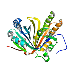
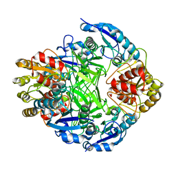
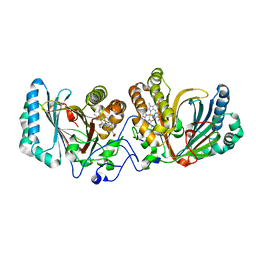
1ORB
 | | ACTIVE SITE STRUCTURAL FEATURES FOR CHEMICALLY MODIFIED FORMS OF RHODANESE | | 分子名称: | ACETATE ION, CARBOXYMETHYLATED RHODANESE | | 著者 | Gliubich, F, Gazerro, M, Zanotti, G, Delbono, S, Berni, R. | | 登録日 | 1995-07-24 | | 公開日 | 1995-10-15 | | 最終更新日 | 2024-06-05 | | 実験手法 | X-RAY DIFFRACTION (2 Å) | | 主引用文献 | Active site structural features for chemically modified forms of rhodanese.
J.Biol.Chem., 271, 1996
|
|
7DIH
 | |
2VPL
 | | The structure of the complex between the first domain of L1 protein from Thermus thermophilus and mRNA from Methanococcus jannaschii | | 分子名称: | 50S RIBOSOMAL PROTEIN L1, FRAGMENT OF MRNA FOR L1-OPERON CONTAINING REGULATOR L1-BINDING SITE, POTASSIUM ION | | 著者 | Kljashtorny, V, Tishchenko, S, Nevskaya, N, Nikonov, S, Garber, M. | | 登録日 | 2008-03-01 | | 公開日 | 2008-09-23 | | 最終更新日 | 2023-12-13 | | 実験手法 | X-RAY DIFFRACTION (2.3 Å) | | 主引用文献 | Domain II of Thermus thermophilus ribosomal protein L1 hinders recognition of its mRNA.
J. Mol. Biol., 383, 2008
|
|
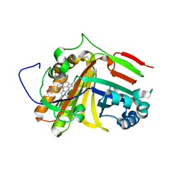
3P0F
 | | Structure of hUPP2 in an inactive conformation with bound 5-benzylacyclouridine | | 分子名称: | 1-((2-HYDROXYETHOXY)METHYL)-5-BENZYLPYRIMIDINE-2,4(1H,3H)-DIONE, COBALT (II) ION, MAGNESIUM ION, ... | | 著者 | Roosild, T.P, Castronovo, S, Villoso, A. | | 登録日 | 2010-09-28 | | 公開日 | 2011-09-07 | | 最終更新日 | 2023-09-06 | | 実験手法 | X-RAY DIFFRACTION (1.54 Å) | | 主引用文献 | A novel structural mechanism for redox regulation of uridine phosphorylase 2 activity.
J.Struct.Biol., 176, 2011
|
|
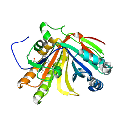
3JSY
 | | N-terminal fragment of ribosomal protein L10 from Methanococcus jannaschii | | 分子名称: | Acidic ribosomal protein P0 homolog | | 著者 | Kravchenko, O, Mitroshin, I, Nikulin, A.D, Piendl, W, Garber, M, Nikonov, S. | | 登録日 | 2009-09-11 | | 公開日 | 2010-05-26 | | 最終更新日 | 2024-03-20 | | 実験手法 | X-RAY DIFFRACTION (1.6 Å) | | 主引用文献 | Structure of a two-domain N-terminal fragment of ribosomal protein L10 from Methanococcus jannaschii reveals a specific piece of the archaeal ribosomal stalk
J.Mol.Biol., 399, 2010
|
|
2AIY
 | | R6 HUMAN INSULIN HEXAMER (SYMMETRIC), NMR, 20 STRUCTURES | | 分子名称: | PHENOL, PROTEIN (INSULIN) | | 著者 | O'Donoghue, S.I, Chang, X, Abseher, R, Nilges, M, Led, J.J. | | 登録日 | 1998-12-28 | | 公開日 | 2000-02-28 | | 最終更新日 | 2024-10-16 | | 実験手法 | SOLUTION NMR | | 主引用文献 | Unraveling the symmetry ambiguity in a hexamer: calculation of the R6 human insulin structure.
J.Biomol.NMR, 16, 2000
|
|
1ZCF
 | |
2BH8
 | | Combinatorial Protein 1b11 | | 分子名称: | 1B11 | | 著者 | De Bono, S, Riechmann, L, Girard, E, Williams, R.L, Winter, G. | | 登録日 | 2005-01-07 | | 公開日 | 2005-02-07 | | 最終更新日 | 2024-05-08 | | 実験手法 | X-RAY DIFFRACTION (1.9 Å) | | 主引用文献 | A Segment of Cold Shock Protein Directs the Folding of a Combinatorial Protein
Proc.Natl.Acad.Sci.USA, 102, 2005
|
|
5AZ3
 | | Crystal structure of heme binding protein HmuT | | 分子名称: | ABC-type transporter, periplasmic component, PROTOPORPHYRIN IX CONTAINING FE | | 著者 | Muraki, N, Aono, S. | | 登録日 | 2015-09-18 | | 公開日 | 2015-11-18 | | 最終更新日 | 2023-11-08 | | 実験手法 | X-RAY DIFFRACTION (1.423 Å) | | 主引用文献 | Structural Basis for Heme Recognition by HmuT Responsible for Heme Transport to the Heme Transporter in Corynebacterium glutamicum
Chem Lett., 2015
|
|
1JUN
 | |
3A16
 | | Crystal Structure of Aldoxime Dehydratase (OxdRE) in Complex with Propionaldoxime | | 分子名称: | (1Z)-propanal oxime, Aldoxime dehydratase, MAGNESIUM ION, ... | | 著者 | Sawai, H, Sugimoto, H, Kato, Y, Asano, Y, Shiro, Y, Aono, S. | | 登録日 | 2009-03-26 | | 公開日 | 2009-09-08 | | 最終更新日 | 2023-11-01 | | 実験手法 | X-RAY DIFFRACTION (1.6 Å) | | 主引用文献 | X-ray crystal structure of michaelis complex of aldoxime dehydratase
J.Biol.Chem., 284, 2009
|
|
4QGB
 | | Crystal structure of mutant ribosomal protein G219V TthL1 | | 分子名称: | 50S ribosomal protein L1, ACETATE ION, CHLORIDE ION | | 著者 | Gabdulkhakov, A.G, Nevskaya, N.A, Nikonov, S.V. | | 登録日 | 2014-05-22 | | 公開日 | 2015-02-11 | | 最終更新日 | 2023-09-20 | | 実験手法 | X-RAY DIFFRACTION (2.6 Å) | | 主引用文献 | Protein-RNA affinity of ribosomal protein L1 mutants does not correlate with the number of intermolecular interactions.
Acta Crystallogr.,Sect.D, 71, 2015
|
|


