4LNP
 
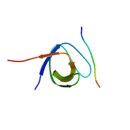 | | The first SH3 domain from CAP/Ponsin in complex with proline rich peptide from Vinculin | | 分子名称: | Sorbin and SH3 domain-containing protein 1, Vinculin | | 著者 | Zhao, D, Li, F, Wu, J, Shi, Y, Zhang, Z, Gong, Q. | | 登録日 | 2013-07-11 | | 公開日 | 2014-05-28 | | 最終更新日 | 2024-03-20 | | 実験手法 | X-RAY DIFFRACTION (1.41 Å) | | 主引用文献 | Structural investigation of the interaction between the tandem SH3 domains of c-Cbl-associated protein and vinculin
J.Struct.Biol., 187, 2014
|
|
4LN2
 
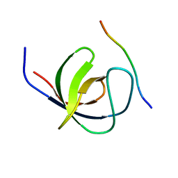 | | The second SH3 domain from CAP/Ponsin in complex with proline rich peptide from Vinculin | | 分子名称: | Sorbin and SH3 domain-containing protein 1, proline rich peptide | | 著者 | Zhao, D, Li, F, Wu, J, Shi, Y, Zhang, Z, Gong, Q. | | 登録日 | 2013-07-11 | | 公開日 | 2014-05-28 | | 最終更新日 | 2023-11-08 | | 実験手法 | X-RAY DIFFRACTION (1 Å) | | 主引用文献 | Structural investigation of the interaction between the tandem SH3 domains of c-Cbl-associated protein and vinculin
J.Struct.Biol., 187, 2014
|
|
5Y1U
 
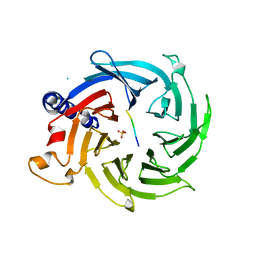 | | Crystal structure of RBBP4 bound to AEBP2 RRK motif | | 分子名称: | Histone-binding protein RBBP4, SULFATE ION, Zinc finger protein AEBP2 | | 著者 | Sun, A, Li, F, Wu, J, Shi, Y. | | 登録日 | 2017-07-21 | | 公開日 | 2018-04-18 | | 最終更新日 | 2023-11-22 | | 実験手法 | X-RAY DIFFRACTION (2.141 Å) | | 主引用文献 | Structural and biochemical insights into human zinc finger protein AEBP2 reveals interactions with RBBP4
Protein Cell, 2017
|
|
6J7L
 
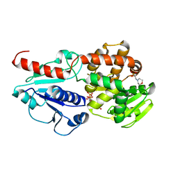 | | Crystal structure of Pseudomonas aeruginosa Earp in complex with TDP | | 分子名称: | 2-(N-MORPHOLINO)-ETHANESULFONIC ACID, Pseudomonas aeruginosa Earp, THYMIDINE-5'-DIPHOSPHATE | | 著者 | He, C, Li, F. | | 登録日 | 2019-01-18 | | 公開日 | 2019-05-15 | | 最終更新日 | 2023-11-22 | | 実験手法 | X-RAY DIFFRACTION (1.851 Å) | | 主引用文献 | Complex Structure ofPseudomonas aeruginosaArginine Rhamnosyltransferase EarP with Its Acceptor Elongation Factor P.
J.Bacteriol., 201, 2019
|
|
5YYZ
 
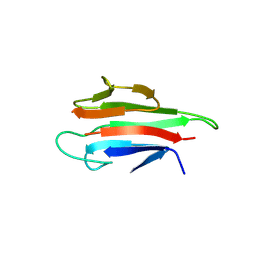 | | Crystal structure of the MEK1 FHA domain in complex with the HOP1 pThr318 peptide. | | 分子名称: | Meiosis-specific protein HOP1, Meiosis-specific serine/threonine-protein kinase MEK1 | | 著者 | Xie, C, Li, F, Jiang, Y, Wu, J, Shi, Y. | | 登録日 | 2017-12-11 | | 公開日 | 2018-10-17 | | 最終更新日 | 2024-10-16 | | 実験手法 | X-RAY DIFFRACTION (1.798 Å) | | 主引用文献 | Structural insights into the recognition of phosphorylated Hop1 by Mek1
Acta Crystallogr D Struct Biol, 74, 2018
|
|
5Z4D
 
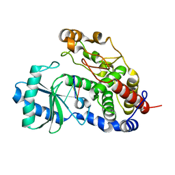 | | Structure of Tailor in complex with AGUU RNA | | 分子名称: | RNA (5'-R(*AP*GP*UP*U)-3'), Terminal uridylyltransferase Tailor | | 著者 | Cheng, L, Li, F, Jiang, Y, Yu, H, Xie, C, Shi, Y, Gong, Q. | | 登録日 | 2018-01-11 | | 公開日 | 2018-10-31 | | 最終更新日 | 2023-11-22 | | 実験手法 | X-RAY DIFFRACTION (1.803 Å) | | 主引用文献 | Structural insights into a unique preference for 3' terminal guanine of mirtron in Drosophila TUTase tailor.
Nucleic Acids Res., 47, 2019
|
|
5Z4M
 
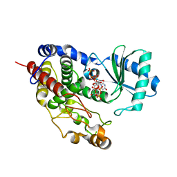 | | Structure of TailorD343A with bound UTP and Mg | | 分子名称: | MAGNESIUM ION, Terminal uridylyltransferase Tailor, URIDINE 5'-TRIPHOSPHATE | | 著者 | Cheng, L, Li, F, Jiang, Y, Yu, H, Xie, C, Shi, Y, Gong, Q. | | 登録日 | 2018-01-11 | | 公開日 | 2018-10-31 | | 最終更新日 | 2023-11-22 | | 実験手法 | X-RAY DIFFRACTION (1.74 Å) | | 主引用文献 | Structural insights into a unique preference for 3' terminal guanine of mirtron in Drosophila TUTase tailor.
Nucleic Acids Res., 47, 2019
|
|
7EIO
 
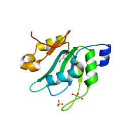 | | Crystal Structure of Mei2 RRM3 | | 分子名称: | GLYCEROL, Meiosis protein mei2, SULFATE ION | | 著者 | Shen, S.Y, Li, F.D. | | 登録日 | 2021-03-31 | | 公開日 | 2022-04-06 | | 最終更新日 | 2024-05-29 | | 実験手法 | X-RAY DIFFRACTION (1.895 Å) | | 主引用文献 | Structural insights reveal the specific recognition of meiRNA by the Mei2 protein.
J Mol Cell Biol, 14, 2022
|
|
5Z4C
 
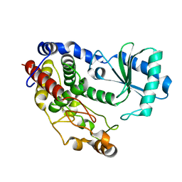 | | Crystal structure of Tailor | | 分子名称: | Terminal uridylyltransferase Tailor | | 著者 | Cheng, L, Li, F, Jiang, Y, Yu, H, Xie, C, Shi, Y, Gong, Q. | | 登録日 | 2018-01-10 | | 公開日 | 2018-10-31 | | 最終更新日 | 2023-11-22 | | 実験手法 | X-RAY DIFFRACTION (1.65 Å) | | 主引用文献 | Structural insights into a unique preference for 3' terminal guanine of mirtron in Drosophila TUTase tailor.
Nucleic Acids Res., 47, 2019
|
|
5Z4J
 
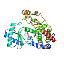 | | Structure of Tailor in complex with U4 RNA | | 分子名称: | RNA (5'-R(*UP*UP*UP*U)-3'), Terminal uridylyltransferase Tailor | | 著者 | Cheng, L, Li, F, Jiang, Y, Yu, H, Xie, C, Shi, Y, Gong, Q. | | 登録日 | 2018-01-11 | | 公開日 | 2018-10-31 | | 最終更新日 | 2023-11-22 | | 実験手法 | X-RAY DIFFRACTION (1.82 Å) | | 主引用文献 | Structural insights into a unique preference for 3' terminal guanine of mirtron in Drosophila TUTase tailor.
Nucleic Acids Res., 47, 2019
|
|
5Z4A
 
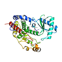 | | Structure of Tailor in complex with AGU RNA | | 分子名称: | RNA (5'-R(*AP*GP*U)-3'), Terminal uridylyltransferase Tailor | | 著者 | Cheng, L, Li, F, Jiang, Y, Yu, H, Xie, C, Shi, Y, Gong, Q. | | 登録日 | 2018-01-10 | | 公開日 | 2018-10-31 | | 最終更新日 | 2023-11-22 | | 実験手法 | X-RAY DIFFRACTION (1.637 Å) | | 主引用文献 | Structural insights into a unique preference for 3' terminal guanine of mirtron in Drosophila TUTase tailor.
Nucleic Acids Res., 47, 2019
|
|
5YYX
 
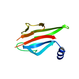 | | Crystal Structure of the MEK1 FHA domain | | 分子名称: | Meiosis-specific serine/threonine-protein kinase MEK1 | | 著者 | Xie, C, Li, F, Jiang, Y, Wu, J, Shi, Y. | | 登録日 | 2017-12-11 | | 公開日 | 2018-10-10 | | 最終更新日 | 2023-11-22 | | 実験手法 | X-RAY DIFFRACTION (1.684 Å) | | 主引用文献 | Structural insights into the recognition of phosphorylated Hop1 by Mek1
Acta Crystallogr D Struct Biol, 74, 2018
|
|
6AJK
 
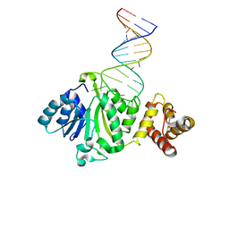 | | Crystal structure of TFB1M and h45 in homo sapiens | | 分子名称: | Dimethyladenosine transferase 1, mitochondrial, RNA (28-MER) | | 著者 | Liu, X, Shen, S, Wu, P, Li, F, Wang, C, Gong, Q, Wu, J, Zhang, H, Shi, Y. | | 登録日 | 2018-08-28 | | 公開日 | 2019-06-05 | | 最終更新日 | 2023-11-22 | | 実験手法 | X-RAY DIFFRACTION (3.001 Å) | | 主引用文献 | Structural insights into dimethylation of 12S rRNA by TFB1M: indispensable role in translation of mitochondrial genes and mitochondrial function.
Nucleic Acids Res., 47, 2019
|
|
5Y5D
 
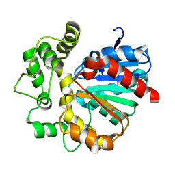 | |
6JP4
 
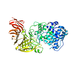 | | Crystal structure of the catalytic domain of a multi-domain alginate lyase Dp0100 from thermophilic bacterium Defluviitalea phaphyphila | | 分子名称: | 1,2-ETHANEDIOL, ACETATE ION, Alginate lyase, ... | | 著者 | Ji, S.Q, Dix, S.R, Aziz, A, Sedelnikova, S.E, Li, F.L, Rice, D.W. | | 登録日 | 2019-03-25 | | 公開日 | 2019-10-30 | | 最終更新日 | 2024-03-27 | | 実験手法 | X-RAY DIFFRACTION (2.069 Å) | | 主引用文献 | The molecular basis of endolytic activity of a multidomain alginate lyase fromDefluviitalea phaphyphila, a representative of a new lyase family, PL39.
J.Biol.Chem., 294, 2019
|
|
6JPH
 
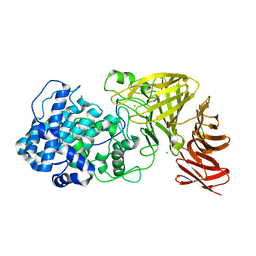 | | Crystal structure of the catalytic domain of a multi-domain alginate lyase Dp0100 from thermophilic bacterium Defluviitalea phaphyphila | | 分子名称: | ACETATE ION, Alginate lyase, CALCIUM ION, ... | | 著者 | Ji, S.Q, Dix, S.R, Aziz, A, Sedelnikova, S.E, Li, F.L, Rice, D.W. | | 登録日 | 2019-03-27 | | 公開日 | 2019-10-30 | | 最終更新日 | 2024-03-27 | | 実験手法 | X-RAY DIFFRACTION (2.759 Å) | | 主引用文献 | The molecular basis of endolytic activity of a multidomain alginate lyase fromDefluviitalea phaphyphila, a representative of a new lyase family, PL39.
J.Biol.Chem., 294, 2019
|
|
6JPN
 
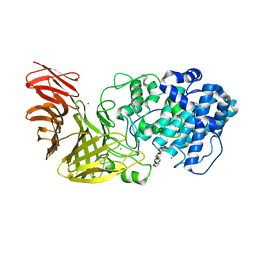 | | Crystal structure of the catalytic domain of a multi-domain alginate lyase Dp0100 from thermophilic bacterium Defluviitalea phaphyphila | | 分子名称: | Alginate lyase, CALCIUM ION, MAGNESIUM ION, ... | | 著者 | Ji, S.Q, Dix, S.R, Aziz, A, Sedelnikova, S.E, Li, F.L, Rice, D.W. | | 登録日 | 2019-03-27 | | 公開日 | 2019-10-30 | | 最終更新日 | 2024-03-27 | | 実験手法 | X-RAY DIFFRACTION (2.85 Å) | | 主引用文献 | The molecular basis of endolytic activity of a multidomain alginate lyase fromDefluviitalea phaphyphila, a representative of a new lyase family, PL39.
J.Biol.Chem., 294, 2019
|
|
6J7M
 
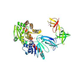 | |
6J7J
 
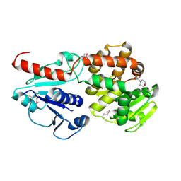 | | Crystal structure of Pseudomonas aeruginosa Earp | | 分子名称: | 2-(N-MORPHOLINO)-ETHANESULFONIC ACID, Pseudomonas aeruginosa Earp | | 著者 | He, C, Li, F. | | 登録日 | 2019-01-18 | | 公開日 | 2019-05-15 | | 最終更新日 | 2019-06-26 | | 実験手法 | X-RAY DIFFRACTION (1.75 Å) | | 主引用文献 | Complex Structure ofPseudomonas aeruginosaArginine Rhamnosyltransferase EarP with Its Acceptor Elongation Factor P.
J.Bacteriol., 201, 2019
|
|
6J7K
 
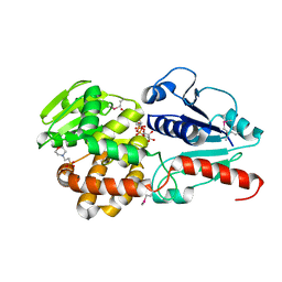 | |
7DMK
 
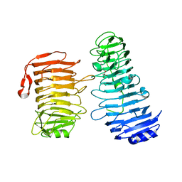 | | PL6 alginate lyase BcAlyPL6 | | 分子名称: | BcAlyPL6, CALCIUM ION, MALONATE ION | | 著者 | Dong, S, Wang, B, Ma, X.Q, Li, F.L, Feng, Y.G. | | 登録日 | 2020-12-04 | | 公開日 | 2021-10-06 | | 最終更新日 | 2023-11-29 | | 実験手法 | X-RAY DIFFRACTION (2.213 Å) | | 主引用文献 | Structural basis for the exolytic activity of polysaccharide lyase family 6 alginate lyase BcAlyPL6 from human gut microbe Bacteroides clarus.
Biochem.Biophys.Res.Commun., 547, 2021
|
|
3PHD
 
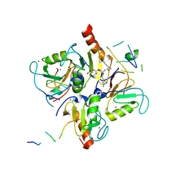 | | Crystal structure of human HDAC6 in complex with ubiquitin | | 分子名称: | Histone deacetylase 6, Polyubiquitin, ZINC ION | | 著者 | Dong, A, Qui, W, Ravichandran, M, Schuetz, A, Loppnau, P, Li, F, Mackenzie, F, Kozieradzki, I, Ouyang, H, Structural Genomics Consortium (SGC) | | 登録日 | 2010-11-03 | | 公開日 | 2011-02-23 | | 最終更新日 | 2023-09-06 | | 実験手法 | X-RAY DIFFRACTION (3 Å) | | 主引用文献 | Protein Aggregates Are Recruited to Aggresome by Histone Deacetylase 6 via Unanchored Ubiquitin C Termini.
J.Biol.Chem., 287, 2012
|
|
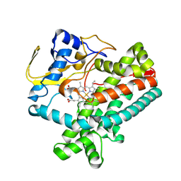
8W7G
 
 | |
5X0W
 
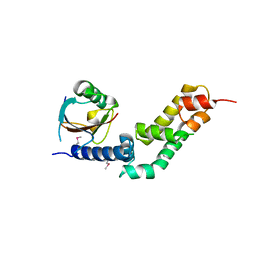 | | Molecular mechanism for the binding between Sharpin and HOIP | | 分子名称: | E3 ubiquitin-protein ligase RNF31, Sharpin | | 著者 | Liu, J, Li, F, Cheng, X, Pan, L. | | 登録日 | 2017-01-23 | | 公開日 | 2017-10-18 | | 実験手法 | X-RAY DIFFRACTION (3 Å) | | 主引用文献 | Structural Insights into SHARPIN-Mediated Activation of HOIP for the Linear Ubiquitin Chain Assembly
Cell Rep, 21, 2017
|
|
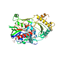
7CGS
 
 | |
