7D3W
 
 | |
4EHM
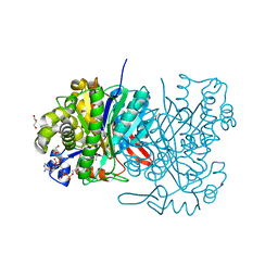
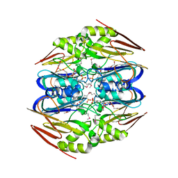
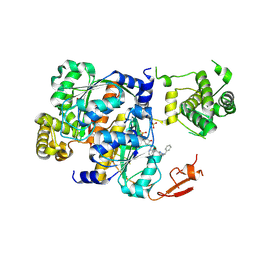
 | | RabGGTase in complex with covalently bound Psoromic acid | | 分子名称: | 4-formyl-3-hydroxy-8-methoxy-1,9-dimethyl-11-oxo-11H-dibenzo[b,e][1,4]dioxepine-6-carboxylic acid, CALCIUM ION, Geranylgeranyl transferase type-2 subunit alpha, ... | | 著者 | Guo, Z, Deraeve, C, Bon, R.S, Alexandrov, K, Waldmann, H, Wu, Y.W, Goody, R.S, Blankenfeldt, W. | | 登録日 | 2012-04-02 | | 公開日 | 2012-05-30 | | 最終更新日 | 2017-08-09 | | 実験手法 | X-RAY DIFFRACTION (2.2 Å) | | 主引用文献 | Psoromic Acid is a Selective and Covalent Rab-Prenylation Inhibitor Targeting Autoinhibited RabGGTase
J.Am.Chem.Soc., 134, 2012
|
|
1U6Q
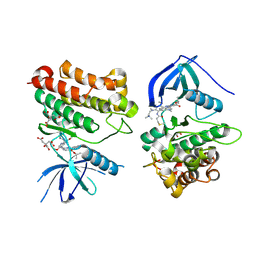
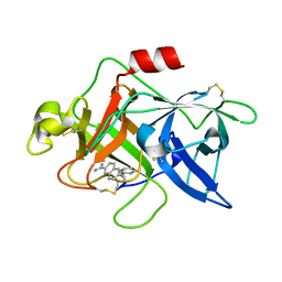 | | Substituted 2-Naphthamadine inhibitors of Urokinase | | 分子名称: | TRANS-6-(2-PHENYLCYCLOPROPYL)-NAPHTHALENE-2-CARBOXAMIDINE, Urokinase-type plasminogen activator | | 著者 | Bruncko, M, McClellan, W, Wendt, M.D, Sauer, D.R, Geyer, A, Dalton, C.R, Kaminski, M.K, Nienaber, V.L, Rockway, T.R, Giranda, V.L. | | 登録日 | 2004-07-30 | | 公開日 | 2004-10-19 | | 最終更新日 | 2011-07-13 | | 実験手法 | X-RAY DIFFRACTION (2.02 Å) | | 主引用文献 | Naphthamidine urokinase plasminogen activator inhibitors with improved pharmacokinetic properties.
Bioorg.Med.Chem.Lett., 15, 2005
|
|
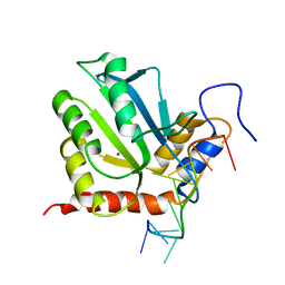
2Q0I
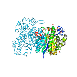
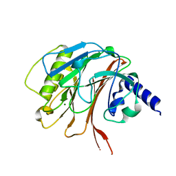
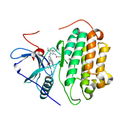
 | | Structure of Pseudomonas Quinolone Signal Response Protein PqsE | | 分子名称: | BENZOIC ACID, FE (III) ION, Quinolone signal response protein | | 著者 | Yu, S, Jensen, V, Feldmann, I, Haussler, S, Blankenfeldt, W. | | 登録日 | 2007-05-22 | | 公開日 | 2008-06-03 | | 最終更新日 | 2024-02-21 | | 実験手法 | X-RAY DIFFRACTION (1.57 Å) | | 主引用文献 | Structure elucidation and preliminary assessment of hydrolase activity of PqsE, the Pseudomonas quinolone signal (PQS) response protein.
Biochemistry, 48, 2009
|
|
1A1R
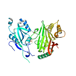
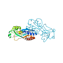
 | | HCV NS3 PROTEASE DOMAIN:NS4A PEPTIDE COMPLEX | | 分子名称: | NS3 PROTEIN, NS4A PROTEIN, ZINC ION | | 著者 | Kim, J.L, Morgenstern, K.A, Lin, C, Fox, T, Dwyer, M.D, Landro, J.A, Chambers, S.P, Markland, W, Lepre, C.A, O'Malley, E.T, Harbeson, S.L, Rice, C.M, Murcko, M.A, Caron, P.R, Thomson, J.A. | | 登録日 | 1997-12-15 | | 公開日 | 1998-06-17 | | 最終更新日 | 2024-02-07 | | 実験手法 | X-RAY DIFFRACTION (2.5 Å) | | 主引用文献 | Crystal structure of the hepatitis C virus NS3 protease domain complexed with a synthetic NS4A cofactor peptide.
Cell(Cambridge,Mass.), 87, 1996
|
|
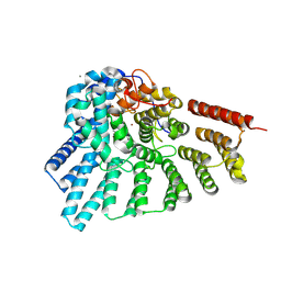
6U87
 | | Pseudomonas aeruginosa HasA mutant - Y75H | | 分子名称: | HasAp, PROTOPORPHYRIN IX CONTAINING FE | | 著者 | Brimberry, M, Lanzilotta, W, Wilks, A, Dent, A. | | 登録日 | 2019-09-04 | | 公開日 | 2020-09-09 | | 最終更新日 | 2023-10-11 | | 実験手法 | X-RAY DIFFRACTION (1.3 Å) | | 主引用文献 | Axial Heme Coordination by the Tyr-His Motif in the Extracellular Hemophore HasAp Is Critical for the Release of Heme to the HasR Receptor of Pseudomonas aeruginosa .
Biochemistry, 60, 2021
|
|
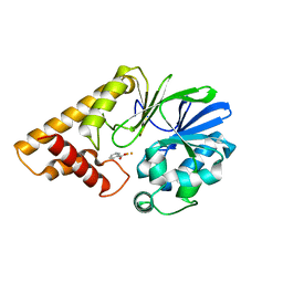
5HWP
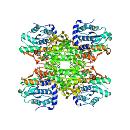
 | | MvaS with acetylated Cys115 in complex with coenzyme A | | 分子名称: | COENZYME A, GLYCEROL, Hydroxymethylglutaryl-CoA synthase, ... | | 著者 | Bock, T, Kasten, J, Blankenfeldt, W. | | 登録日 | 2016-01-29 | | 公開日 | 2016-05-11 | | 最終更新日 | 2016-07-13 | | 実験手法 | X-RAY DIFFRACTION (2.2 Å) | | 主引用文献 | Crystal Structure of the HMG-CoA Synthase MvaS from the Gram-Negative Bacterium Myxococcus xanthus.
Chembiochem, 17, 2016
|
|
5HWO
 | | MvaS in complex with 3-hydroxy-3-methylglutaryl coenzyme A | | 分子名称: | 3-HYDROXY-3-METHYLGLUTARYL-COENZYME A, GLYCEROL, Hydroxymethylglutaryl-CoA synthase, ... | | 著者 | Bock, T, Kasten, J, Blankenfeldt, W. | | 登録日 | 2016-01-29 | | 公開日 | 2016-05-11 | | 最終更新日 | 2024-01-10 | | 実験手法 | X-RAY DIFFRACTION (1.48 Å) | | 主引用文献 | Crystal Structure of the HMG-CoA Synthase MvaS from the Gram-Negative Bacterium Myxococcus xanthus.
Chembiochem, 17, 2016
|
|
5HWQ
 | | MvaS in complex with acetoacetyl coenzyme A | | 分子名称: | ACETOACETYL-COENZYME A, GLYCEROL, Hydroxymethylglutaryl-CoA synthase, ... | | 著者 | Bock, T, Kasten, J, Blankenfeldt, W. | | 登録日 | 2016-01-29 | | 公開日 | 2016-05-11 | | 最終更新日 | 2023-11-15 | | 実験手法 | X-RAY DIFFRACTION (1.5 Å) | | 主引用文献 | Crystal Structure of the HMG-CoA Synthase MvaS from the Gram-Negative Bacterium Myxococcus xanthus.
Chembiochem, 17, 2016
|
|
5HWR
 | | MvaS in complex with coenzyme A | | 分子名称: | COENZYME A, GLYCEROL, Hydroxymethylglutaryl-CoA synthase, ... | | 著者 | Bock, T, Kasten, J, Blankenfeldt, W. | | 登録日 | 2016-01-29 | | 公開日 | 2016-05-11 | | 最終更新日 | 2016-07-13 | | 実験手法 | X-RAY DIFFRACTION (1.5 Å) | | 主引用文献 | Crystal Structure of the HMG-CoA Synthase MvaS from the Gram-Negative Bacterium Myxococcus xanthus.
Chembiochem, 17, 2016
|
|
5W49
 | | The crystal structure of human S-adenosylhomocysteine hydrolase (AHCY) bound to oxadiazole inhibitor | | 分子名称: | (4-amino-1,2,5-oxadiazol-3-yl)[(3R)-3-{4-[(3-methoxyphenyl)amino]-6-methylpyridin-2-yl}pyrrolidin-1-yl]methanone, 1,2-ETHANEDIOL, Adenosylhomocysteinase, ... | | 著者 | Dougan, D.R, Lawson, J.D, Lane, W. | | 登録日 | 2017-06-09 | | 公開日 | 2017-06-28 | | 最終更新日 | 2024-03-13 | | 実験手法 | X-RAY DIFFRACTION (2.4 Å) | | 主引用文献 | Identification of AHCY inhibitors using novel high-throughput mass spectrometry.
Biochem. Biophys. Res. Commun., 491, 2017
|
|
5W4B
 | | The crystal structure of human S-adenosylhomocysteine hydrolase (AHCY) bound to benzothiazole inhibitor | | 分子名称: | 1,2-ETHANEDIOL, 4-[(2,5-dioxo-2,5-dihydro-1H-imidazol-1-yl)methyl]-N-[2-(morpholin-4-yl)-1,3-benzothiazol-6-yl]benzamide, Adenosylhomocysteinase, ... | | 著者 | Dougan, D.R, Lawson, J.D, Lane, W. | | 登録日 | 2017-06-09 | | 公開日 | 2017-06-28 | | 最終更新日 | 2024-03-13 | | 実験手法 | X-RAY DIFFRACTION (2.65 Å) | | 主引用文献 | Identification of AHCY inhibitors using novel high-throughput mass spectrometry.
Biochem. Biophys. Res. Commun., 491, 2017
|
|
4ZFK
 | | Ergothioneine-biosynthetic Ntn hydrolase EgtC with glutamine | | 分子名称: | 1,2-ETHANEDIOL, Amidohydrolase EgtC, GLUTAMINE | | 著者 | Vit, A, Seebeck, F.P, Blankenfeldt, W. | | 登録日 | 2015-04-21 | | 公開日 | 2015-07-01 | | 最終更新日 | 2024-01-10 | | 実験手法 | X-RAY DIFFRACTION (1.82 Å) | | 主引用文献 | Structure of the Ergothioneine-Biosynthesis Amidohydrolase EgtC.
Chembiochem, 16, 2015
|
|
4ZFJ
 | | Ergothioneine-biosynthetic Ntn hydrolase EgtC, apo form | | 分子名称: | 2-(N-MORPHOLINO)-ETHANESULFONIC ACID, 3,6,9,12,15,18,21,24,27-NONAOXANONACOSANE-1,29-DIOL, Amidohydrolase EgtC | | 著者 | Vit, A, Seebeck, F.P, Blankenfeldt, W. | | 登録日 | 2015-04-21 | | 公開日 | 2015-07-01 | | 最終更新日 | 2017-10-18 | | 実験手法 | X-RAY DIFFRACTION (1.75 Å) | | 主引用文献 | Structure of the Ergothioneine-Biosynthesis Amidohydrolase EgtC.
Chembiochem, 16, 2015
|
|
2LFF
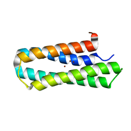 | | Solution structure of Diiron protein in presence of 8 eq Zn2+, Northeast Structural Genomics consortium target OR21 | | 分子名称: | Diiron protein, ZINC ION | | 著者 | Pires, M, Wu, Y, Mills, J.L, Reig, A, Englander, W, Degrado, W, Montelione, G.T, Szyperski, T, Northeast Structural Genomics Consortium (NESG) | | 登録日 | 2011-06-29 | | 公開日 | 2011-08-24 | | 最終更新日 | 2024-05-15 | | 実験手法 | SOLUTION NMR | | 主引用文献 | Solution structure of Diiron protein in presence of 8 eq Zn2+
To be Published
|
|
6YHN
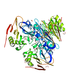
 | | Crystal structure of domains 4-5 of CNFy from Yersinia pseudotuberculosis | | 分子名称: | (R,R)-2,3-BUTANEDIOL, CHLORIDE ION, Cytotoxic necrotizing factor, ... | | 著者 | Lukat, P, Gazdag, E.M, Heidler, T.V, Blankenfeldt, W. | | 登録日 | 2020-03-30 | | 公開日 | 2020-12-30 | | 最終更新日 | 2024-05-01 | | 実験手法 | X-RAY DIFFRACTION (1.8 Å) | | 主引用文献 | Crystal structure of bacterial cytotoxic necrotizing factor CNF Y reveals molecular building blocks for intoxication.
Embo J., 40, 2021
|
|
6YHM
 | | Crystal structure of the C-terminal domain of CNFy from Yersinia pseudotuberculosis | | 分子名称: | Cytotoxic necrotizing factor, MAGNESIUM ION | | 著者 | Lukat, P, Gazdag, E.M, Heidler, T.V, Blankenfeldt, W. | | 登録日 | 2020-03-30 | | 公開日 | 2020-12-30 | | 最終更新日 | 2024-01-24 | | 実験手法 | X-RAY DIFFRACTION (1.13 Å) | | 主引用文献 | Crystal structure of bacterial cytotoxic necrotizing factor CNF Y reveals molecular building blocks for intoxication.
Embo J., 40, 2021
|
|
6YHL
 | |
6YHK
 | | Crystal structure of full-length CNFy (C866S) from Yersinia pseudotuberculosis | | 分子名称: | CHLORIDE ION, Cytotoxic necrotizing factor, SULFATE ION | | 著者 | Lukat, P, Gazdag, E.M, Heidler, T.V, Blankenfeldt, W. | | 登録日 | 2020-03-30 | | 公開日 | 2020-12-30 | | 最終更新日 | 2024-05-01 | | 実験手法 | X-RAY DIFFRACTION (2.7 Å) | | 主引用文献 | Crystal structure of bacterial cytotoxic necrotizing factor CNF Y reveals molecular building blocks for intoxication.
Embo J., 40, 2021
|
|
6XOG
 | | Structure of SUMO1-ML786519 adduct bound to SAE | | 分子名称: | SULFATE ION, SUMO-activating enzyme subunit 1, SUMO-activating enzyme subunit 2, ... | | 著者 | Sintchak, M, Lane, W, Bump, N. | | 登録日 | 2020-07-07 | | 公開日 | 2021-03-10 | | 最終更新日 | 2023-10-18 | | 実験手法 | X-RAY DIFFRACTION (1.98 Å) | | 主引用文献 | Discovery of TAK-981, a First-in-Class Inhibitor of SUMO-Activating Enzyme for the Treatment of Cancer.
J.Med.Chem., 64, 2021
|
|
6XOH
 | | Structure of SUMO1-ML00789344 adduct bound to SAE | | 分子名称: | SULFATE ION, SUMO-activating enzyme subunit 1, SUMO-activating enzyme subunit 2, ... | | 著者 | Sintchak, M, Lane, W, Bump, N. | | 登録日 | 2020-07-07 | | 公開日 | 2021-03-24 | | 最終更新日 | 2023-10-18 | | 実験手法 | X-RAY DIFFRACTION (2.226 Å) | | 主引用文献 | Discovery of TAK-981, a First-in-Class Inhibitor of SUMO-Activating Enzyme for the Treatment of Cancer.
J.Med.Chem., 64, 2021
|
|
6XOI
 | | Structure of SUMO1-ML00752641 adduct bound to SAE | | 分子名称: | SULFATE ION, SUMO-activating enzyme subunit 1, SUMO-activating enzyme subunit 2, ... | | 著者 | Sintchak, M, Lane, W, Bump, N. | | 登録日 | 2020-07-07 | | 公開日 | 2021-03-24 | | 最終更新日 | 2023-10-18 | | 実験手法 | X-RAY DIFFRACTION (2 Å) | | 主引用文献 | Discovery of TAK-981, a First-in-Class Inhibitor of SUMO-Activating Enzyme for the Treatment of Cancer.
J.Med.Chem., 64, 2021
|
|
7T4J
 | | Crystal Structure of EGFR_D770_N771insNPG/V948R in complex with TAK-788 | | 分子名称: | 1,2-ETHANEDIOL, CITRIC ACID, Epidermal growth factor receptor, ... | | 著者 | Skene, R.J, Lane, W, Hu, Y. | | 登録日 | 2021-12-10 | | 公開日 | 2022-12-07 | | 最終更新日 | 2023-10-25 | | 実験手法 | X-RAY DIFFRACTION (2.2 Å) | | 主引用文献 | Discovery of mobocertinib, a potent, oral inhibitor of EGFR exon 20 insertion mutations in non-small cell lung cancer.
Bioorg.Med.Chem.Lett., 80, 2022
|
|
7T4I
 | | Crystal Structure of wild type EGFR in complex with TAK-788 | | 分子名称: | Epidermal growth factor receptor, propan-2-yl 2-[[4-[2-(dimethylamino)ethyl-methyl-amino]-2-methoxy-5-(propanoylamino)phenyl]amino]-4-(1-methylindol-3-yl)pyrimidine-5-carboxylate | | 著者 | Skene, R.J, Lane, W. | | 登録日 | 2021-12-10 | | 公開日 | 2022-12-07 | | 最終更新日 | 2023-10-25 | | 実験手法 | X-RAY DIFFRACTION (2.61 Å) | | 主引用文献 | Discovery of mobocertinib, a potent, oral inhibitor of EGFR exon 20 insertion mutations in non-small cell lung cancer.
Bioorg.Med.Chem.Lett., 80, 2022
|
|
7P4U
 | |


