4OUJ
 
 | | Crystal structure of HA33B-Lac | | 分子名称: | Hemagglutinin component HA33, beta-D-galactopyranose-(1-4)-alpha-D-glucopyranose | | 著者 | Lee, K, Lam, K.-H, Perry, K, Rummel, A, Jin, R. | | 登録日 | 2014-02-17 | | 公開日 | 2014-06-04 | | 最終更新日 | 2023-09-20 | | 実験手法 | X-RAY DIFFRACTION (1.46 Å) | | 主引用文献 | High-resolution crystal structure of HA33 of botulinum neurotoxin type B progenitor toxin complex.
Biochem.Biophys.Res.Commun., 446, 2014
|
|
5JLV
 | | Receptor binding domain of Botulinum neurotoxin A in complex with human glycosylated SV2C | | 分子名称: | 2-acetamido-2-deoxy-beta-D-glucopyranose, 2-acetamido-2-deoxy-beta-D-glucopyranose-(1-4)-[alpha-L-fucopyranose-(1-6)]2-acetamido-2-deoxy-beta-D-glucopyranose, ACETATE ION, ... | | 著者 | Yao, G, Zhang, S, Mahrhold, S, Lam, K, Stern, D, Bagramyan, K, Perry, K, Kalkum, M, Rummel, A, Dong, M, Jin, R. | | 登録日 | 2016-04-27 | | 公開日 | 2016-06-15 | | 最終更新日 | 2023-09-27 | | 実験手法 | X-RAY DIFFRACTION (2 Å) | | 主引用文献 | N-linked glycosylation of SV2 is required for binding and uptake of botulinum neurotoxin A.
Nat.Struct.Mol.Biol., 23, 2016
|
|
5JMC
 | | Receptor binding domain of Botulinum neurotoxin A in complex with rat SV2C | | 分子名称: | Botulinum neurotoxin type A, Synaptic vesicle glycoprotein 2C | | 著者 | Yao, G, Zhang, S, Mahrhold, S, Lam, K, Stern, D, Bagramyan, K, Perry, K, Kalkum, M, Rummel, A, Dong, M, Jin, R. | | 登録日 | 2016-04-28 | | 公開日 | 2016-06-15 | | 最終更新日 | 2023-09-27 | | 実験手法 | X-RAY DIFFRACTION (2.64 Å) | | 主引用文献 | N-linked glycosylation of SV2 is required for binding and uptake of botulinum neurotoxin A.
Nat.Struct.Mol.Biol., 23, 2016
|
|
6MHJ
 | | Structure of BoNT mutant | | 分子名称: | Botulinum neurotoxin type A, PHOSPHATE ION | | 著者 | Lam, K, Jin, R. | | 登録日 | 2018-09-18 | | 公開日 | 2018-12-26 | | 最終更新日 | 2023-10-11 | | 実験手法 | X-RAY DIFFRACTION (3.019 Å) | | 主引用文献 | A viral-fusion-peptide-like molecular switch drives membrane insertion of botulinum neurotoxin A1.
Nat Commun, 9, 2018
|
|
7UIB
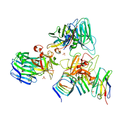 | | Crystal structure of BoNT/E receptor binding domain in complex with SV2, VHH, and sialic acid | | 分子名称: | 2-acetamido-2-deoxy-beta-D-glucopyranose, 2-acetamido-2-deoxy-beta-D-glucopyranose-(1-4)-2-acetamido-2-deoxy-beta-D-glucopyranose, N-acetyl-beta-neuraminic acid, ... | | 著者 | Liu, Z, Jin, R, Chen, P. | | 登録日 | 2022-03-29 | | 公開日 | 2023-04-05 | | 最終更新日 | 2023-10-25 | | 実験手法 | X-RAY DIFFRACTION (2.77 Å) | | 主引用文献 | Structural basis for botulinum neurotoxin E recognition of synaptic vesicle protein 2.
Nat Commun, 14, 2023
|
|
7UIA
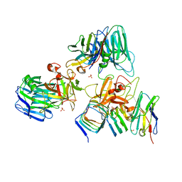 | | Crystal structure of BoNT/E receptor binding domain in complex with SV2 and VHH | | 分子名称: | 2-acetamido-2-deoxy-beta-D-glucopyranose, 2-acetamido-2-deoxy-beta-D-glucopyranose-(1-4)-2-acetamido-2-deoxy-beta-D-glucopyranose, DI(HYDROXYETHYL)ETHER, ... | | 著者 | Liu, Z, Jin, R, Chen, P. | | 登録日 | 2022-03-28 | | 公開日 | 2023-04-05 | | 最終更新日 | 2023-10-25 | | 実験手法 | X-RAY DIFFRACTION (2.59 Å) | | 主引用文献 | Structural basis for botulinum neurotoxin E recognition of synaptic vesicle protein 2.
Nat Commun, 14, 2023
|
|
3R4S
 | | Cell entry of botulinum neurotoxin type C is dependent upon interaction with two ganglioside molecules | | 分子名称: | Botulinum neurotoxin type C1, N-acetyl-alpha-neuraminic acid, N-acetyl-beta-neuraminic acid | | 著者 | Strotmeier, J, Gu, S, Jutzi, S, Mahrhold, S, Zhou, J, Pich, A, Bigalke, H, Rummel, A, Jin, R, Binz, T. | | 登録日 | 2011-03-17 | | 公開日 | 2011-06-08 | | 最終更新日 | 2024-02-21 | | 実験手法 | X-RAY DIFFRACTION (2.15 Å) | | 主引用文献 | The biological activity of botulinum neurotoxin type C is dependent upon novel types of ganglioside binding sites.
Mol.Microbiol., 81, 2011
|
|
3R4U
 | | Cell entry of botulinum neurotoxin type C is dependent upon interaction with two ganglioside molecules | | 分子名称: | Botulinum neurotoxin type C1 | | 著者 | Strotmeier, J, Gu, S, Jutzi, S, Mahrhold, S, Zhou, J, Pich, A, Bigalke, H, Rummel, A, Jin, R, Binz, T. | | 登録日 | 2011-03-17 | | 公開日 | 2011-06-08 | | 最終更新日 | 2024-02-21 | | 実験手法 | X-RAY DIFFRACTION (2.2 Å) | | 主引用文献 | The biological activity of botulinum neurotoxin type C is dependent upon novel types of ganglioside binding sites.
Mol.Microbiol., 81, 2011
|
|
6C0B
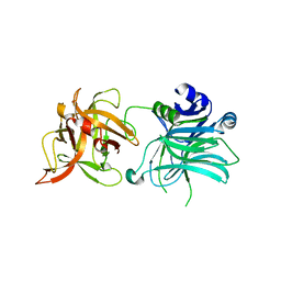
 | | Structural basis for recognition of frizzled proteins by Clostridium difficile toxin B | | 分子名称: | 2-acetamido-2-deoxy-beta-D-glucopyranose, Frizzled-2, MALONATE ION, ... | | 著者 | Chen, P, Lam, K, Jin, R. | | 登録日 | 2017-12-28 | | 公開日 | 2018-05-16 | | 最終更新日 | 2020-07-29 | | 実験手法 | X-RAY DIFFRACTION (2.5 Å) | | 主引用文献 | Structural basis for recognition of frizzled proteins byClostridium difficiletoxin B.
Science, 360, 2018
|
|
5WIX
 | |
8GYD
 | | Structure of Schistosoma japonicum Glutathione S-transferase bound with the ligand complex of 16 | | 分子名称: | (2R)-2-[[2-(5-chloranylthiophen-2-yl)-4-oxidanylidene-6-[2-(1H-1,2,3,4-tetrazol-5-yl)phenyl]quinazolin-3-yl]methyl]-3-(4-chlorophenyl)propanoic acid, ETHANOL, Glutathione S-transferase class-mu 26 kDa isozyme | | 著者 | Wen, X, Jin, R, Hu, H, Zhu, J, Song, W, Lu, X. | | 登録日 | 2022-09-22 | | 公開日 | 2023-08-23 | | 最終更新日 | 2023-09-13 | | 実験手法 | X-RAY DIFFRACTION (1.7 Å) | | 主引用文献 | Discovery, SAR Study of GST Inhibitors from a Novel Quinazolin-4(1 H )-one Focused DNA-Encoded Library.
J.Med.Chem., 66, 2023
|
|
7OHF
 | | Cryo-EM structure of pyrococcus furiosus apoferritin in nanofluidic channels | | 分子名称: | Ferritin | | 著者 | Huber, S.T, Sarajlic, E, Huijink, R, Evers, W.H, Jakobi, A.J. | | 登録日 | 2021-05-10 | | 公開日 | 2021-08-11 | | 最終更新日 | 2024-07-10 | | 実験手法 | ELECTRON MICROSCOPY (3 Å) | | 主引用文献 | Nanofluidic chips for cryo-EM structure determination from picoliter sample volumes.
Elife, 11, 2022
|
|
4GH4
 | | Crystal Structure of Foot and Mouth Disease Virus A22 Serotype | | 分子名称: | capsid protein VP1, capsid protein VP2, capsid protein VP3, ... | | 著者 | Kotecha, A, Jinshan, R, Curry, S, Fry, E, Stuart, D. | | 登録日 | 2012-08-07 | | 公開日 | 2013-02-20 | | 最終更新日 | 2024-02-28 | | 実験手法 | X-RAY DIFFRACTION (3 Å) | | 主引用文献 | Perturbations in the surface structure of A22 Iraq foot-and-mouth disease virus accompanying coupled changes in host cell specificity and antigenicity.
Structure, 4, 1996
|
|
1R4H
 | | NMR Solution structure of the IIIc domain of GB Virus B IRES Element | | 分子名称: | 5'-R(*GP*GP*GP*CP*AP*AP*GP*CP*CP*C)-3' | | 著者 | Kaluarachchi, K, Thiviyanathan, V, Rijinbrand, R, Lemon, S.M, Gorenstein, D.G. | | 登録日 | 2003-10-06 | | 公開日 | 2004-10-19 | | 最終更新日 | 2024-05-22 | | 実験手法 | SOLUTION NMR | | 主引用文献 | Mutational and structural analysis of stem-loop IIIC of the hepatitis C virus and GB virus B internal ribosome entry sites.
J.Mol.Biol., 343, 2004
|
|


