Loading
PDBj
MenuPDBj@FacebookPDBj@TwitterPDBj@YouTubewwPDB FoundationwwPDB
RCSB PDBPDBeBMRBAdv. SearchSearch help
PDB: 190 results

2SGA
DownloadVisualize
BU of 2sga by Molmil
ELECTRON DENSITY CALCULATIONS AS AN EXTENSION OF PROTEIN STRUCTURE REFINEMENT. STREPTOMYCES GRISEUS PROTEASE AT 1.5 ANGSTROMS RESOLUTION
Descriptor: PROTEINASE A
Authors:James, M.N.G, Sielecki, A.R.
Deposit date:1983-01-21
Release date:1983-04-21
Last modified:2011-07-13
Method:X-RAY DIFFRACTION (1.5 Å)
Cite:Electron density calculations as an extension of protein structure refinement. Streptomyces griseus protease A at 1.5 A resolution.
J.Mol.Biol., 182, 1985
2PSG
DownloadVisualize
BU of 2psg by Molmil
REFINED STRUCTURE OF PORCINE PEPSINOGEN AT 1.8 ANGSTROMS RESOLUTION
Descriptor: PEPSINOGEN
Authors:James, M.N.G, Sielecki, A.R.
Deposit date:1991-01-23
Release date:1992-10-15
Last modified:2011-07-13
Method:X-RAY DIFFRACTION (1.8 Å)
Cite:Refined structure of porcine pepsinogen at 1.8 A resolution.
J.Mol.Biol., 219, 1991
4SGA
DownloadVisualize
BU of 4sga by Molmil
STRUCTURES OF PRODUCT AND INHIBITOR COMPLEXES OF STREPTOMYCES GRISEUS PROTEASE A AT 1.8 ANGSTROMS RESOLUTION. A MODEL FOR SERINE PROTEASE CATALYSIS
Descriptor: PROTEINASE A (SGPA), TETRAPEPTIDE ACE-PRO-ALA-PRO-PHE
Authors:Sielecki, A.R, James, M.N.G.
Deposit date:1990-05-29
Release date:1991-10-15
Last modified:2013-03-13
Method:X-RAY DIFFRACTION (1.8 Å)
Cite:Structures of product and inhibitor complexes of Streptomyces griseus protease A at 1.8 A resolution. A model for serine protease catalysis.
J.Mol.Biol., 144, 1980
3APP
DownloadVisualize
BU of 3app by Molmil
STRUCTURE AND REFINEMENT OF PENICILLOPEPSIN AT 1.8 ANGSTROMS RESOLUTION
Descriptor: PENICILLOPEPSIN
Authors:Sielecki, A.R, James, M.N.G.
Deposit date:1990-11-27
Release date:1991-01-15
Last modified:2011-07-13
Method:X-RAY DIFFRACTION (1.8 Å)
Cite:Structure and refinement of penicillopepsin at 1.8 A resolution.
J.Mol.Biol., 163, 1983
6AQ7
DownloadVisualize
BU of 6aq7 by Molmil
Structure of POM6 FAB fragment complexed with mouse PrPc
Descriptor: 3,3',3''-phosphanetriyltripropanoic acid, GLYCEROL, Major prion protein, ...
Authors:Baral, P.K, Swayampakula, M, James, M.N.G.
Deposit date:2017-08-18
Release date:2018-04-04
Last modified:2023-10-04
Method:X-RAY DIFFRACTION (1.83 Å)
Cite:Structural characterization of POM6 Fab and mouse prion protein complex identifies key regions for prions conformational conversion.
FEBS J., 285, 2018
5TNC
DownloadVisualize
BU of 5tnc by Molmil
REFINED CRYSTAL STRUCTURE OF TROPONIN C FROM TURKEY SKELETAL MUSCLE AT 2.0 ANGSTROMS RESOLUTION
Descriptor: CALCIUM ION, TROPONIN-C
Authors:Herzberg, O, James, M.N.G.
Deposit date:1988-05-27
Release date:1988-10-09
Last modified:2024-03-06
Method:X-RAY DIFFRACTION (2 Å)
Cite:Refined crystal structure of troponin C from turkey skeletal muscle at 2.0 A resolution.
J.Mol.Biol., 203, 1988
3SGA
DownloadVisualize
BU of 3sga by Molmil
STRUCTURES OF PRODUCT AND INHIBITOR COMPLEXES OF STREPTOMYCES GRISEUS PROTEASE A AT 1.8 ANGSTROMS RESOLUTION. A MODEL FOR SERINE PROTEASE CATALYSIS
Descriptor: ACE-PRO-ALA-PRO-PHE-ALDEHYDE, PROTEINASE A (SGPA)
Authors:Sielecki, A.R, James, M.N.G.
Deposit date:1990-05-29
Release date:1991-10-15
Last modified:2012-12-12
Method:X-RAY DIFFRACTION (1.8 Å)
Cite:Structures of product and inhibitor complexes of Streptomyces griseus protease A at 1.8 A resolution. A model for serine protease catalysis.
J.Mol.Biol., 144, 1980
5SGA
DownloadVisualize
BU of 5sga by Molmil
Structures of product and inhibitor complexes of Streptomyces griseus protease a at 1.8 Angstroms resolution. a model for serine protease catalysis
Descriptor: PROTEINASE A (SGPA), TETRAPEPTIDE ACE-PRO-ALA-PRO-TYR
Authors:Sielecki, A.R, James, M.N.G.
Deposit date:1990-05-29
Release date:1991-10-15
Last modified:2013-03-13
Method:X-RAY DIFFRACTION (1.8 Å)
Cite:Structures of product and inhibitor complexes of Streptomyces griseus protease A at 1.8 A resolution. A model for serine protease catalysis.
J.Mol.Biol., 144, 1980
3SGB
DownloadVisualize
BU of 3sgb by Molmil
STRUCTURE OF THE COMPLEX OF STREPTOMYCES GRISEUS PROTEASE B AND THE THIRD DOMAIN OF THE TURKEY OVOMUCOID INHIBITOR AT 1.8 ANGSTROMS RESOLUTION
Descriptor: PROTEINASE B (SGPB), TURKEY OVOMUCOID INHIBITOR (OMTKY3)
Authors:Read, R.J, Fujinaga, M, Sielecki, A.R, James, M.N.G.
Deposit date:1983-01-21
Release date:1983-07-12
Last modified:2017-11-29
Method:X-RAY DIFFRACTION (1.8 Å)
Cite:Structure of the complex of Streptomyces griseus protease B and the third domain of the turkey ovomucoid inhibitor at 1.8-A resolution.
Biochemistry, 22, 1983
1APW
DownloadVisualize
BU of 1apw by Molmil
CRYSTALLOGRAPHIC ANALYSIS OF TRANSITION STATE MIMICS BOUND TO PENICILLOPEPSIN: DIFLUOROSTATINE-AND DIFLUOROSTATONE-CONTAINING PEPTIDES
Descriptor: DIMETHYLFORMAMIDE, INHIBITOR ISOVALERYL (IVA)-VAL-VAL-DIFLUOROSTATINE-N-METHYLAMINE, PENICILLOPEPSIN, ...
Authors:Sielecki, A.R, James, M.N.G.
Deposit date:1991-12-16
Release date:1994-01-31
Last modified:2020-07-29
Method:X-RAY DIFFRACTION (1.8 Å)
Cite:Crystallographic analysis of transition state mimics bound to penicillopepsin: difluorostatine- and difluorostatone-containing peptides.
Biochemistry, 31, 1992
1APV
DownloadVisualize
BU of 1apv by Molmil
CRYSTALLOGRAPHIC ANALYSIS OF TRANSITION STATE MIMICS BOUND TO PENICILLOPEPSIN: DIFLUOROSTATINE-AND DIFLUOROSTATONE-CONTAINING PEPTIDES
Descriptor: DIMETHYLFORMAMIDE, INHIBITOR ISOVALERYL (IVA)-VAL-VAL-HYDRATED DIFLUOROSTATONE-N-METHYLAMINE, PENICILLOPEPSIN, ...
Authors:Sielecki, A.R, James, M.N.G.
Deposit date:1991-12-16
Release date:1994-01-31
Last modified:2020-07-29
Method:X-RAY DIFFRACTION (1.8 Å)
Cite:Crystallographic analysis of transition state mimics bound to penicillopepsin: difluorostatine- and difluorostatone-containing peptides.
Biochemistry, 31, 1992
1APT
DownloadVisualize
BU of 1apt by Molmil
CRYSTALLOGRAPHIC ANALYSIS OF A PEPSTATIN ANALOGUE BINDING TO THE ASPARTYL PROTEINASE PENICILLOPEPSIN AT 1.8 ANGSTROMS RESOLUTION
Descriptor: INHIBITOR ISOVALERYL (IVA)-VAL-VAL-LYSTA-O-ET (LYSTA IS A LYSYL SIDE CHAIN ANALOGUE OF STATIN, PENICILLOPEPSIN, alpha-D-mannopyranose
Authors:Sielecki, A.R, James, M.N.G.
Deposit date:1991-12-16
Release date:1994-01-31
Last modified:2020-07-29
Method:X-RAY DIFFRACTION (1.8 Å)
Cite:Crystallographic Analysis of a Pepstatin Analogue Binding to the Aspartyl Proteinase Penicillopepsin at 1.8 Angstroms Resolution
Peptides: Structure and Function, Proceedings of the of the Eighth American Peptide Symposium, 1, 1983
1APU
DownloadVisualize
BU of 1apu by Molmil
Crystallographic analysis of a pepstatin analogue binding to the aspartyl proteinase penicillopepsin at 1.8 angstroms resolution
Descriptor: PEPSTATIN ANALOGUE ISOVALERYL-VAL-VAL-STA-O-ET, PROTEIN (PENICILLOPEPSIN), alpha-D-mannopyranose
Authors:Sielecki, A.R, James, M.N.G.
Deposit date:1991-12-16
Release date:1994-01-31
Last modified:2020-07-29
Method:X-RAY DIFFRACTION (1.8 Å)
Cite:Crystallographic Analysis of Transition State Mimics Bound to Penicillopepsin: Difluorostatine-and Difluorostatone-Containing Peptides
Biochemistry, 31, 1992
2ALP
DownloadVisualize
BU of 2alp by Molmil
REFINED STRUCTURE OF ALPHA-LYTIC PROTEASE AT 1.7 ANGSTROMS RESOLUTION. ANALYSIS OF HYDROGEN BONDING AND SOLVENT STRUCTURE
Descriptor: ALPHA-LYTIC PROTEASE, SULFATE ION
Authors:Fujinaga, M, Delbaere, L.T.J, Brayer, G.D, James, M.N.G.
Deposit date:1985-03-07
Release date:1985-07-17
Last modified:2011-07-13
Method:X-RAY DIFFRACTION (1.7 Å)
Cite:Refined structure of alpha-lytic protease at 1.7 A resolution. Analysis of hydrogen bonding and solvent structure.
J.Mol.Biol., 184, 1985
1PPL
DownloadVisualize
BU of 1ppl by Molmil
CRYSTALLOGRAPHIC ANALYSIS OF TRANSITION-STATE MIMICS BOUND TO PENICILLOPEPSIN: PHOSPHORUS-CONTAINING PEPTIDE ANALOGUES
Descriptor: N-(3-methylbutanoyl)-L-valyl-N-{(1S)-1-[(R)-[(1R)-1-benzyl-2-methoxy-2-oxoethoxy](hydroxy)phosphoryl]-3-methylbutyl}-L- valinamide, PENICILLOPEPSIN, SULFATE ION, ...
Authors:Fraser, M.E, James, M.N.G.
Deposit date:1992-06-01
Release date:1993-10-31
Last modified:2020-07-29
Method:X-RAY DIFFRACTION (1.7 Å)
Cite:Crystallographic analysis of transition-state mimics bound to penicillopepsin: phosphorus-containing peptide analogues.
Biochemistry, 31, 1992
1PPM
DownloadVisualize
BU of 1ppm by Molmil
CRYSTALLOGRAPHIC ANALYSIS OF TRANSITION-STATE MIMICS BOUND TO PENICILLOPEPSIN: PHOSPHORUS-CONTAINING PEPTIDE ANALOGUES
Descriptor: N-[(benzyloxy)carbonyl]-L-alanyl-N-{(1S)-1-[(R)-[(1R)-1-benzyl-2-methoxy-2-oxoethoxy](hydroxy)phosphoryl]-3-methylbutyl }-L-alaninamide, PENICILLOPEPSIN, SULFATE ION, ...
Authors:Fraser, M.E, James, M.N.G.
Deposit date:1992-06-01
Release date:1993-10-31
Last modified:2020-07-29
Method:X-RAY DIFFRACTION (1.7 Å)
Cite:Crystallographic analysis of transition-state mimics bound to penicillopepsin: phosphorus-containing peptide analogues.
Biochemistry, 31, 1992
3SGQ
DownloadVisualize
BU of 3sgq by Molmil
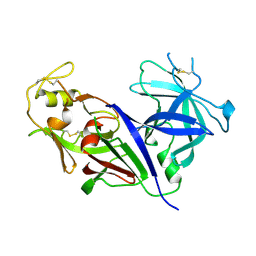
GLN 18 VARIANT OF TURKEY OVOMUCOID INHIBITOR THIRD DOMAIN COMPLEXED WITH STREPTOMYCES GRISEUS PROTEINASE B AT PH 10.7
Descriptor: Ovomucoid, PHOSPHATE ION, Streptogrisin B
Authors:Huang, K, Lu, W, Anderson, S, Laskowski Jr, M, James, M.N.G.
Deposit date:1999-03-25
Release date:2003-08-26
Last modified:2023-09-13
Method:X-RAY DIFFRACTION (1.8 Å)
Cite:Recruitment of a Buried K+ Ion to Stabilize the Negative Charge of Ionized P1 in the Hydrophobic Pocket: Crystal Structures of Glu18, Gln18, Asp18 and Asn18 Variants of Turkey Ovomucoid Inhibitor Third Domain Complexed with Streptomyces griseus Protease B at Various pH's
To be Published
4OBS
DownloadVisualize
BU of 4obs by Molmil
Crystal structure of human alpha-L-iduronidase in the P212121 form
Descriptor: 2-acetamido-2-deoxy-beta-D-glucopyranose, Alpha-L-iduronidase, CHLORIDE ION, ...
Authors:Bie, H, Yin, J, He, X, Kermode, A.R, James, M.N.G.
Deposit date:2014-01-07
Release date:2015-01-14
Last modified:2023-09-20
Method:X-RAY DIFFRACTION (2.26 Å)
Cite:Crystal structure of human alpha-L-iduronidase in the P212121 form
To be Published
4OBR
DownloadVisualize
BU of 4obr by Molmil
Crystal structure of human alpha-L-iduronidase complex with alpha-L-iduronic acid
Descriptor: 2-acetamido-2-deoxy-beta-D-glucopyranose, 2-acetamido-2-deoxy-beta-D-glucopyranose-(1-4)-2-acetamido-2-deoxy-beta-D-glucopyranose, Alpha-L-iduronidase, ...
Authors:Bie, H.Y, Yin, J, He, X, Kermode, A.R, James, M.N.G.
Deposit date:2014-01-07
Release date:2015-01-14
Last modified:2023-09-20
Method:X-RAY DIFFRACTION (2.46 Å)
Cite:Crystal structure of human alpha-L-iduronidase complex with alpha-L-iduronic acid
To be Published
4Q0G
DownloadVisualize
BU of 4q0g by Molmil
Crystal structure of beta subunit of acyl-CoA carboxylase AccD1 from Mycobacterium tuberculosis
Descriptor: GLYCEROL, Probable acetyl-/propionyl-CoA carboxylase (Beta subunit) AccD1
Authors:Bie, H.Y, Yin, J, James, M.N.G, TB Structural Genomics Consortium (TBSGC)
Deposit date:2014-04-02
Release date:2016-04-20
Last modified:2023-09-20
Method:X-RAY DIFFRACTION (2.31 Å)
Cite:Crystal structure of Beta subunit of acyl-CoA carboxylase AccD1 from Mycobacterium tuberculosis
To be Published
9LYZ
DownloadVisualize
BU of 9lyz by Molmil
X-RAY CRYSTALLOGRAPHY OF THE BINDING OF THE BACTERIAL CELL WALL TRISACCHARIDE NAM-NAG-NAM TO LYSOZYME
Descriptor: HEN EGG WHITE LYSOZYME, N-acetyl-beta-muramic acid-(1-4)-2-acetamido-2-deoxy-beta-D-glucopyranose-(1-4)-N-acetyl-beta-muramic acid
Authors:Kelly, J.A, James, M.N.G.
Deposit date:1979-12-06
Release date:1980-02-27
Last modified:2020-07-29
Method:X-RAY DIFFRACTION (2.5 Å)
Cite:X-ray crystallography of the binding of the bacterial cell wall trisaccharide NAM-NAG-NAM to lysozyme.
Nature, 282, 1979
1PPK
DownloadVisualize
BU of 1ppk by Molmil
CRYSTALLOGRAPHIC ANALYSIS OF TRANSITION STATE MIMICS BOUND TO PENICILLOPEPSIN: PHOSPHOROUS-CONTAINING PEPTIDE ANALOGUES
Descriptor: DIMETHYLFORMAMIDE, N-(3-methylbutanoyl)-L-valyl-N-{(1R)-1-[(R)-(2-ethoxy-2-oxoethyl)(hydroxy)phosphoryl]-3-methylbutyl}-L-valinamide, PENICILLOPEPSIN, ...
Authors:Strynadka, N.C.J, James, M.N.G.
Deposit date:1994-01-20
Release date:1994-05-31
Last modified:2020-07-29
Method:X-RAY DIFFRACTION (1.8 Å)
Cite:Crystallographic analysis of transition-state mimics bound to penicillopepsin: phosphorus-containing peptide analogues.
Biochemistry, 31, 1992
3LMY
DownloadVisualize
BU of 3lmy by Molmil
The Crystal Structure of beta-hexosaminidase B in complex with Pyrimethamine
Descriptor: 2-acetamido-2-deoxy-alpha-D-glucopyranose, 2-acetamido-2-deoxy-beta-D-glucopyranose, 2-acetamido-2-deoxy-beta-D-glucopyranose-(1-4)-2-acetamido-2-deoxy-beta-D-glucopyranose, ...
Authors:Bateman, K.S, Cherney, M.M, Withers, S.G, Mahuran, D.J, Tropak, M, James, M.N.G.
Deposit date:2010-02-01
Release date:2011-02-16
Last modified:2023-09-06
Method:X-RAY DIFFRACTION (2.8 Å)
Cite:The Crystal Structure of beta-hexosaminidase B in complex with Pyrimethamine
To be Published
2CI2
DownloadVisualize
BU of 2ci2 by Molmil
CRYSTAL AND MOLECULAR STRUCTURE OF THE SERINE PROTEINASE INHIBITOR CI-2 FROM BARLEY SEEDS
Descriptor: CHYMOTRYPSIN INHIBITOR 2
Authors:Mcphalen, C.A, James, M.N.G.
Deposit date:1988-09-05
Release date:1988-09-07
Last modified:2024-02-14
Method:X-RAY DIFFRACTION (2 Å)
Cite:Crystal and molecular structure of the serine proteinase inhibitor CI-2 from barley seeds.
Biochemistry, 26, 1987
1CQJ
DownloadVisualize
BU of 1cqj by Molmil
CRYSTAL STRUCTURE OF DEPHOSPHORYLATED E. COLI SUCCINYL-COA SYNTHETASE
Descriptor: COENZYME A, PHOSPHATE ION, SUCCINYL-COA SYNTHETASE ALPHA CHAIN, ...
Authors:Joyce, M.A, Fraser, M.E, James, M.N.G, Bridger, W.A, Wolodko, W.T.
Deposit date:1999-08-06
Release date:2000-01-10
Last modified:2017-10-04
Method:X-RAY DIFFRACTION (2.9 Å)
Cite:ADP-binding site of Escherichia coli succinyl-CoA synthetase revealed by x-ray crystallography.
Biochemistry, 39, 2000

219869

PDB entries from 2024-05-15

PDB statisticsPDBj update infoContact PDBjnumon