6TT2
 
 | |
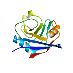
2BHY
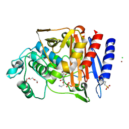
 | | Crystal structure of Deinococcus radiodurans maltooligosyltrehalose trehalohydrolase in complex with trehalose | | 分子名称: | 2-AMINO-2-HYDROXYMETHYL-PROPANE-1,3-DIOL, BETA-MERCAPTOETHANOL, MAGNESIUM ION, ... | | 著者 | Timmins, J, Leiros, H.-K.S, Leonard, G, Leiros, I, McSweeney, S. | | 登録日 | 2005-01-20 | | 公開日 | 2005-03-31 | | 最終更新日 | 2020-07-29 | | 実験手法 | X-RAY DIFFRACTION (1.5 Å) | | 主引用文献 | Crystal Structure of Maltooligosyltrehalose Trehalohydrolase from Deinococcus Radiodurans in Complex with Disaccharides
J.Mol.Biol., 347, 2005
|
|
4YU0
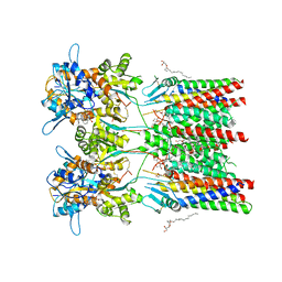
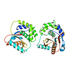
 | | Crystal structure of a tetramer of GluA2 TR mutant ligand binding domains bound with glutamate at 1.26 Angstrom resolution | | 分子名称: | DI(HYDROXYETHYL)ETHER, GLUTAMIC ACID, Glutamate receptor 2,Glutamate receptor 2, ... | | 著者 | Chebli, M, Salazar, H, Baranovic, J, Carbone, A.L, Ghisi, V, Faelber, K, Lau, A.Y, Daumke, O, Plested, A.J.R. | | 登録日 | 2015-03-18 | | 公開日 | 2016-01-13 | | 最終更新日 | 2024-01-10 | | 実験手法 | X-RAY DIFFRACTION (1.26 Å) | | 主引用文献 | Crystal structure of the tetrameric GluA2 ligand-binding domain in complex with glutamate at 1.26 Angstroms resolution
To Be Published
|
|
8XU4
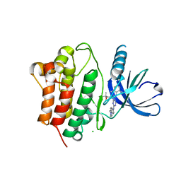
 | | The Crystal Structure of MAPK2 from Biortus. | | 分子名称: | MALONIC ACID, MAP kinase-activated protein kinase 2 | | 著者 | Wang, F, Cheng, W, Yuan, Z, Qi, J, Shen, Z. | | 登録日 | 2024-01-12 | | 公開日 | 2024-01-24 | | 実験手法 | X-RAY DIFFRACTION (3.4 Å) | | 主引用文献 | The Crystal Structure of MAPK2 from Biortus.
To Be Published
|
|
8XOX
 | | The Crystal Structure of FAK2 from Biortus. | | 分子名称: | 1,2-ETHANEDIOL, N-methyl-N-{3-[({2-[(2-oxo-2,3-dihydro-1H-indol-5-yl)amino]-5-(trifluoromethyl)pyrimidin-4-yl}amino)methyl]pyridin-2-yl}methanesulfonamide, Protein-tyrosine kinase 2-beta | | 著者 | Wang, F, Cheng, W, Lv, Z, Ju, C, Wang, J. | | 登録日 | 2024-01-02 | | 公開日 | 2024-01-24 | | 実験手法 | X-RAY DIFFRACTION (1.9 Å) | | 主引用文献 | The Crystal Structure of FAK2 from Biortus.
To Be Published
|
|
4YUP
 | | Multiconformer fixed-target X-ray free electron (XFEL) model of CypA at 273 K | | 分子名称: | Peptidyl-prolyl cis-trans isomerase A | | 著者 | Keedy, D.A, Kenner, L.R, Warkentin, M, Woldeyes, R.A, Thompson, M.C, Brewster, A.S, Van Benschoten, A.H, Baxter, E.L, Hopkins, J.B, Uervirojnangkoorn, M, McPhillips, S.E, Song, J, Mori, R.A, Holton, J.M, Weis, W.I, Brunger, A.T, Soltis, M, Lemke, H, Gonzalez, A, Sauter, N.K, Cohen, A.E, van den Bedem, H, Thorne, R.E, Fraser, J.S. | | 登録日 | 2015-03-18 | | 公開日 | 2015-10-14 | | 最終更新日 | 2023-09-27 | | 実験手法 | X-RAY DIFFRACTION (1.75 Å) | | 主引用文献 | Mapping the conformational landscape of a dynamic enzyme by multitemperature and XFEL crystallography.
Elife, 4, 2015
|
|
7VIL
 | |
8XN6
 | | The Crystal Structure of GSK3b from Biortus. | | 分子名称: | 1,2-ETHANEDIOL, DI(HYDROXYETHYL)ETHER, Glycogen synthase kinase-3 beta, ... | | 著者 | Wang, F, Cheng, W, Yuan, Z, Qi, J, Wu, B. | | 登録日 | 2023-12-29 | | 公開日 | 2024-01-24 | | 実験手法 | X-RAY DIFFRACTION (2.4 Å) | | 主引用文献 | The Crystal Structure of GSK3b from Biortus.
To Be Published
|
|
4YVD
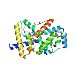
 | | Crytsal structure of human Pleiotropic Regulator 1 (PRL1) | | 分子名称: | CHLORIDE ION, Pleiotropic regulator 1, SODIUM ION, ... | | 著者 | Dong, A, Zeng, H, Xu, C, Tempel, W, Li, Y, He, H, Bountra, C, Arrowsmith, C.H, Edwards, A.M, Brown, P.J, Min, J, Wu, H, Structural Genomics Consortium (SGC) | | 登録日 | 2015-03-19 | | 公開日 | 2015-04-15 | | 最終更新日 | 2023-09-27 | | 実験手法 | X-RAY DIFFRACTION (1.7 Å) | | 主引用文献 | Crytsal structure of human Pleiotropic Regulator 1 (PRL1).
to be published
|
|
8XU5
 | |
7VKR
 | |
4YNY
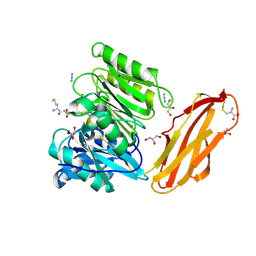
 | | Crystal structure of monoclonal anti-human podoplanin antibody NZ-1 | | 分子名称: | Heavy chain of antigen binding fragment, Fab, Light chain of antigen binding fragment | | 著者 | Fujii, Y, Kitago, Y, Arimori, T, Takagi, J. | | 登録日 | 2015-03-11 | | 公開日 | 2016-03-02 | | 最終更新日 | 2023-11-08 | | 実験手法 | X-RAY DIFFRACTION (1.584 Å) | | 主引用文献 | Tailored placement of a turn-forming PA tag into the structured domain of a protein to probe its conformational state
J.Cell.Sci., 129, 2016
|
|
6QLF
 | | Structure of inner kinetochore CCAN complex with mask1 | | 分子名称: | Inner kinetochore subunit AME1, Inner kinetochore subunit CHL4, Inner kinetochore subunit CTF19, ... | | 著者 | Yan, K, Yang, J, Zhang, Z, McLaughlin, S.H, Chang, L, Fasci, D, Heck, A.J.R, Barford, D. | | 登録日 | 2019-01-31 | | 公開日 | 2019-10-02 | | 最終更新日 | 2024-05-15 | | 実験手法 | ELECTRON MICROSCOPY (3.45 Å) | | 主引用文献 | Structure of the inner kinetochore CCAN complex assembled onto a centromeric nucleosome.
Nature, 574, 2019
|
|
7VKQ
 | |
6Q56
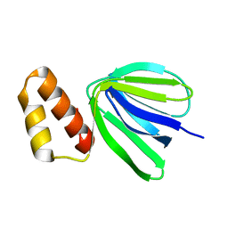
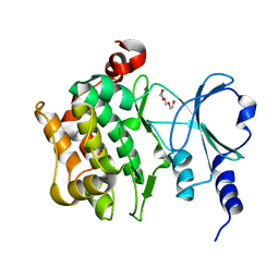
 | | Crystal structure of the B. subtilis M1A22 tRNA methyltransferase TrmK | | 分子名称: | NICKEL (II) ION, tRNA (adenine(22)-N(1))-methyltransferase | | 著者 | Degut, C, Roovers, M, Barraud, P, Brachet, F, Feller, A, Larue, V, Al Refaii, A, Caillet, J, Droogmans, L, Tisne, C. | | 登録日 | 2018-12-07 | | 公開日 | 2019-03-27 | | 最終更新日 | 2024-01-24 | | 実験手法 | X-RAY DIFFRACTION (2 Å) | | 主引用文献 | Structural characterization of B. subtilis m1A22 tRNA methyltransferase TrmK: insights into tRNA recognition.
Nucleic Acids Res., 47, 2019
|
|
8XHR
 | |
6QKC
 | | GluA1/2 In complex with auxiliary subunit gamma-8 | | 分子名称: | (2R)-2,3-dihydroxypropyl (9Z)-octadec-9-enoate, 6-nitro-2,3-bis(oxidanylidene)-1,4-dihydrobenzo[f]quinoxaline-7-sulfonamide, Glutamate receptor 1, ... | | 著者 | Herguedas, B, Garcia-Nafria, J, Greger, I.G. | | 登録日 | 2019-01-28 | | 公開日 | 2019-03-27 | | 最終更新日 | 2020-07-29 | | 実験手法 | ELECTRON MICROSCOPY (4.1 Å) | | 主引用文献 | Architecture of the heteromeric GluA1/2 AMPA receptor in complex with the auxiliary subunit TARP gamma 8.
Science, 364, 2019
|
|
3EQR
 | | Crystal Structure of Ack1 with compound T74 | | 分子名称: | Activated CDC42 kinase 1, CHLORIDE ION, N~3~-(2,6-dimethylphenyl)-1-(3-methoxy-3-methylbutyl)-N~6~-(4-piperazin-1-ylphenyl)-1H-pyrazolo[3,4-d]pyrimidine-3,6-diamine | | 著者 | Liu, J, Wang, Z, Walker, N.P.C. | | 登録日 | 2008-10-01 | | 公開日 | 2008-12-02 | | 最終更新日 | 2023-12-27 | | 実験手法 | X-RAY DIFFRACTION (2 Å) | | 主引用文献 | Identification and optimization of N3,N6-diaryl-1H-pyrazolo[3,4-d]pyrimidine-3,6-diamines as a novel class of ACK1 inhibitors.
Bioorg.Med.Chem.Lett., 18, 2008
|
|
4YTG
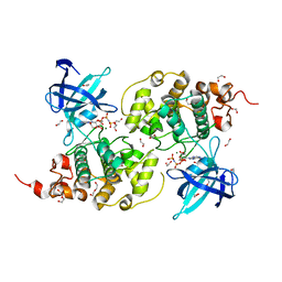
 | | Crystal structure of Porphyromonas gingivalis peptidylarginine deiminase (PPAD) mutant C351A in complex with dipeptide Met-Arg. | | 分子名称: | ARGININE, AZIDE ION, CHLORIDE ION, ... | | 著者 | Goulas, T, Mizgalska, D, Garcia-Ferrer, I, Kantyka, T, Guevara, T, Szmigielski, B, Sroka, A, Millan, C, Uson, I, Veillard, F, Potempa, B, Mydel, P, Sola, M, Potempa, J, Gomis-Ruth, F.X. | | 登録日 | 2015-03-17 | | 公開日 | 2015-07-01 | | 最終更新日 | 2024-01-10 | | 実験手法 | X-RAY DIFFRACTION (1.8 Å) | | 主引用文献 | Structure and mechanism of a bacterial host-protein citrullinating virulence factor, Porphyromonas gingivalis peptidylarginine deiminase.
Sci Rep, 5, 2015
|
|
8YHK
 | | The Crystal Structure of P21-Activated Kinases Pak4 from Biortus | | 分子名称: | Serine/threonine-protein kinase PAK 4, TRIETHYLENE GLYCOL | | 著者 | Wang, F, Cheng, W, Lv, Z, Qi, J, Wu, B. | | 登録日 | 2024-02-28 | | 公開日 | 2024-03-13 | | 実験手法 | X-RAY DIFFRACTION (3.3 Å) | | 主引用文献 | The Crystal Structure of P21-Activated Kinases Pak4 from Biortus
To Be Published
|
|
6T7L
 | | Crystal structure of AmpC from E.coli with Nacubactam (OP0595) | | 分子名称: | (2S,5R)-N-(2-aminoethoxy)-1-formyl-5-[(sulfooxy)amino]piperidine-2-carboxamide, 2-(N-MORPHOLINO)-ETHANESULFONIC ACID, Beta-lactamase, ... | | 著者 | Lang, P.A, Leissing, T.M, Schofield, C.J, Brem, J. | | 登録日 | 2019-10-22 | | 公開日 | 2020-11-18 | | 最終更新日 | 2024-01-24 | | 実験手法 | X-RAY DIFFRACTION (1.47 Å) | | 主引用文献 | Structural Investigations of the Inhibition of Escherichia coli AmpC beta-Lactamase by Diazabicyclooctanes.
Antimicrob.Agents Chemother., 65, 2021
|
|
8XI7
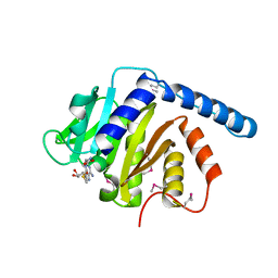
 | | The Crystal Structure of UCHL1 from Biortus. | | 分子名称: | 1,2-ETHANEDIOL, SULFATE ION, Ubiquitin carboxyl-terminal hydrolase isozyme L1 | | 著者 | Wang, F, Cheng, W, Yuan, Z, Qi, J, Shen, Z. | | 登録日 | 2023-12-19 | | 公開日 | 2024-03-06 | | 実験手法 | X-RAY DIFFRACTION (1.95 Å) | | 主引用文献 | The Crystal Structure of UCHL1 from Biortus.
To Be Published
|
|
6QJH
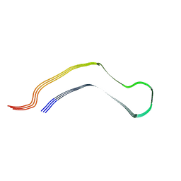 | | Cryo-EM structure of heparin-induced 2N4R tau snake filaments | | 分子名称: | Microtubule-associated protein tau | | 著者 | Zhang, W, Falcon, B, Murzin, A.G, Fan, J, Crowther, R.A, Goedert, M, Scheres, S.H.W. | | 登録日 | 2019-01-24 | | 公開日 | 2019-02-20 | | 最終更新日 | 2024-05-15 | | 実験手法 | ELECTRON MICROSCOPY (3.3 Å) | | 主引用文献 | Heparin-induced tau filaments are polymorphic and differ from those in Alzheimer's and Pick's diseases.
Elife, 8, 2019
|
|
6QJM
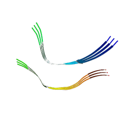 | | Cryo-EM structure of heparin-induced 2N4R tau twister filaments | | 分子名称: | Microtubule-associated protein tau | | 著者 | Zhang, W, Falcon, B, Murzin, A.G, Fan, J, Crowther, R.A, Goedert, M, Scheres, S.H.W. | | 登録日 | 2019-01-24 | | 公開日 | 2019-02-27 | | 最終更新日 | 2024-05-15 | | 実験手法 | ELECTRON MICROSCOPY (3.3 Å) | | 主引用文献 | Heparin-induced tau filaments are polymorphic and differ from those in Alzheimer's and Pick's diseases.
Elife, 8, 2019
|
|
4YUG
 | | Multiconformer synchrotron model of CypA at 100 K | | 分子名称: | Peptidyl-prolyl cis-trans isomerase A | | 著者 | Keedy, D.A, Kenner, L.R, Warkentin, M, Woldeyes, R.A, Thompson, M.C, Brewster, A.S, Van Benschoten, A.H, Baxter, E.L, Hopkins, J.B, Uervirojnangkoorn, M, McPhillips, S.E, Song, J, Mori, R.A, Holton, J.M, Weis, W.I, Brunger, A.T, Soltis, M, Lemke, H, Gonzalez, A, Sauter, N.K, Cohen, A.E, van den Bedem, H, Thorne, R.E, Fraser, J.S. | | 登録日 | 2015-03-18 | | 公開日 | 2015-10-14 | | 最終更新日 | 2024-02-28 | | 実験手法 | X-RAY DIFFRACTION (1.48 Å) | | 主引用文献 | Mapping the conformational landscape of a dynamic enzyme by multitemperature and XFEL crystallography.
Elife, 4, 2015
|
|


