Loading
PDBj
MenuPDBj@FacebookPDBj@TwitterPDBj@YouTubewwPDB FoundationwwPDB
RCSB PDBPDBeBMRBAdv. SearchSearch help
PDB: 160 results

5V8Z
DownloadVisualize
BU of 5v8z by Molmil
Crystal structure of ERp29 D-domain in complex with the P-domain of calmegin
Descriptor: Calmegin, Endoplasmic reticulum resident protein 29
Authors:Kozlov, G, Munoz-Escobar, J, Gehring, K.
Deposit date:2017-03-22
Release date:2017-06-21
Last modified:2023-10-04
Method:X-RAY DIFFRACTION (2.105 Å)
Cite:Mapping the ER Interactome: The P Domains of Calnexin and Calreticulin as Plurivalent Adapters for Foldases and Chaperones.
Structure, 25, 2017
5V90
DownloadVisualize
BU of 5v90 by Molmil
Crystal structure of ERp29 D-domain in complex with the P-domain of calreticulin
Descriptor: Calreticulin, Endoplasmic reticulum resident protein 29, GLYCEROL
Authors:Kozlov, G, Munoz-Escobar, J, Gehring, K.
Deposit date:2017-03-22
Release date:2017-06-21
Last modified:2023-10-04
Method:X-RAY DIFFRACTION (3.255 Å)
Cite:Mapping the ER Interactome: The P Domains of Calnexin and Calreticulin as Plurivalent Adapters for Foldases and Chaperones.
Structure, 25, 2017
3NTW
DownloadVisualize
BU of 3ntw by Molmil
Structure of the MLLE domain of EDD in complex with a PAM2 peptide from Paip1
Descriptor: E3 ubiquitin-protein ligase UBR5, Polyadenylate-binding protein-interacting protein 1
Authors:Kozlov, G, Gehring, K.
Deposit date:2010-07-05
Release date:2011-07-06
Last modified:2023-09-06
Method:X-RAY DIFFRACTION (2.6 Å)
Cite:The MLLE domain of the ubiquitin ligase UBR5 binds to its catalytic domain to regulate substrate binding.
J. Biol. Chem., 290, 2015
3NY1
DownloadVisualize
BU of 3ny1 by Molmil
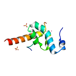
Structure of the ubr-box of the UBR1 ubiquitin ligase
Descriptor: E3 ubiquitin-protein ligase UBR1, ZINC ION
Authors:Matta-Camacho, E, Kozlov, G, Li, F, Gehring, K.
Deposit date:2010-07-14
Release date:2010-08-11
Last modified:2024-02-21
Method:X-RAY DIFFRACTION (2.085 Å)
Cite:Structural basis of substrate recognition and specificity in the N-end rule pathway.
Nat.Struct.Mol.Biol., 17, 2010
3O0X
DownloadVisualize
BU of 3o0x by Molmil
Structural basis of carbohydrate recognition by calreticulin
Descriptor: CALCIUM ION, Calreticulin, alpha-D-glucopyranose-(1-3)-alpha-D-mannopyranose-(1-2)-alpha-D-mannopyranose-(1-2)-alpha-D-mannopyranose
Authors:Kozlov, G, Gehring, K.
Deposit date:2010-07-20
Release date:2010-09-29
Last modified:2023-12-06
Method:X-RAY DIFFRACTION (2.01 Å)
Cite:Structural basis of carbohydrate recognition by calreticulin.
J.Biol.Chem., 285, 2010
3NY2
DownloadVisualize
BU of 3ny2 by Molmil
Structure of the ubr-box of UBR2 ubiquitin ligase
Descriptor: E3 ubiquitin-protein ligase UBR2, ZINC ION
Authors:Matta-Camacho, E, Kozlov, G, Li, F, Gehring, K.
Deposit date:2010-07-14
Release date:2010-08-11
Last modified:2024-02-21
Method:X-RAY DIFFRACTION (2.61 Å)
Cite:Structural basis of substrate recognition and specificity in the N-end rule pathway.
Nat.Struct.Mol.Biol., 17, 2010
3O0W
DownloadVisualize
BU of 3o0w by Molmil
Structural basis of carbohydrate recognition by calreticulin
Descriptor: CALCIUM ION, Calreticulin, alpha-D-glucopyranose-(1-3)-alpha-D-mannopyranose-(1-2)-alpha-D-mannopyranose-(1-2)-alpha-D-mannopyranose
Authors:Kozlov, G, Gehring, K.
Deposit date:2010-07-20
Release date:2010-09-29
Last modified:2020-07-29
Method:X-RAY DIFFRACTION (1.95 Å)
Cite:Structural basis of carbohydrate recognition by calreticulin.
J.Biol.Chem., 285, 2010
3NY3
DownloadVisualize
BU of 3ny3 by Molmil
Structure of the ubr-box of UBR2 in complex with N-degron
Descriptor: E3 ubiquitin-protein ligase UBR2, N-degron, ZINC ION
Authors:Matta-Camacho, E, Kozlov, G, Li, F, Gehring, K.
Deposit date:2010-07-14
Release date:2010-08-11
Last modified:2024-02-21
Method:X-RAY DIFFRACTION (1.6 Å)
Cite:Structural basis of substrate recognition and specificity in the N-end rule pathway.
Nat.Struct.Mol.Biol., 17, 2010
3O0V
DownloadVisualize
BU of 3o0v by Molmil
Crystal structure of the calreticulin lectin domain
Descriptor: CALCIUM ION, Calreticulin
Authors:Kozlov, G, Gehring, K.
Deposit date:2010-07-20
Release date:2010-09-29
Last modified:2023-09-06
Method:X-RAY DIFFRACTION (2.3 Å)
Cite:Structural basis of carbohydrate recognition by calreticulin.
J.Biol.Chem., 285, 2010
3O10
DownloadVisualize
BU of 3o10 by Molmil
Crystal structure of the HEPN domain from human sacsin
Descriptor: MALONATE ION, Sacsin
Authors:Kozlov, G, Gehring, K.
Deposit date:2010-07-20
Release date:2011-03-30
Last modified:2014-07-30
Method:X-RAY DIFFRACTION (1.9 Å)
Cite:Structural basis of defects in the sacsin HEPN domain responsible for autosomal recessive spastic ataxia of Charlevoix-Saguenay (ARSACS).
J.Biol.Chem., 286, 2011
3PKN
DownloadVisualize
BU of 3pkn by Molmil
Crystal structure of MLLE domain of poly(A) binding protein in complex with PAM2 motif of La-related protein 4 (LARP4)
Descriptor: IODIDE ION, La-related protein 4, Polyadenylate-binding protein 1, ...
Authors:Xie, J, Kozlov, G, Gehring, K.
Deposit date:2010-11-11
Release date:2011-01-12
Last modified:2023-09-06
Method:X-RAY DIFFRACTION (1.8 Å)
Cite:La-Related Protein 4 Binds Poly(A), Interacts with the Poly(A)-Binding Protein MLLE Domain via a Variant PAM2w Motif, and Can Promote mRNA Stability.
Mol.Cell.Biol., 31, 2011
3PT3
DownloadVisualize
BU of 3pt3 by Molmil
Crystal structure of the C-terminal lobe of the human UBR5 HECT domain
Descriptor: E3 ubiquitin-protein ligase UBR5
Authors:Matta-Camacho, E, Kozlov, G, Menade, M, Gehring, K.
Deposit date:2010-12-02
Release date:2012-01-25
Last modified:2023-09-06
Method:X-RAY DIFFRACTION (1.97 Å)
Cite:Structure of the HECT C-lobe of the UBR5 E3 ubiquitin ligase.
Acta Crystallogr.,Sect.F, 68, 2012
3PDZ
DownloadVisualize
BU of 3pdz by Molmil
SOLUTION STRUCTURE OF THE PDZ2 DOMAIN FROM HUMAN PHOSPHATASE HPTP1E
Descriptor: PROTEIN (TYROSINE PHOSPHATASE (PTP-BAS, TYPE 1))
Authors:Kozlov, G, Gehring, K, Ekiel, I.
Deposit date:1999-05-10
Release date:2000-03-17
Last modified:2023-12-27
Method:SOLUTION NMR
Cite:Solution structure of the PDZ2 domain from human phosphatase hPTP1E and its interactions with C-terminal peptides from the Fas receptor.
Biochemistry, 39, 2000
1DU6
DownloadVisualize
BU of 1du6 by Molmil
SOLUTION STRUCTURE OF THE TRUNCATED PBX HOMEODOMAIN
Descriptor: HOMEOBOX PROTEIN PBX1
Authors:Sprules, T, Green, N, Featherstone, M, Gehring, K.
Deposit date:2000-01-14
Release date:2000-08-16
Last modified:2024-05-22
Method:SOLUTION NMR
Cite:Conformational changes in the PBX homeodomain and C-terminal extension upon binding DNA and HOX-derived YPWM peptides.
Biochemistry, 39, 2000
3ICH
DownloadVisualize
BU of 3ich by Molmil
Crystal structure of cyclophilin B at 1.2 A resolution
Descriptor: Peptidyl-prolyl cis-trans isomerase B
Authors:Kozlov, G, Gehring, K.
Deposit date:2009-07-17
Release date:2010-07-21
Last modified:2023-09-06
Method:X-RAY DIFFRACTION (1.2 Å)
Cite:Structural Basis of Cyclophilin B Binding by the Calnexin/Calreticulin P-domain.
J.Biol.Chem., 285, 2010
3ICI
DownloadVisualize
BU of 3ici by Molmil
Crystal structure of cyclophilin B in complex with calmegin fragment
Descriptor: 2-(N-MORPHOLINO)-ETHANESULFONIC ACID, Calnexin, Peptidyl-prolyl cis-trans isomerase B, ...
Authors:Kozlov, G, Gehring, K.
Deposit date:2009-07-17
Release date:2010-07-21
Last modified:2023-09-06
Method:X-RAY DIFFRACTION (1.7 Å)
Cite:Structural Basis of Cyclophilin B Binding by the Calnexin/Calreticulin P-domain.
J.Biol.Chem., 285, 2010
3KTP
DownloadVisualize
BU of 3ktp by Molmil
Structural basis of GW182 recognition by poly(A)-binding protein
Descriptor: Polyadenylate-binding protein 1, Trinucleotide repeat-containing gene 6C protein
Authors:Kozlov, G, Gehring, K.
Deposit date:2009-11-25
Release date:2010-02-23
Last modified:2023-09-06
Method:X-RAY DIFFRACTION (1.5 Å)
Cite:Structural basis of binding of P-body-associated proteins GW182 and ataxin-2 by the Mlle domain of poly(A)-binding protein.
J.Biol.Chem., 285, 2010
3KUJ
DownloadVisualize
BU of 3kuj by Molmil
Crystal structure of C-terminal domain of PABPC1 in complex with binding region of eRF3a
Descriptor: GSPT1 protein, Polyadenylate-binding protein 1, SULFATE ION
Authors:Kozlov, G, Gehring, K.
Deposit date:2009-11-27
Release date:2010-05-12
Last modified:2023-09-06
Method:X-RAY DIFFRACTION (1.4 Å)
Cite:Molecular basis of eRF3 recognition by the MLLE domain of poly(A)-binding protein.
Plos One, 5, 2010
3KUS
DownloadVisualize
BU of 3kus by Molmil
Crystal structure of the MLLE domain of poly(A)-binding protein in complex with the binding region of Paip2
Descriptor: 4-(2-HYDROXYETHYL)-1-PIPERAZINE ETHANESULFONIC ACID, GLYCEROL, PAIP2 protein, ...
Authors:Kozlov, G, Gehring, K.
Deposit date:2009-11-27
Release date:2010-02-09
Last modified:2023-09-06
Method:X-RAY DIFFRACTION (1.4 Å)
Cite:Molecular Determinants of PAM2 Recognition by the MLLE Domain of Poly(A)-Binding Protein.
J.Mol.Biol., 397, 2010
3KUI
DownloadVisualize
BU of 3kui by Molmil
Crystal structure of C-terminal domain of PABPC1 in complex with binding region of eRF3a
Descriptor: GSPT1 protein, Polyadenylate-binding protein 1, SULFATE ION, ...
Authors:Kozlov, G, Gehring, K.
Deposit date:2009-11-27
Release date:2010-05-12
Last modified:2023-09-06
Method:X-RAY DIFFRACTION (2.3 Å)
Cite:Molecular basis of eRF3 recognition by the MLLE domain of poly(A)-binding protein.
Plos One, 5, 2010
3KUR
DownloadVisualize
BU of 3kur by Molmil
Crystal structure of the MLLE domain of poly(A)-binding protein
Descriptor: CHLORIDE ION, Polyadenylate-binding protein 1
Authors:Kozlov, G, Gehring, K.
Deposit date:2009-11-27
Release date:2010-02-09
Last modified:2023-09-06
Method:X-RAY DIFFRACTION (2.5 Å)
Cite:Molecular Determinants of PAM2 Recognition by the MLLE Domain of Poly(A)-Binding Protein.
J.Mol.Biol., 397, 2010
3IDV
DownloadVisualize
BU of 3idv by Molmil
Crystal structure of the a0a fragment of ERp72
Descriptor: CHLORIDE ION, Protein disulfide-isomerase A4, ZINC ION
Authors:Kozlov, G, Gehring, K.
Deposit date:2009-07-21
Release date:2010-07-07
Last modified:2023-09-06
Method:X-RAY DIFFRACTION (1.95 Å)
Cite:Structure of the Catalytic a(0)a Fragment of the Protein Disulfide Isomerase ERp72.
J.Mol.Biol., 401, 2010
3KUT
DownloadVisualize
BU of 3kut by Molmil
Crystal structure of the MLLE domain of poly(A)-binding protein in complex with the binding region of Paip2
Descriptor: CHLORIDE ION, PAIP2 protein, Polyadenylate-binding protein 1
Authors:Kozlov, G, Gehring, K.
Deposit date:2009-11-27
Release date:2010-02-09
Last modified:2023-09-06
Method:X-RAY DIFFRACTION (1.5 Å)
Cite:Molecular Determinants of PAM2 Recognition by the MLLE Domain of Poly(A)-Binding Protein.
J.Mol.Biol., 397, 2010
3KTR
DownloadVisualize
BU of 3ktr by Molmil
Structural basis of ataxin-2 recognition by poly(A)-binding protein
Descriptor: Ataxin-2, CADMIUM ION, Polyadenylate-binding protein 1, ...
Authors:Kozlov, G, Gehring, K.
Deposit date:2009-11-25
Release date:2010-02-23
Last modified:2023-09-06
Method:X-RAY DIFFRACTION (1.7 Å)
Cite:Structural basis of binding of P-body-associated proteins GW182 and ataxin-2 by the Mlle domain of poly(A)-binding protein.
J.Biol.Chem., 285, 2010
2ILX
DownloadVisualize
BU of 2ilx by Molmil
Solution structure of catalytic domain of rat 2',3'-cyclic-nucleotide 3'-phosphodiesterase (CNP) protein
Descriptor: 2',3'-cyclic-nucleotide 3'-phosphodiesterase
Authors:Denisov, A.Y, Kozlov, G, Gehring, K.
Deposit date:2006-10-03
Release date:2007-03-06
Last modified:2024-05-29
Method:SOLUTION NMR
Cite:Solution structure of the catalytic domain of RICH protein from goldfish.
Febs J., 274, 2007

222624

PDB entries from 2024-07-17

PDB statisticsPDBj update infoContact PDBjnumon