8EFX
 
 | |
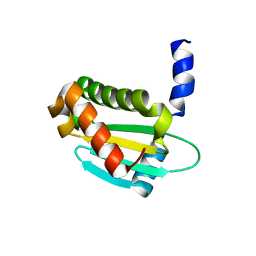
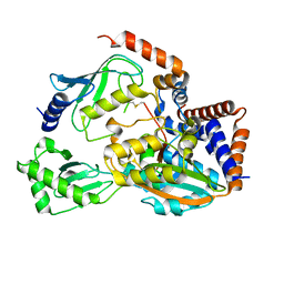
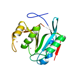
8EDE
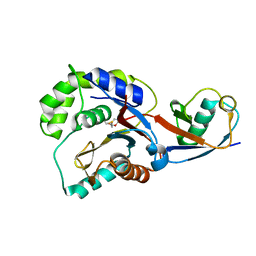
 | | Crystal structure of covalent inhibitor 2-chloro-N'-(N-(4-chlorophenyl)-N-methylglycyl)acetohydrazide bound to Ubiquitin C-terminal Hydrolase-L1 | | 分子名称: | 2-[(4-chlorophenyl)-methyl-amino]-~{N}'-ethanoyl-ethanehydrazide, SULFATE ION, Ubiquitin carboxyl-terminal hydrolase isozyme L1 | | 著者 | Patel, R, Imhoff, R, Flaherty, D, Das, C. | | 登録日 | 2022-09-04 | | 公開日 | 2023-09-20 | | 最終更新日 | 2024-04-10 | | 実験手法 | X-RAY DIFFRACTION (1.799 Å) | | 主引用文献 | Covalent Fragment Screening and Optimization Identifies the Chloroacetohydrazide Scaffold as Inhibitors for Ubiquitin C-terminal Hydrolase L1.
J.Med.Chem., 67, 2024
|
|
8DY8
 | |
8FEK
 | |
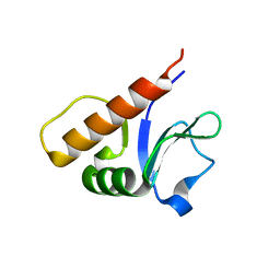
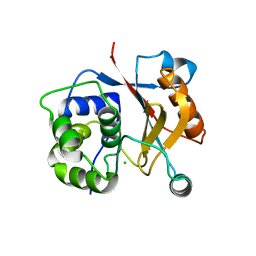
6OAM
 | | Crystal Structure of ChlaDUB2 DUB domain | | 分子名称: | Deubiquitinase and deneddylase Dub2, Ubiquitin | | 著者 | Hausman, J.M, Das, C. | | 登録日 | 2019-03-17 | | 公開日 | 2020-04-22 | | 最終更新日 | 2023-10-11 | | 実験手法 | X-RAY DIFFRACTION (2.503 Å) | | 主引用文献 | The Two Deubiquitinating Enzymes fromChlamydia trachomatisHave Distinct Ubiquitin Recognition Properties.
Biochemistry, 59, 2020
|
|
6OV1
 | | Structure of Staphylococcus aureus RNase P protein mutant with defective mRNA degradation activity | | 分子名称: | Ribonuclease P protein component | | 著者 | Ha, L, Colquhoun, J, Noinaj, N, Das, C, Dunman, P, Flaherty, D.P. | | 登録日 | 2019-05-06 | | 公開日 | 2020-12-02 | | 最終更新日 | 2023-10-11 | | 実験手法 | X-RAY DIFFRACTION (1.66 Å) | | 主引用文献 | Genetic and biochemical characterization of Staphylococcus aureus RnpA
To Be Published
|
|
6P5H
 | |
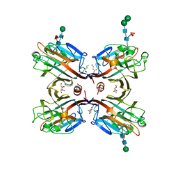
6P5B
 | | Crystal Structure of MavC in Complex with Ub-UbE2N | | 分子名称: | MavC, Ubiquitin, Ubiquitin-conjugating enzyme E2 N | | 著者 | Puvar, K, Iyer, S, Negron Teron, K.I, Das, C. | | 登録日 | 2019-05-30 | | 公開日 | 2020-05-27 | | 最終更新日 | 2023-10-11 | | 実験手法 | X-RAY DIFFRACTION (2.099 Å) | | 主引用文献 | Legionella effector MavC targets the Ube2N~Ub conjugate for noncanonical ubiquitination.
Nat Commun, 11, 2020
|
|
6ULH
 | |
6UMS
 | |
6UMP
 | | Crystal structure of MavC in complex with substrate mimic in P65 space group | | 分子名称: | MavC, Ubiquitin, Ubiquitin-conjugating enzyme E2 N | | 著者 | Puvar, K, Iyer, S, Luo, Z.Q, Das, C. | | 登録日 | 2019-10-10 | | 公開日 | 2020-05-27 | | 最終更新日 | 2023-10-11 | | 実験手法 | X-RAY DIFFRACTION (2.8 Å) | | 主引用文献 | Legionella effector MavC targets the Ube2N~Ub conjugate for noncanonical ubiquitination.
Nat Commun, 11, 2020
|
|
6WTG
 | | SdeA DUB Domain in complex with Ubiquitin | | 分子名称: | Ubiquitin, Ubiquitinating/deubiquitinating enzyme SdeA | | 著者 | Kenny, S, Sheedlo, M, Das, C. | | 登録日 | 2020-05-02 | | 公開日 | 2021-03-03 | | 最終更新日 | 2023-10-18 | | 実験手法 | X-RAY DIFFRACTION (2.63 Å) | | 主引用文献 | Insights into Ubiquitin Product Release in Hydrolysis Catalyzed by the Bacterial Deubiquitinase SdeA.
Biochemistry, 60, 2021
|
|
8UX2
 | | Chromobacterium violaceum mono-ADP-ribosyltransferase CteC in complex with NAD+ | | 分子名称: | 1,2-ETHANEDIOL, CALCIUM ION, NAD(+)--protein-threonine ADP-ribosyltransferase, ... | | 著者 | Zhang, Z, Rondon, H, Das, C. | | 登録日 | 2023-11-08 | | 公開日 | 2024-01-17 | | 最終更新日 | 2024-02-07 | | 実験手法 | X-RAY DIFFRACTION (1.87 Å) | | 主引用文献 | Crystal structure of bacterial ubiquitin ADP-ribosyltransferase CteC reveals a substrate-recruiting insertion.
J.Biol.Chem., 300, 2023
|
|
6D1R
 | | Structure of Staphylococcus aureus RNase P protein at 2.0 angstrom | | 分子名称: | Ribonuclease P protein component | | 著者 | Ha, L, Colquhoun, J, Noinaj, N, Das, C, Dunman, P, Flaherty, D.P. | | 登録日 | 2018-04-12 | | 公開日 | 2018-09-26 | | 最終更新日 | 2024-03-13 | | 実験手法 | X-RAY DIFFRACTION (1.995 Å) | | 主引用文献 | Crystal structure of the ribonuclease-P-protein subunit from Staphylococcus aureus.
Acta Crystallogr F Struct Biol Commun, 74, 2018
|
|
5KKV
 | |
4O1Y
 | | Crystal structure of Porcine Pancreatic Phospholipase A2 in complex with 1-Naphthaleneacetic acid | | 分子名称: | CALCIUM ION, NAPHTHALEN-1-YL-ACETIC ACID, Phospholipase A2, ... | | 著者 | Dileep, K.V, Remya, C, Tintu, I, Mandal, P.K, Karthe, P, Haridas, M, Sadasivan, C. | | 登録日 | 2013-12-16 | | 公開日 | 2014-01-29 | | 最終更新日 | 2023-11-08 | | 実験手法 | X-RAY DIFFRACTION (2.5 Å) | | 主引用文献 | Crystal structure of Porcine Pancreatic Phospholipase A2 in complex with 1-Naphthaleneacetic acid
To be published
|
|
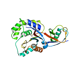
6MRN
 | | Crystal Structure of ChlaDUB2 DUB domain | | 分子名称: | Deubiquitinase and deneddylase Dub2 | | 著者 | Hausman, J.M, Das, C. | | 登録日 | 2018-10-15 | | 公開日 | 2019-10-30 | | 最終更新日 | 2023-10-11 | | 実験手法 | X-RAY DIFFRACTION (2.29 Å) | | 主引用文献 | The Two Deubiquitinating Enzymes fromChlamydia trachomatisHave Distinct Ubiquitin Recognition Properties.
Biochemistry, 59, 2020
|
|
7LM3
 | |
3USU
 | | Crystal structure of Butea monosperma seed lectin | | 分子名称: | 2-acetamido-2-deoxy-beta-D-glucopyranose, 2-acetamido-2-deoxy-beta-D-glucopyranose-(1-4)-2-acetamido-2-deoxy-beta-D-glucopyranose, CALCIUM ION, ... | | 著者 | Abhilash, J, Geethanandan, K, Bharath, S.R, Sadasivan, C, Haridas, M. | | 登録日 | 2011-11-24 | | 公開日 | 2012-01-04 | | 最終更新日 | 2023-11-15 | | 実験手法 | X-RAY DIFFRACTION (2.46 Å) | | 主引用文献 | Crystal structure of Butea monosperma seed lectin
To be Published
|
|
3IRT
 | |
3KVF
 | |
3KW5
 | |
5CRC
 | | Structure of the SdeA DUB Domain | | 分子名称: | SdeA | | 著者 | Sheedlo, M.J, Qiu, J, Tan, Y, Paul, L.N, Luo, Z.Q, Das, C. | | 登録日 | 2015-07-22 | | 公開日 | 2015-11-25 | | 最終更新日 | 2019-12-04 | | 実験手法 | X-RAY DIFFRACTION (2.853 Å) | | 主引用文献 | Structural basis of substrate recognition by a bacterial deubiquitinase important for dynamics of phagosome ubiquitination.
Proc.Natl.Acad.Sci.USA, 112, 2015
|
|
5CRB
 | |
5CRA
 | | Structure of the SdeA DUB Domain | | 分子名称: | METHYL 4-AMINOBUTANOATE, Polyubiquitin-B, SULFATE ION, ... | | 著者 | Sheedlo, M.J, Qiu, J, Luo, Z.Q, Das, C. | | 登録日 | 2015-07-22 | | 公開日 | 2015-11-25 | | 最終更新日 | 2023-11-15 | | 実験手法 | X-RAY DIFFRACTION (2.64 Å) | | 主引用文献 | Structural basis of substrate recognition by a bacterial deubiquitinase important for dynamics of phagosome ubiquitination.
Proc.Natl.Acad.Sci.USA, 112, 2015
|
|


