7SXM
 
 | |
6CIO
 | |
4LZO
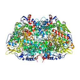
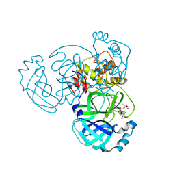
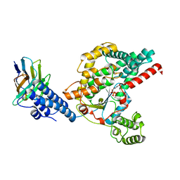
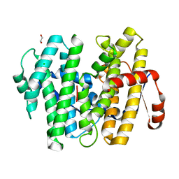
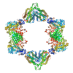
 | | Crystal structure of human PRS1 A87T mutant | | 分子名称: | Ribose-phosphate pyrophosphokinase 1, SULFATE ION | | 著者 | Chen, P, Teng, M, Li, X. | | 登録日 | 2013-07-31 | | 公開日 | 2015-02-04 | | 最終更新日 | 2024-03-20 | | 実験手法 | X-RAY DIFFRACTION (3.31 Å) | | 主引用文献 | Crystal and EM Structures of Human Phosphoribosyl Pyrophosphate Synthase I (PRS1) Provide Novel Insights into the Disease-Associated Mutations
Plos One, 10, 2015
|
|
4LYG
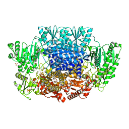
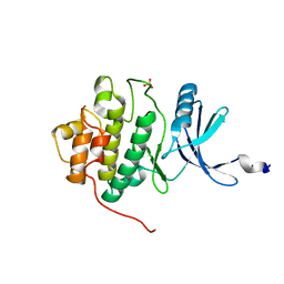
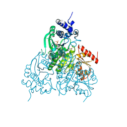
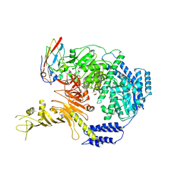
 | | Crystal structure of human PRS1 E43T mutant | | 分子名称: | Ribose-phosphate pyrophosphokinase 1, SULFATE ION | | 著者 | Chen, P, Teng, M, Li, X. | | 登録日 | 2013-07-31 | | 公開日 | 2015-02-04 | | 最終更新日 | 2024-03-20 | | 実験手法 | X-RAY DIFFRACTION (3 Å) | | 主引用文献 | Crystal and EM Structures of Human Phosphoribosyl Pyrophosphate Synthase I (PRS1) Provide Novel Insights into the Disease-Associated Mutations
Plos One, 10, 2015
|
|
4LZN
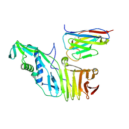
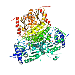
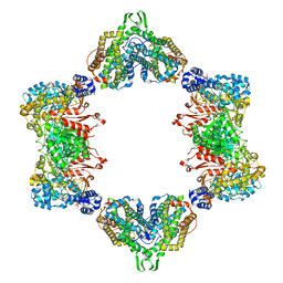
 | | Crystal structure of human PRS1 D65N mutant | | 分子名称: | Ribose-phosphate pyrophosphokinase 1, SULFATE ION | | 著者 | Chen, P, Teng, M, Li, X. | | 登録日 | 2013-07-31 | | 公開日 | 2015-02-04 | | 最終更新日 | 2023-11-08 | | 実験手法 | X-RAY DIFFRACTION (2.14 Å) | | 主引用文献 | Crystal and EM Structures of Human Phosphoribosyl Pyrophosphate Synthase I (PRS1) Provide Novel Insights into the Disease-Associated Mutations
Plos One, 10, 2015
|
|
4M0P
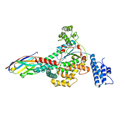
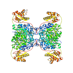
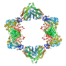
 | | Crystal structure of human PRS1 M115T mutant | | 分子名称: | Ribose-phosphate pyrophosphokinase 1, SULFATE ION | | 著者 | Chen, P, Teng, M, Li, X. | | 登録日 | 2013-08-01 | | 公開日 | 2015-02-04 | | 最終更新日 | 2024-03-20 | | 実験手法 | X-RAY DIFFRACTION (2.11 Å) | | 主引用文献 | Crystal and EM Structures of Human Phosphoribosyl Pyrophosphate Synthase I (PRS1) Provide Novel Insights into the Disease-Associated Mutations
Plos One, 10, 2015
|
|
8X7N
 | |
9BPF
 | |
9BOO
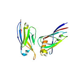
 | | Crystal structure of MERS-CoV Nsp5 in complex with PF-07817883 | | 分子名称: | 3C-like proteinase nsp5, N-(methoxycarbonyl)-3-methyl-L-valyl-(4R)-N-{(1Z,2S)-1-imino-3-[(3S)-2-oxopyrrolidin-3-yl]propan-2-yl}-4-(trifluoromethyl)-L-prolinamide | | 著者 | Chen, P, Arutyunova, E, Lemieux, M.J. | | 登録日 | 2024-05-05 | | 公開日 | 2024-08-07 | | 最終更新日 | 2024-09-11 | | 実験手法 | X-RAY DIFFRACTION (1.5 Å) | | 主引用文献 | A Structural Comparison of Oral SARS-CoV-2 Drug Candidate Ibuzatrelvir Complexed with the Main Protease (M pro ) of SARS-CoV-2 and MERS-CoV.
Jacs Au, 4, 2024
|
|
1IA8
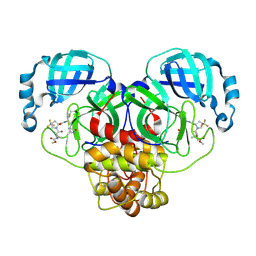
 | | THE 1.7 A CRYSTAL STRUCTURE OF HUMAN CELL CYCLE CHECKPOINT KINASE CHK1 | | 分子名称: | CHK1 CHECKPOINT KINASE, SULFATE ION | | 著者 | Chen, P, Luo, C, Deng, Y, Ryan, K, Register, J, Margosiak, S, Tempczyk-Russell, A, Nguyen, B, Myers, P, Lundgren, K, Chen Kan, C.-C, O'Connor, P.M. | | 登録日 | 2001-03-22 | | 公開日 | 2001-04-18 | | 最終更新日 | 2024-04-03 | | 実験手法 | X-RAY DIFFRACTION (1.7 Å) | | 主引用文献 | The 1.7 A crystal structure of human cell cycle checkpoint kinase Chk1: implications for Chk1 regulation.
Cell(Cambridge,Mass.), 100, 2000
|
|
6OQ5
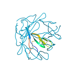
 | | Structure of the full-length Clostridium difficile toxin B in complex with 3 VHHs | | 分子名称: | 5D, 7F, E3, ... | | 著者 | Chen, P, Lam, K, Jin, R. | | 登録日 | 2019-04-25 | | 公開日 | 2019-07-10 | | 最終更新日 | 2023-10-11 | | 実験手法 | X-RAY DIFFRACTION (3.87 Å) | | 主引用文献 | Structure of the full-length Clostridium difficile toxin B.
Nat.Struct.Mol.Biol., 26, 2019
|
|
6OQ6
 | |
6OQ8
 | |
6OQ7
 | | Structure of the GTD domain of Clostridium difficile toxin B in complex with VHH E3 | | 分子名称: | E3, MAGNESIUM ION, MANGANESE (II) ION, ... | | 著者 | Chen, P, Lam, K, Jin, R. | | 登録日 | 2019-04-25 | | 公開日 | 2019-07-10 | | 最終更新日 | 2023-10-11 | | 実験手法 | X-RAY DIFFRACTION (2.39 Å) | | 主引用文献 | Structure of the full-length Clostridium difficile toxin B.
Nat.Struct.Mol.Biol., 26, 2019
|
|
6OUW
 | |
6OUV
 | |
7S2X
 | | Structure of SalC, a gamma-lactam-beta-lactone bicyclase for salinosporamide biosynthesis | | 分子名称: | 4-(2-HYDROXYETHYL)-1-PIPERAZINE ETHANESULFONIC ACID, POTASSIUM ION, SalC | | 著者 | Chen, P.Y.-T, Trivella, D.B.B, Bauman, K.D, Moore, B.S. | | 登録日 | 2021-09-04 | | 公開日 | 2022-04-06 | | 最終更新日 | 2023-10-18 | | 実験手法 | X-RAY DIFFRACTION (2.85 Å) | | 主引用文献 | Enzymatic assembly of the salinosporamide gamma-lactam-beta-lactone anticancer warhead.
Nat.Chem.Biol., 18, 2022
|
|
7S5L
 | |
1PI2
 | |
2BV6
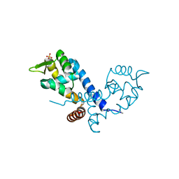 | | Crystal structure of MgrA, a global regulator and major virulence determinant in Staphylococcus aureus | | 分子名称: | HTH-TYPE TRANSCRIPTIONAL REGULATOR MGRA, SULFATE ION | | 著者 | Chen, P.R, Bae, T, Williams, W.A, Duguid, E.M, Rice, P.A, Schneewind, O, He, C. | | 登録日 | 2005-06-22 | | 公開日 | 2006-09-20 | | 最終更新日 | 2011-07-13 | | 実験手法 | X-RAY DIFFRACTION (2.8 Å) | | 主引用文献 | An Oxidation-Sensing Mechanism is Used by the Global Regulator Mgra in Staphylococcus Aureus.
Nat.Chem.Biol., 2, 2006
|
|
1GRC
 | |
5CNV
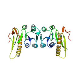
 | | Crystal structure of the dATP inhibited E. coli class Ia ribonucleotide reductase complex bound to GDP and TTP at 3.20 Angstroms resolution | | 分子名称: | 2'-DEOXYADENOSINE-5'-DIPHOSPHATE, GUANOSINE-5'-DIPHOSPHATE, MAGNESIUM ION, ... | | 著者 | Chen, P.Y.-T, Zimanyi, C.M, Funk, M.A, Drennan, C.L. | | 登録日 | 2015-07-18 | | 公開日 | 2016-01-20 | | 最終更新日 | 2023-09-27 | | 実験手法 | X-RAY DIFFRACTION (3.2 Å) | | 主引用文献 | Molecular basis for allosteric specificity regulation in class Ia ribonucleotide reductase from Escherichia coli.
Elife, 5, 2016
|
|
7ML7
 | |
5CNS
 | | Crystal structure of the dATP inhibited E. coli class Ia ribonucleotide reductase complex bound to CDP and dATP at 2.97 Angstroms resolution | | 分子名称: | 2'-DEOXYADENOSINE 5'-TRIPHOSPHATE, 2'-DEOXYADENOSINE-5'-DIPHOSPHATE, CYTIDINE-5'-DIPHOSPHATE, ... | | 著者 | Chen, P.Y.-T, Zimanyi, C.M, Funk, M.A, Drennan, C.L. | | 登録日 | 2015-07-18 | | 公開日 | 2016-01-20 | | 最終更新日 | 2023-09-27 | | 実験手法 | X-RAY DIFFRACTION (2.975 Å) | | 主引用文献 | Molecular basis for allosteric specificity regulation in class Ia ribonucleotide reductase from Escherichia coli.
Elife, 5, 2016
|
|
5CNT
 | | Crystal structure of the dATP inhibited E. coli class Ia ribonucleotide reductase complex bound to UDP and dATP at 3.25 Angstroms resolution | | 分子名称: | 2'-DEOXYADENOSINE 5'-TRIPHOSPHATE, MAGNESIUM ION, MU-OXO-DIIRON, ... | | 著者 | Chen, P.Y.-T, Zimanyi, C.M, Funk, M.A, Drennan, C.L. | | 登録日 | 2015-07-18 | | 公開日 | 2016-01-20 | | 最終更新日 | 2023-09-27 | | 実験手法 | X-RAY DIFFRACTION (3.25 Å) | | 主引用文献 | Molecular basis for allosteric specificity regulation in class Ia ribonucleotide reductase from Escherichia coli.
Elife, 5, 2016
|
|


