2VX0
 
 | | ephB4 kinase domain inhibitor complex | | 分子名称: | EPHRIN TYPE-B RECEPTOR 4, MAGNESIUM ION, N'-(5-CHLORO-1,3-BENZODIOXOL-4-YL)-N-(3-MORPHOLIN-4-YLPHENYL)PYRIMIDINE-2,4-DIAMINE | | 著者 | Read, J, Brassington, C.A, Green, I, McCall, E.J, Valentine, A.L, Barratt, D, Leach, A.G, Kettle, J.G. | | 登録日 | 2008-06-30 | | 公開日 | 2008-10-28 | | 最終更新日 | 2023-12-13 | | 実験手法 | X-RAY DIFFRACTION (2.1 Å) | | 主引用文献 | Inhibitors of the Tyrosine Kinase Ephb4. Part 1: Structure-Based Design and Optimization of a Series of 2,4-Bis-Anilinopyrimidines.
Bioorg.Med.Chem.Lett., 18, 2008
|
|
2VWX
 | | ephB4 kinase domain inhibitor complex | | 分子名称: | 3-({4-[(5-chloro-1,3-benzodioxol-4-yl)amino]pyrimidin-2-yl}amino)benzenesulfonamide, EPHRIN TYPE-B RECEPTOR 4, MAGNESIUM ION | | 著者 | Read, J, Brassington, C.A, Green, I, McCall, E.J, Valentine, A.L, Barratt, D, Leach, A.G, Kettle, J.G. | | 登録日 | 2008-06-27 | | 公開日 | 2008-10-28 | | 最終更新日 | 2023-12-13 | | 実験手法 | X-RAY DIFFRACTION (1.65 Å) | | 主引用文献 | Inhibitors of the Tyrosine Kinase Ephb4. Part 2: Structure-Based Discovery and Optimisation of 3,5-Bis Substituted Anilinopyrimidines.
Bioorg.Med.Chem.Lett., 18, 2008
|
|
5OC0
 | | Structure of E. coli superoxide oxidase | | 分子名称: | Cytochrome b561, GLYCEROL, MAGNESIUM ION, ... | | 著者 | Lundgren, C.A.K, Sjostrand, D, Biner, O, Bennett, M, von Ballmoos, C, Hogbom, M. | | 登録日 | 2017-06-29 | | 公開日 | 2018-06-20 | | 最終更新日 | 2018-11-07 | | 実験手法 | X-RAY DIFFRACTION (1.97 Å) | | 主引用文献 | Scavenging of superoxide by a membrane-bound superoxide oxidase.
Nat. Chem. Biol., 14, 2018
|
|
2VWV
 | | ephB4 kinase domain inhibitor complex | | 分子名称: | EPHRIN TYPE-B RECEPTOR 4, N'-(3-CHLORO-4-METHOXY-PHENYL)-N-(3,4,5-TRIMETHOXYPHENYL)-1,3,5-TRIAZINE-2,4-DIAMINE | | 著者 | Read, J, Brassington, C.A, Green, I, McCall, E.J, Valentine, A.L, Kettle, J.G, Leach, A.G. | | 登録日 | 2008-06-27 | | 公開日 | 2008-07-08 | | 最終更新日 | 2023-12-13 | | 実験手法 | X-RAY DIFFRACTION (1.9 Å) | | 主引用文献 | Inhibitors of the Tyrosine Kinase Ephb4. Part 1: Structure-Based Design and Optimization of a Series of 2,4-Bis-Anilinopyrimidines
Bioorg.Med.Chem.Lett., 18, 2008
|
|
3M4Z
 | | Crystal Structure of B. subtilis ferrochelatase with Cobalt bound at the active site | | 分子名称: | CHLORIDE ION, COBALT (II) ION, Ferrochelatase, ... | | 著者 | Soderberg, C.A.G, Hansson, M.D, Sreekanth, R, Al-Karadaghi, S, Hansson, M. | | 登録日 | 2010-03-12 | | 公開日 | 2010-11-10 | | 最終更新日 | 2023-11-01 | | 実験手法 | X-RAY DIFFRACTION (1.94 Å) | | 主引用文献 | Bacterial ferrochelatase goes human: Tyr13 determines the apparent metal specificity of Bacillus subtilis ferrochelatase
To be Published
|
|
2VV9
 | | CDK2 in complex with an imidazole piperazine | | 分子名称: | 2-{4-[4-({4-[2-methyl-1-(1-methylethyl)-1H-imidazol-5-yl]pyrimidin-2-yl}amino)phenyl]piperazin-1-yl}-2-oxoethanol, CELL DIVISION PROTEIN KINASE 2 | | 著者 | Acton, D.G, Anderson, M, Andrews, D.M, Barker, A.J, Brassington, C.A, Finlay, M.R, Fisher, E, Gerhardt, S, Graham, M.A, Green, C.P, Heaton, D.W, Loddick, S.A, Morgentin, R, Read, J, Roberts, A, Stanway, J, Tucker, J.A, Weir, H.M. | | 登録日 | 2008-06-04 | | 公開日 | 2008-08-05 | | 最終更新日 | 2024-05-01 | | 実験手法 | X-RAY DIFFRACTION (1.9 Å) | | 主引用文献 | Imidazole Piperazines: Sar and Development of a Potent Class of Cyclin-Dependent Kinase Inhibitors with a Novel Binding Mode.
Bioorg.Med.Chem.Lett., 18, 2008
|
|
1Y7N
 | | Solution structure of the second PDZ domain of the human neuronal adaptor X11alpha | | 分子名称: | Amyloid beta A4 precursor protein-binding family A member 1 | | 著者 | Duquesne, A.E, de Ruijter, M, Brouwer, J, Drijfhout, J.W, Nabuurs, S.B, Spronk, C.A.E.M, Vuister, G.W, Ubbink, M, Canters, G.W. | | 登録日 | 2004-12-09 | | 公開日 | 2005-11-22 | | 最終更新日 | 2024-05-29 | | 実験手法 | SOLUTION NMR | | 主引用文献 | Solution structure of the second PDZ domain of the neuronal adaptor X11alpha and its interaction with the C-terminal peptide of the human copper chaperone for superoxide dismutase
J.Biomol.Nmr, 32, 2005
|
|
5OEO
 | | Solution structure of the complex of TRPV5(655-725) with a Calmodulin E32Q/E68Q double mutant | | 分子名称: | CALCIUM ION, Calmodulin-1, Transient receptor potential cation channel subfamily V member 5 | | 著者 | Vuister, G.W, Bokhovchuk, F.M, Bate, N, Kovalevskaya, N, Goult, B.T, Spronk, C.A.E.M. | | 登録日 | 2017-07-09 | | 公開日 | 2018-04-25 | | 最終更新日 | 2024-05-15 | | 実験手法 | SOLUTION NMR | | 主引用文献 | The Structural Basis of Calcium-Dependent Inactivation of the Transient Receptor Potential Vanilloid 5 Channel.
Biochemistry, 57, 2018
|
|
5OH5
 | | Legionella pneumophila RidL N-terminal retromer binding domain | | 分子名称: | RidL | | 著者 | Baerlocher, K, Hutter, C.A.J, Swart, A.L, Steiner, B, Welin, A, Hohl, M, Letourneur, F, Seeger, M.A, Hilbi, H. | | 登録日 | 2017-07-14 | | 公開日 | 2017-11-22 | | 最終更新日 | 2024-05-08 | | 実験手法 | X-RAY DIFFRACTION (1.9 Å) | | 主引用文献 | Structural insights into Legionella RidL-Vps29 retromer subunit interaction reveal displacement of the regulator TBC1D5.
Nat Commun, 8, 2017
|
|
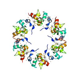
1C94
 | | REVERSING THE SEQUENCE OF THE GCN4 LEUCINE ZIPPER DOES NOT AFFECT ITS FOLD. | | 分子名称: | RETRO-GCN4 LEUCINE ZIPPER | | 著者 | Mittl, P.R.E, Deillon, C.A, Sargent, D, Liu, N, Klauser, S, Thomas, R.M, Gutte, B, Gruetter, M.G. | | 登録日 | 1999-07-30 | | 公開日 | 2000-03-22 | | 最終更新日 | 2024-02-07 | | 実験手法 | X-RAY DIFFRACTION (2.08 Å) | | 主引用文献 | The retro-GCN4 leucine zipper sequence forms a stable three-dimensional structure.
Proc.Natl.Acad.Sci.USA, 97, 2000
|
|
3PAZ
 | | REDUCED NATIVE PSEUDOAZURIN FROM A. FAECALIS | | 分子名称: | COPPER (II) ION, PSEUDOAZURIN | | 著者 | Adman, E.T, Libeu, C.A.P. | | 登録日 | 1997-02-20 | | 公開日 | 1997-08-20 | | 最終更新日 | 2024-02-21 | | 実験手法 | X-RAY DIFFRACTION (1.73 Å) | | 主引用文献 | Site-directed mutants of pseudoazurin: explanation of increased redox potentials from X-ray structures and from calculation of redox potential differences.
Biochemistry, 36, 1997
|
|
7JJA
 | | Crystal structure of the ZinT-like domain of Streptococcus pneumoniae AdcA in the apo form | | 分子名称: | SODIUM ION, Zinc-binding lipoprotein AdcA, {[-(BIS-CARBOXYMETHYL-AMINO)-ETHYL]-CARBOXYMETHYL-AMINO}-ACETIC ACID | | 著者 | Luo, Z, More, J.R, Kobe, B, McDevitt, C.A. | | 登録日 | 2020-07-24 | | 公開日 | 2020-12-23 | | 最終更新日 | 2023-10-18 | | 実験手法 | X-RAY DIFFRACTION (1.01 Å) | | 主引用文献 | A Trap-Door Mechanism for Zinc Acquisition by Streptococcus pneumoniae AdcA.
Mbio, 12, 2021
|
|
3MLM
 | | Crystal structure of Bn IV in complex with myristic acid: A Lys49 myotoxic phospholipase A2 from Bothrops neuwiedi venom | | 分子名称: | BN-IV Lys-49 Phospholipase A2, MYRISTIC ACID, SULFATE ION | | 著者 | Delatorre, P, Rocha, B.A.M, Cavada, B.S, Toyama, M.H, Toyama, D, Gadelha, C.A.A. | | 登録日 | 2010-04-17 | | 公開日 | 2011-05-18 | | 最終更新日 | 2023-09-06 | | 実験手法 | X-RAY DIFFRACTION (2.21 Å) | | 主引用文献 | Crystal structure of Bn IV in complex with myristic acid: a Lys49 myotoxic phospholipase A2 from Bothrops neuwiedi venom.
Biochimie, 93, 2011
|
|
2X9F
 | | ephB4 kinase domain inhibitor complex | | 分子名称: | EPHRIN TYPE-B RECEPTOR 4, MAGNESIUM ION, N^4^-1H-INDAZOL-4-YL-N^2^-[3-(METHYLSULFONYL)PHENYL]PYRIMIDINE-2,4-DIAMINE | | 著者 | Read, J, Brassington, C.A, Green, I, McCall, E.J, Valentine, A.L, Barratt, D. | | 登録日 | 2010-03-17 | | 公開日 | 2010-09-29 | | 最終更新日 | 2023-12-20 | | 実験手法 | X-RAY DIFFRACTION (1.75 Å) | | 主引用文献 | Inhibitors of the Tyrosine Kinase Ephb4. Part 3: Identification of Non-Benzodioxole-Based Kinase Inhibitors.
Bioorg.Med.Chem.Lett., 20, 2010
|
|
7JJ9
 | | Crystal structure of Zn(II)-bound AdcA from Streptococcus pneumoniae | | 分子名称: | CHLORIDE ION, ZINC ION, Zinc-binding lipoprotein AdcA | | 著者 | Luo, Z, More, J.R, Kobe, B, McDevitt, C.A. | | 登録日 | 2020-07-24 | | 公開日 | 2020-12-23 | | 最終更新日 | 2023-10-18 | | 実験手法 | X-RAY DIFFRACTION (1.58 Å) | | 主引用文献 | A Trap-Door Mechanism for Zinc Acquisition by Streptococcus pneumoniae AdcA.
Mbio, 12, 2021
|
|
7JJ8
 | | Crystal structure of the Zn(II)-bound ZnuA-like domain of Streptococcus pneumoniae AdcA | | 分子名称: | ZINC ION, Zinc-binding lipoprotein AdcA | | 著者 | Luo, Z, More, J.R, Kobe, B, McDevitt, C.A. | | 登録日 | 2020-07-24 | | 公開日 | 2020-12-23 | | 最終更新日 | 2023-10-18 | | 実験手法 | X-RAY DIFFRACTION (2.03 Å) | | 主引用文献 | A Trap-Door Mechanism for Zinc Acquisition by Streptococcus pneumoniae AdcA.
Mbio, 12, 2021
|
|
7JIS
 | | HUMAN PI3KDELTA IN COMPLEX WITH COMPOUND 2F | | 分子名称: | (3S)-3-benzyl-5-[9-ethyl-8-(2-methylpyrimidin-5-yl)-9H-purin-6-yl]-3-methyl-1,3-dihydro-2H-indol-2-one, Phosphatidylinositol 3-kinase regulatory subunit alpha, Phosphatidylinositol 4,5-bisphosphate 3-kinase catalytic subunit delta isoform | | 著者 | Lesburg, C.A, Augustin, M. | | 登録日 | 2020-07-23 | | 公開日 | 2020-12-30 | | 最終更新日 | 2024-04-03 | | 実験手法 | X-RAY DIFFRACTION (2.42 Å) | | 主引用文献 | Optimization of Versatile Oxindoles as Selective PI3K delta Inhibitors.
Acs Med.Chem.Lett., 11, 2020
|
|
7JJB
 | | Crystal structure of Zn(II)-bound ZinT-like domain of Streptococcus pneumoniae AdcA | | 分子名称: | MAGNESIUM ION, SODIUM ION, ZINC ION, ... | | 著者 | Luo, Z, More, J.R, Kobe, B, McDevitt, C.A. | | 登録日 | 2020-07-24 | | 公開日 | 2020-12-23 | | 最終更新日 | 2023-10-18 | | 実験手法 | X-RAY DIFFRACTION (1.1 Å) | | 主引用文献 | A Trap-Door Mechanism for Zinc Acquisition by Streptococcus pneumoniae AdcA.
Mbio, 12, 2021
|
|
7JIU
 | | HUMAN PI3KDELTA IN COMPLEX WITH COMPOUND 2F | | 分子名称: | (3S)-3-benzyl-5-[9-ethyl-8-(2-methylpyrimidin-5-yl)-9H-purin-6-yl]-3-methyl-1,3-dihydro-2H-indol-2-one, Phosphatidylinositol 4,5-bisphosphate 3-kinase catalytic subunit alpha isoform | | 著者 | Lesburg, C.A, Augustin, M. | | 登録日 | 2020-07-23 | | 公開日 | 2021-06-30 | | 最終更新日 | 2024-04-03 | | 実験手法 | X-RAY DIFFRACTION (2.12 Å) | | 主引用文献 | Optimization of Versatile Oxindoles as Selective PI3K delta Inhibitors.
Acs Med.Chem.Lett., 11, 2020
|
|
3OER
 | | Crystal structure of trimeric frataxin from the yeast saccharomyces cerevisiae, complexed with cobalt | | 分子名称: | COBALT (II) ION, Frataxin homolog, mitochondrial | | 著者 | Soderberg, C.A.G, Rajan, S, Gakh, O, Ta, C, Isaya, G, Al-Karadaghi, S. | | 登録日 | 2010-08-13 | | 公開日 | 2011-08-24 | | 最終更新日 | 2023-09-06 | | 実験手法 | X-RAY DIFFRACTION (3.2 Å) | | 主引用文献 | Oligomerization Propensity and Flexibility of Yeast Frataxin Studied by X-ray Crystallography and Small-Angle X-ray Scattering.
J.Mol.Biol., 414, 2011
|
|
3OEQ
 | | Crystal structure of trimeric frataxin from the yeast Saccharomyces cerevisiae, with full length n-terminus | | 分子名称: | Frataxin homolog, mitochondrial | | 著者 | Soderberg, C.A.G, Rajan, S, Gakh, O, Ta, C, Isaya, G, Al-Karadaghi, S. | | 登録日 | 2010-08-13 | | 公開日 | 2011-08-24 | | 最終更新日 | 2023-09-06 | | 実験手法 | X-RAY DIFFRACTION (2.96 Å) | | 主引用文献 | Oligomerization Propensity and Flexibility of Yeast Frataxin Studied by X-ray Crystallography and Small-Angle X-ray Scattering.
J.Mol.Biol., 414, 2011
|
|
1XB1
 | | The Structure of the BIR domain of IAP-like protein 2 | | 分子名称: | Baculoviral IAP repeat-containing protein 8, Diablo homolog, mitochondrial, ... | | 著者 | Shin, H, Renatus, M, Eckelman, B.P, Nunes, V.A, Sampaio, C.A.M, Salvesen, G.S. | | 登録日 | 2004-08-27 | | 公開日 | 2004-11-02 | | 最終更新日 | 2011-07-13 | | 実験手法 | X-RAY DIFFRACTION (2.7 Å) | | 主引用文献 | The BIR domain of IAP-like protein 2 is conformationally unstable: implications for caspase inhibition
Biochem.J., 385, 2005
|
|
2A74
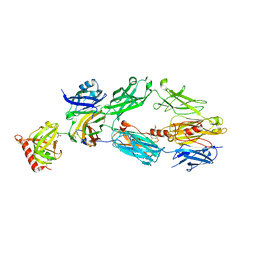 | | Human Complement Component C3c | | 分子名称: | 2-acetamido-2-deoxy-alpha-D-glucopyranose-(1-4)-2-acetamido-2-deoxy-alpha-D-glucopyranose, Complement Component C3c, GLYCEROL, ... | | 著者 | Janssen, B.J.C, Huizinga, E.G, Raaijmakers, H.C.A, Roos, A, Daha, M.R, Nilsson-Ekdahl, K, Nilsson, B, Gros, P. | | 登録日 | 2005-07-04 | | 公開日 | 2005-09-27 | | 最終更新日 | 2020-07-29 | | 実験手法 | X-RAY DIFFRACTION (2.4 Å) | | 主引用文献 | Structures of complement component C3 provide insights into the function and evolution of immunity.
Nature, 437, 2005
|
|
2A73
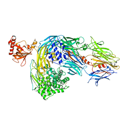 | | Human Complement Component C3 | | 分子名称: | 2-acetamido-2-deoxy-beta-D-glucopyranose-(1-4)-2-acetamido-2-deoxy-beta-D-glucopyranose, Complement C3, alpha-D-mannopyranose-(1-3)-[beta-D-mannopyranose-(1-6)]alpha-D-mannopyranose-(1-4)-2-acetamido-2-deoxy-beta-D-glucopyranose-(1-4)-2-acetamido-2-deoxy-beta-D-glucopyranose | | 著者 | Janssen, B.J.C, Huizinga, E.G, Raaijmakers, H.C.A, Roos, A, Daha, M.R, Nilsson-Ekdahl, K, Nilsson, B, Gros, P. | | 登録日 | 2005-07-04 | | 公開日 | 2005-09-27 | | 最終更新日 | 2023-08-23 | | 実験手法 | X-RAY DIFFRACTION (3.3 Å) | | 主引用文献 | Structures of complement component C3 provide insights into the function and evolution of immunity.
Nature, 437, 2005
|
|
1VJH
 | | Crystal structure of gene product of At1g24000 from Arabidopsis thaliana | | 分子名称: | Bet v I allergen family | | 著者 | Wesenberg, G.E, Smith, D.W, Phillips Jr, G.N, Johnson, K.A, Bingman, C.A, Center for Eukaryotic Structural Genomics (CESG) | | 登録日 | 2004-02-20 | | 公開日 | 2004-03-16 | | 最終更新日 | 2023-12-27 | | 実験手法 | X-RAY DIFFRACTION (2.1 Å) | | 主引用文献 | 1H, 15N and 13C resonance assignments of the putative Bet v 1 family protein At1g24000.1 from Arabidopsis thaliana.
J.Biomol.Nmr, 32, 2005
|
|


