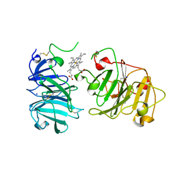
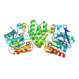
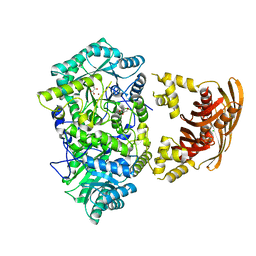
1OQH
 
 | | Crystal Structure of the R124A mutant of the N-lobe human transferrin | | 分子名称: | CARBONATE ION, FE (III) ION, POTASSIUM ION, ... | | 著者 | Baker, H.M, He, Q.-Y, Brigg, S.K, Mason, A.B, N Baker, E. | | 登録日 | 2003-03-10 | | 公開日 | 2003-03-18 | | 最終更新日 | 2023-08-16 | | 実験手法 | X-RAY DIFFRACTION (2.4 Å) | | 主引用文献 | Structural and functional consequences of binding site mutations in transferrin: crystal structures of the Asp63Glu and Arg124Ala mutants of the N-lobe of human transferrin
Biochemistry, 42, 2003
|
|
6OVT
 | | Crystal Structure of IlvD from Mycobacterium tuberculosis | | 分子名称: | DI(HYDROXYETHYL)ETHER, Dihydroxy-acid dehydratase, FE2/S2 (INORGANIC) CLUSTER, ... | | 著者 | Almo, S.C, Grove, T.L, Bonanno, J.B, Baker, E.N, Bashiri, G. | | 登録日 | 2019-05-08 | | 公開日 | 2019-08-07 | | 最終更新日 | 2019-12-18 | | 実験手法 | X-RAY DIFFRACTION (1.88 Å) | | 主引用文献 | The active site of theMycobacterium tuberculosisbranched-chain amino acid biosynthesis enzyme dihydroxyacid dehydratase contains a 2Fe-2S cluster.
J.Biol.Chem., 294, 2019
|
|
3RMJ
 | | Crystal structure of truncated alpha-Isopropylmalate Synthase from Neisseria meningitidis | | 分子名称: | 2-isopropylmalate synthase, GLYCEROL, MAGNESIUM ION, ... | | 著者 | Huisman, F.H.A, Baker, H.M, Koon, N, Baker, E.N, Parker, E.J. | | 登録日 | 2011-04-20 | | 公開日 | 2012-03-14 | | 最終更新日 | 2023-11-01 | | 実験手法 | X-RAY DIFFRACTION (1.95 Å) | | 主引用文献 | Removal of the C-terminal regulatory domain of alpha-isopropylmalate synthase disrupts functional substrate binding
Biochemistry, 51, 2012
|
|
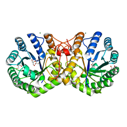
3NUE
 | | The structure of 3-deoxy-d-arabino-heptulosonate 7-phosphate synthase from mycobacterium tuberculosis complexed with tryptophan | | 分子名称: | CHLORIDE ION, GLYCEROL, MANGANESE (II) ION, ... | | 著者 | Parker, E.J, Jameson, G.B, Jiao, W, Webby, C.J, Baker, E.N, Baker, H.M. | | 登録日 | 2010-07-06 | | 公開日 | 2010-07-28 | | 最終更新日 | 2023-11-01 | | 実験手法 | X-RAY DIFFRACTION (2.5 Å) | | 主引用文献 | Synergistic allostery, a sophisticated regulatory network for the control of aromatic amino acid biosynthesis in Mycobacterium tuberculosis
J.Biol.Chem., 285, 2010
|
|
6N0A
 | | Structure of the major pilin protein (T-18.1) from Streptococcus pyogenes serotype MGAS8232 | | 分子名称: | CALCIUM ION, Major pilin backbone protein T-antigen | | 著者 | Young, P.G, Raynes, J.M, Loh, J.M, Proft, T, Baker, E.N, Moreland, N.J. | | 登録日 | 2018-11-06 | | 公開日 | 2019-04-17 | | 最終更新日 | 2023-10-11 | | 実験手法 | X-RAY DIFFRACTION (1.75 Å) | | 主引用文献 | Group AStreptococcusT Antigens Have a Highly Conserved Structure Concealed under a Heterogeneous Surface That Has Implications for Vaccine Design.
Infect.Immun., 87, 2019
|
|
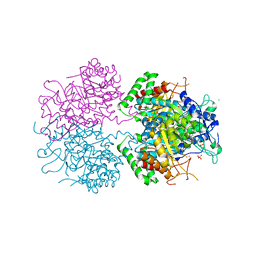
3NUD
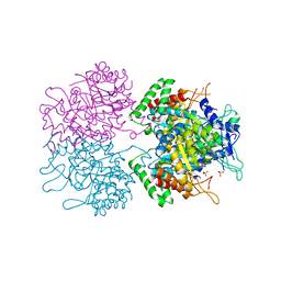 | | The structure of 3-deoxy-d-arabino-heptulosonate 7-phosphate synthase from mycobacterium tuberculosis complexed with phenylalanine | | 分子名称: | PHENYLALANINE, PHOSPHATE ION, Probable 3-deoxy-D-arabino-heptulosonate 7-phosphate synthase AroG | | 著者 | Parker, E.J, Jameson, G.B, Jiao, W, Webby, C.J, Baker, E.N, Baker, H.M. | | 登録日 | 2010-07-06 | | 公開日 | 2010-07-28 | | 最終更新日 | 2023-11-01 | | 実験手法 | X-RAY DIFFRACTION (3 Å) | | 主引用文献 | Synergistic allostery, a sophisticated regulatory network for the control of aromatic amino acid biosynthesis in Mycobacterium tuberculosis
J.Biol.Chem., 285, 2010
|
|
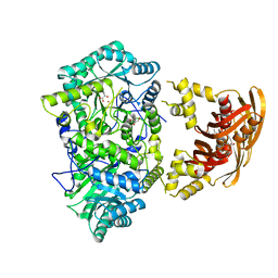
1QHU
 | | MAMMALIAN BLOOD SERUM HAEMOPEXIN DEGLYCOSYLATED AND IN COMPLEX WITH ITS LIGAND HAEM | | 分子名称: | CHLORIDE ION, PHOSPHATE ION, PROTEIN (HEMOPEXIN), ... | | 著者 | Paoli, M, Baker, H.M, Morgan, W.T, Smith, A, Baker, E.N. | | 登録日 | 1999-05-27 | | 公開日 | 1999-10-06 | | 最終更新日 | 2023-08-16 | | 実験手法 | X-RAY DIFFRACTION (2.3 Å) | | 主引用文献 | Crystal structure of hemopexin reveals a novel high-affinity heme site formed between two beta-propeller domains.
Nat.Struct.Biol., 6, 1999
|
|
3U6W
 | | Truncated M. tuberculosis LeuA (1-425) complexed with KIV | | 分子名称: | 2-isopropylmalate synthase, 3-METHYL-2-OXOBUTANOIC ACID, GLYCEROL, ... | | 著者 | Koon, N, Baker, E.N, Squire, C.J. | | 登録日 | 2011-10-13 | | 公開日 | 2012-03-14 | | 最終更新日 | 2023-09-13 | | 実験手法 | X-RAY DIFFRACTION (2.21 Å) | | 主引用文献 | Removal of the C-terminal regulatory domain of alpha-isopropylmalate synthase disrupts functional substrate binding.
Biochemistry, 51, 2012
|
|
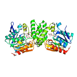
1OQG
 | | Crystal structure of the D63E mutant of the N-lobe human transferrin | | 分子名称: | CARBONATE ION, FE (III) ION, Serotransferrin | | 著者 | Baker, H.M, He, Q.-Y, Brigg, S.K, Mason, A.B, Baker, E.N. | | 登録日 | 2003-03-09 | | 公開日 | 2003-03-18 | | 最終更新日 | 2023-08-16 | | 実験手法 | X-RAY DIFFRACTION (1.9 Å) | | 主引用文献 | Structural and functional consequences of binding site mutations in transferrin: crystal structures of the Asp63Glu and Arg124Ala mutants of the N-lobe of human transferrin
Biochemistry, 42, 2003
|
|
1PGS
 | | THE THREE-DIMENSIONAL STRUCTURE OF PNGASE F, A GLYCOSYLASPARAGINASE FROM FLAVOBACTERIUM MENINGOSEPTICUM | | 分子名称: | PEPTIDE-N(4)-(N-ACETYL-BETA-D-GLUCOSAMINYL)ASPARAGINE AMIDASE F | | 著者 | Norris, G.E, Stillman, T.J, Anderson, B.F, Baker, E.N. | | 登録日 | 1994-10-06 | | 公開日 | 1995-01-26 | | 最終更新日 | 2024-06-05 | | 実験手法 | X-RAY DIFFRACTION (1.8 Å) | | 主引用文献 | The three-dimensional structure of PNGase F, a glycosylasparaginase from Flavobacterium meningosepticum.
Structure, 2, 1994
|
|
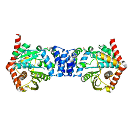
3HYS
 | | Structure of Rv0554 from Mycobacterium tuberculosis complexed with Malonic Acid | | 分子名称: | 1,2-ETHANEDIOL, 2-AMINO-2-HYDROXYMETHYL-PROPANE-1,3-DIOL, MALONIC ACID, ... | | 著者 | Johnston, J.M, Baker, E.N. | | 登録日 | 2009-06-23 | | 公開日 | 2010-06-02 | | 最終更新日 | 2024-02-21 | | 実験手法 | X-RAY DIFFRACTION (2.3 Å) | | 主引用文献 | Structural and functional analysis of Rv0554 from Mycobacterium tuberculosis: testing a putative role in menaquinone biosynthesis.
Acta Crystallogr.,Sect.D, 66, 2010
|
|
3HZO
 | |
3HPS
 | |
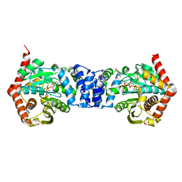
3HSS
 | | A higher resolution structure of Rv0554 from Mycobacterium tuberculosis complexed with malonic acid | | 分子名称: | 1,2-ETHANEDIOL, 2-AMINO-2-HYDROXYMETHYL-PROPANE-1,3-DIOL, ACETATE ION, ... | | 著者 | Johnston, J.M, Baker, E.N. | | 登録日 | 2009-06-10 | | 公開日 | 2010-05-26 | | 最終更新日 | 2024-02-21 | | 実験手法 | X-RAY DIFFRACTION (1.9 Å) | | 主引用文献 | Structural and functional analysis of Rv0554 from Mycobacterium tuberculosis: testing a putative role in menaquinone biosynthesis.
Acta Crystallogr.,Sect.D, 66, 2010
|
|
3HQ1
 | |
3HPZ
 | |
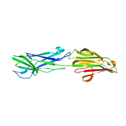
3KLQ
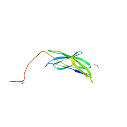 | | Crystal Structure of the Minor Pilin FctB from Streptococcus pyogenes 90/306S | | 分子名称: | GLYCEROL, Putative pilus anchoring protein | | 著者 | Linke, C, Young, P.G, Bunker, R.D, Caradoc-Davies, T.T, Baker, E.N. | | 登録日 | 2009-11-08 | | 公開日 | 2010-04-28 | | 最終更新日 | 2024-03-20 | | 実験手法 | X-RAY DIFFRACTION (1.9 Å) | | 主引用文献 | Crystal structure of the minor pilin FctB reveals determinants of Group A streptococcal pilus anchoring
J.Biol.Chem., 285, 2010
|
|
6GWX
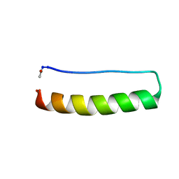 | | Stabilising and Understanding a Miniprotein by Rational Design. | | 分子名称: | Optimised PPa-TYR | | 著者 | Porter Goff, K.L, Williams, C, Baker, E.G, Nicol, D, Samphire, J.L, Zieleniewski, F.L, Crump, M.P, Woolfson, D.N. | | 登録日 | 2018-06-26 | | 公開日 | 2019-07-10 | | 最終更新日 | 2023-06-14 | | 実験手法 | SOLUTION NMR | | 主引用文献 | Stabilizing and Understanding a Miniprotein by Rational Redesign.
Biochemistry, 58, 2019
|
|
3HST
 | |
4N8Q
 | | Alternative substrates of Mycobacterium tuberculosis anthranilate phosphoribosyl transferase | | 分子名称: | 2-amino-4-fluorobenzoic acid, Anthranilate phosphoribosyltransferase | | 著者 | Castell, A, Cookson, T.V.M, Bulloch, E, Evans, G.L, Baker, E.N, Lott, J.S, Parker, E.J. | | 登録日 | 2013-10-17 | | 公開日 | 2014-04-23 | | 最終更新日 | 2023-09-20 | | 実験手法 | X-RAY DIFFRACTION (2.08 Å) | | 主引用文献 | Alternative substrates reveal catalytic cycle and key binding events in the reaction catalysed by anthranilate phosphoribosyltransferase from Mycobacterium tuberculosis.
Biochem.J., 461, 2014
|
|
4N5V
 | | Alternative substrates of Mycobacterium tuberculosis anthranilate phosphoribosyl transferase | | 分子名称: | 1-O-pyrophosphono-5-O-phosphono-alpha-D-ribofuranose, 2-amino-4-fluorobenzoic acid, Anthranilate phosphoribosyltransferase, ... | | 著者 | Castell, A, Cookson, T.V.M, Parker, E.J, Baker, E.N, Lott, J.S. | | 登録日 | 2013-10-10 | | 公開日 | 2014-04-23 | | 最終更新日 | 2023-09-20 | | 実験手法 | X-RAY DIFFRACTION (1.9 Å) | | 主引用文献 | Alternative substrates reveal catalytic cycle and key binding events in the reaction catalysed by anthranilate phosphoribosyltransferase from Mycobacterium tuberculosis.
Biochem.J., 461, 2014
|
|
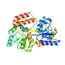
5CEV
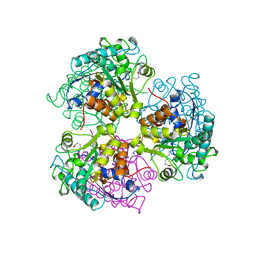 | | ARGINASE FROM BACILLUS CALDEVELOX, L-LYSINE COMPLEX | | 分子名称: | GUANIDINE, LYSINE, MANGANESE (II) ION, ... | | 著者 | Bewley, M.C, Jeffrey, P.D, Patchett, M.L, Kanyo, Z.F, Baker, E.N. | | 登録日 | 1999-03-16 | | 公開日 | 1999-04-16 | | 最終更新日 | 2023-09-20 | | 実験手法 | X-RAY DIFFRACTION (2.5 Å) | | 主引用文献 | Crystal structures of Bacillus caldovelox arginase in complex with substrate and inhibitors reveal new insights into activation, inhibition and catalysis in the arginase superfamily.
Structure Fold.Des., 7, 1999
|
|
5CWA
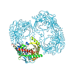 | | Structure of Anthranilate Synthase Component I (TrpE) from Mycobacterium tuberculosis with inhibitor bound | | 分子名称: | 3-{[(1Z)-1-carboxyprop-1-en-1-yl]oxy}-2-hydroxybenzoic acid, Anthranilate synthase component 1, GLYCEROL, ... | | 著者 | Johnston, J.M, Bashiri, G, Evans, G.L, Lott, J.S, Baker, E.N. | | 登録日 | 2015-07-28 | | 公開日 | 2015-08-12 | | 最終更新日 | 2023-09-27 | | 実験手法 | X-RAY DIFFRACTION (2.1 Å) | | 主引用文献 | Structure and inhibition of subunit I of the anthranilate synthase complex of Mycobacterium tuberculosis and expression of the active complex.
Acta Crystallogr.,Sect.D, 71, 2015
|
|
3HTL
 | | Structure of the Corynebacterium diphtheriae major pilin SpaA points to a modular pilus assembly with stabilizing isopeptide bonds | | 分子名称: | CALCIUM ION, Putative surface-anchored fimbrial subunit, SODIUM ION | | 著者 | Kang, H.J, Paterson, N.G, Gaspar, A.H, Ton-That, H, Baker, E.N. | | 登録日 | 2009-06-11 | | 公開日 | 2009-09-15 | | 最終更新日 | 2011-07-13 | | 実験手法 | X-RAY DIFFRACTION (1.8 Å) | | 主引用文献 | The Corynebacterium diphtheriae shaft pilin SpaA is built of tandem Ig-like modules with stabilizing isopeptide and disulfide bonds
Proc.Natl.Acad.Sci.USA, 106, 2009
|
|
3HR6
 | | Structure of the Corynebacterium diphtheriae major pilin SpaA points to a modular pilus assembly stabilizing isopeptide bonds | | 分子名称: | CALCIUM ION, IODIDE ION, Putative surface-anchored fimbrial subunit | | 著者 | Kang, H.J, Paterson, N.G, Gaspar, A.H, Ton-That, H, Baker, E.N. | | 登録日 | 2009-06-08 | | 公開日 | 2009-09-15 | | 最終更新日 | 2011-07-13 | | 実験手法 | X-RAY DIFFRACTION (1.6 Å) | | 主引用文献 | The Corynebacterium diphtheriae shaft pilin SpaA is built of tandem Ig-like modules with stabilizing isopeptide and disulfide bonds
Proc.Natl.Acad.Sci.USA, 106, 2009
|
|


