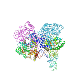
3E3J
 
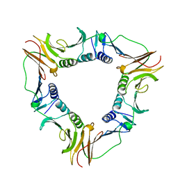
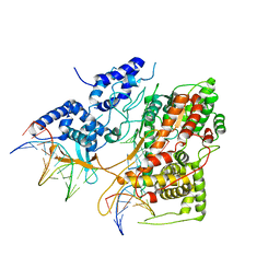 | | Crystal Structure of an Intermediate Complex of T7 RNAP and 8nt of RNA | | 分子名称: | DNA (28-MER), DNA (5'-D(*DTP*DAP*DAP*DTP*DAP*DCP*DGP*DAP*DCP*DTP*DCP*DAP*DCP*DTP*DAP*DTP*DAP*DTP*DTP*DTP*DCP*DTP*DGP*DCP*DCP*DAP*DAP*DAP*DCP*DGP*DGP*DC)-3'), DNA-directed RNA polymerase, ... | | 著者 | Durniak, K.J, Bailey, S, Steitz, T.A. | | 登録日 | 2008-08-07 | | 公開日 | 2008-11-04 | | 最終更新日 | 2023-08-30 | | 実験手法 | X-RAY DIFFRACTION (6.7 Å) | | 主引用文献 | The structure of a transcribing t7 RNA polymerase in transition from initiation to elongation
Science, 322, 2008
|
|
3E2E
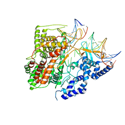 | | Crystal Structure of an Intermediate Complex of T7 RNAP and 7nt of RNA | | 分子名称: | DNA (28-MER), DNA (31-MER), DNA-directed RNA polymerase, ... | | 著者 | Durniak, K.J, Bailey, S, Steitz, T.A. | | 登録日 | 2008-08-05 | | 公開日 | 2008-11-04 | | 最終更新日 | 2024-02-21 | | 実験手法 | X-RAY DIFFRACTION (3 Å) | | 主引用文献 | The structure of a transcribing t7 RNA polymerase in transition from initiation to elongation
Science, 322, 2008
|
|
1FG0
 | | LARGE RIBOSOMAL SUBUNIT COMPLEXED WITH A 13 BP MINIHELIX-PUROMYCIN COMPOUND | | 分子名称: | 23S RIBOSOMAL RNA, 5'-R(CCGGCGGGCUGGUUCAAACCGGCCCGCCGGACC)-3'-5'-R(P-PUROMYCIN)-3' | | 著者 | Nissen, P, Hansen, J, Ban, N, Moore, P.B, Steitz, T.A. | | 登録日 | 2000-07-26 | | 公開日 | 2000-08-28 | | 最終更新日 | 2024-02-07 | | 実験手法 | X-RAY DIFFRACTION (3 Å) | | 主引用文献 | The structural basis of ribosome activity in peptide bond synthesis.
Science, 289, 2000
|
|
1FFZ
 | | LARGE RIBOSOMAL SUBUNIT COMPLEXED WITH R(CC)-DA-PUROMYCIN | | 分子名称: | 23S RIBOSOMAL RNA, R(P*CP*C*)-D(P*A)-R(P*(PU)) | | 著者 | Nissen, P, Hansen, J, Ban, N, Moore, P.B, Steitz, T.A. | | 登録日 | 2000-07-26 | | 公開日 | 2000-08-28 | | 最終更新日 | 2024-02-07 | | 実験手法 | X-RAY DIFFRACTION (3.2 Å) | | 主引用文献 | The structural basis of ribosome activity in peptide bond synthesis.
Science, 289, 2000
|
|
3EDU
 | | Crystal structure of the ankyrin-binding domain of human erythroid spectrin | | 分子名称: | Spectrin beta chain, erythrocyte | | 著者 | Simonovic, M, Stabach, P, Simonovic, I, Steitz, T.A, Morrow, J.S. | | 登録日 | 2008-09-03 | | 公開日 | 2009-02-10 | | 最終更新日 | 2024-02-21 | | 実験手法 | X-RAY DIFFRACTION (2.1 Å) | | 主引用文献 | The structure of the ankyrin-binding site of {beta}-spectrin reveals how tandem spectrin-repeats generate unique ligand-binding properties
Blood, 113, 2009
|
|
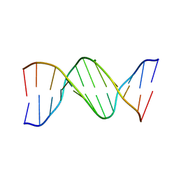
354D
 | | Structure of loop E FROM E. coli 5S RRNA | | 分子名称: | MAGNESIUM ION, RNA (5'-R(*CP*CP*GP*AP*UP*GP*GP*UP*AP*GP*UP*G)-3'), RNA-DNA (5'-R(*GP*CP*GP*AP*GP*AP*GP*UP*AP*)-D(*DGP(S)*)-R(*GP*C)-3') | | 著者 | Correll, C.C, Freeborn, B, Moore, P.B, Steitz, T.A. | | 登録日 | 1997-10-01 | | 公開日 | 1997-11-26 | | 最終更新日 | 2024-02-21 | | 実験手法 | X-RAY DIFFRACTION (1.5 Å) | | 主引用文献 | Metals, motifs, and recognition in the crystal structure of a 5S rRNA domain.
Cell(Cambridge,Mass.), 91, 1997
|
|
357D
 | | 3.5 A structure of fragment I from E. coli 5S RRNA | | 分子名称: | MAGNESIUM ION, MERCURY (II) ION, RNA (5'-R(*CP*CP*CP*CP*AP*UP*GP*CP*GP*AP*GP*AP*GP*UP*AP*GP*G P*GP*AP*AP*CP*UP* GP*CP*CP*AP*GP*GP*CP*AP*U)-3'), ... | | 著者 | Correll, C.C, Freeborn, B, Moore, P.B, Steitz, T.A. | | 登録日 | 1997-10-09 | | 公開日 | 1997-12-01 | | 最終更新日 | 2024-02-21 | | 実験手法 | X-RAY DIFFRACTION (3.5 Å) | | 主引用文献 | Metals, motifs, and recognition in the crystal structure of a 5S rRNA domain.
Cell(Cambridge,Mass.), 91, 1997
|
|
364D
 | | 3.0 A STRUCTURE OF FRAGMENT I FROM E. COLI 5S RRNA | | 分子名称: | MAGNESIUM ION, RNA (5'-R(*CP*CP*CP*CP*AP*UP*GP*CP*GP*AP*GP*AP*GP*UP*AP*GP*G P*GP*AP*AP*CP*UP*GP*CP*CP*AP*GP*GP*CP*AP*U)-3'), RNA (5'-R(*CP*CP*GP*AP*UP*GP*GP*UP*AP*GP*UP*GP*UP*GP*GP*GP*G *UP*C)-3'), ... | | 著者 | Correll, C.C, Freeborn, B, Moore, P.B, Steitz, T.A. | | 登録日 | 1997-12-08 | | 公開日 | 1998-01-02 | | 最終更新日 | 2024-02-21 | | 実験手法 | X-RAY DIFFRACTION (3 Å) | | 主引用文献 | Metals, motifs, and recognition in the crystal structure of a 5S rRNA domain.
Cell(Cambridge,Mass.), 91, 1997
|
|
1GSG
 | | Structure of E.coli glutaminyl-tRNA synthetase complexed with trnagln and ATP at 2.8 Angstroms resolution | | 分子名称: | GLUTAMINYL-TRNA SYNTHETASE, TRNAGLN | | 著者 | Rould, M.A, Perona, J.J, Soell, D, Steitz, T.A. | | 登録日 | 1990-04-03 | | 公開日 | 1992-02-24 | | 最終更新日 | 2024-02-07 | | 実験手法 | X-RAY DIFFRACTION (2.8 Å) | | 主引用文献 | Structure of E. coli glutaminyl-tRNA synthetase complexed with tRNA(Gln) and ATP at 2.8 A resolution.
Science, 246, 1989
|
|
3G6E
 | | Co-crystal structure of Homoharringtonine bound to the large ribosomal subunit | | 分子名称: | (3beta)-O~3~-[(2R)-2,6-dihydroxy-2-(2-methoxy-2-oxoethyl)-6-methylheptanoyl]cephalotaxine, 23S ribosomal RNA, 50S ribosomal protein L10E, ... | | 著者 | Gurel, G, Blaha, G, Moore, P.B, Steitz, T.A. | | 登録日 | 2009-02-06 | | 公開日 | 2009-04-28 | | 最終更新日 | 2023-09-06 | | 実験手法 | X-RAY DIFFRACTION (2.7 Å) | | 主引用文献 | U2504 determines the species specificity of the A-site cleft antibiotics: the structures of tiamulin, homoharringtonine, and bruceantin bound to the ribosome.
J.Mol.Biol., 389, 2009
|
|
3G71
 | | Co-crystal structure of Bruceantin bound to the large ribosomal subunit | | 分子名称: | 23S ribosomal RNA, 50S ribosomal protein L10E, 50S ribosomal protein L10e, ... | | 著者 | Gurel, G, Blaha, G, Moore, P.B, Steitz, T.A. | | 登録日 | 2009-02-09 | | 公開日 | 2009-04-28 | | 最終更新日 | 2023-09-06 | | 実験手法 | X-RAY DIFFRACTION (2.85 Å) | | 主引用文献 | U2504 determines the species specificity of the A-site cleft antibiotics: the structures of tiamulin, homoharringtonine, and bruceantin bound to the ribosome.
J.Mol.Biol., 389, 2009
|
|
3G4S
 | | Co-crystal structure of Tiamulin bound to the large ribosomal subunit | | 分子名称: | 23S ribosomal RNA, 50S ribosomal protein L10, 50S ribosomal protein L10e, ... | | 著者 | Gurel, G, Blaha, G, Moore, P.B, Steitz, T.A. | | 登録日 | 2009-02-04 | | 公開日 | 2009-04-28 | | 最終更新日 | 2023-09-06 | | 実験手法 | X-RAY DIFFRACTION (3.2 Å) | | 主引用文献 | U2504 determines the species specificity of the A-site cleft antibiotics: the structures of tiamulin, homoharringtonine, and bruceantin bound to the ribosome.
J.Mol.Biol., 389, 2009
|
|
3GNA
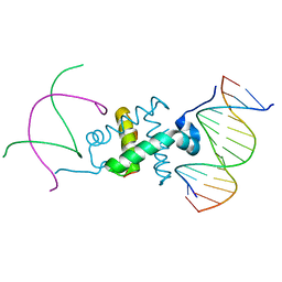 | | Crystal structure of the RAG1 nonamer-binding domain with DNA | | 分子名称: | 5'-D(*AP*CP*TP*TP*AP*AP*CP*AP*AP*AP*AP*AP*CP*C)-3', 5'-D(*TP*GP*GP*TP*TP*TP*TP*TP*GP*TP*TP*AP*AP*G)-3', V(D)J recombination-activating protein 1 | | 著者 | Yin, F.F, Bailey, S, Innis, C.A, Steitz, T.A, Schatz, D.G. | | 登録日 | 2009-03-16 | | 公開日 | 2009-04-28 | | 最終更新日 | 2024-02-21 | | 実験手法 | X-RAY DIFFRACTION (2.4 Å) | | 主引用文献 | Structure of the RAG1 nonamer binding domain with DNA reveals a dimer that mediates DNA synapsis.
Nat.Struct.Mol.Biol., 16, 2009
|
|
3GNB
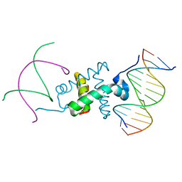 | | Crystal structure of the RAG1 nonamer-binding domain with DNA | | 分子名称: | 5'-D(*AP*AP*TP*TP*TP*TP*CP*AP*GP*AP*AP*AP*CP*C)-3', 5'-D(*AP*GP*GP*TP*TP*TP*CP*TP*GP*AP*AP*AP*AP*C)-3', V(D)J recombination-activating protein 1 | | 著者 | Yin, F.F, Bailey, S, Innis, C.A, Steitz, T.A, Schatz, D.G. | | 登録日 | 2009-03-16 | | 公開日 | 2009-04-28 | | 最終更新日 | 2023-09-06 | | 実験手法 | X-RAY DIFFRACTION (3 Å) | | 主引用文献 | Structure of the RAG1 nonamer binding domain with DNA reveals a dimer that mediates DNA synapsis.
Nat.Struct.Mol.Biol., 16, 2009
|
|
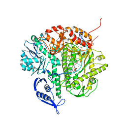
1IH7
 | | High-Resolution Structure of Apo RB69 DNA Polymerase | | 分子名称: | DNA POLYMERASE, GUANOSINE, POTASSIUM ION | | 著者 | Franklin, M.C, Wang, J, Steitz, T.A. | | 登録日 | 2001-04-18 | | 公開日 | 2001-06-13 | | 最終更新日 | 2023-08-16 | | 実験手法 | X-RAY DIFFRACTION (2.21 Å) | | 主引用文献 | Structure of the Replicating Complex of a Pol alpha Family DNA Polymerase
Cell(Cambridge,Mass.), 105, 2001
|
|
3HL2
 | | The crystal structure of the human SepSecS-tRNASec complex | | 分子名称: | (5-HYDROXY-4,6-DIMETHYLPYRIDIN-3-YL)METHYL DIHYDROGEN PHOSPHATE, Monothiophosphate, O-phosphoseryl-tRNA(Sec) selenium transferase, ... | | 著者 | Palioura, S, Steitz, T.A, Soll, D, Simonovic, M. | | 登録日 | 2009-05-26 | | 公開日 | 2009-10-06 | | 最終更新日 | 2023-09-06 | | 実験手法 | X-RAY DIFFRACTION (2.81 Å) | | 主引用文献 | The human SepSecS-tRNASec complex reveals the mechanism of selenocysteine formation.
Science, 325, 2009
|
|
3I55
 | | Co-crystal structure of Mycalamide A Bound to the Large Ribosomal Subunit | | 分子名称: | 23S ribosomal RNA, 50S ribosomal protein L10E, 50S ribosomal protein L10e, ... | | 著者 | Gurel, G, Blaha, G, Steitz, T.A, Moore, P.B. | | 登録日 | 2009-07-03 | | 公開日 | 2010-03-09 | | 最終更新日 | 2024-02-21 | | 実験手法 | X-RAY DIFFRACTION (3.11 Å) | | 主引用文献 | Structures of triacetyloleandomycin and mycalamide A bind to the large ribosomal subunit of Haloarcula marismortui.
Antimicrob.Agents Chemother., 53, 2009
|
|
3I56
 | | Co-crystal structure of Triacetyloleandomcyin Bound to the Large Ribosomal Subunit | | 分子名称: | 23S ribosomal RNA, 50S ribosomal protein L10E, 50S ribosomal protein L10e, ... | | 著者 | Gurel, G, Blaha, G, Steitz, T.A, Moore, P.B. | | 登録日 | 2009-07-03 | | 公開日 | 2010-03-09 | | 最終更新日 | 2024-02-21 | | 実験手法 | X-RAY DIFFRACTION (2.9 Å) | | 主引用文献 | Structures of triacetyloleandomycin and mycalamide A bind to the large ribosomal subunit of Haloarcula marismortui.
Antimicrob.Agents Chemother., 53, 2009
|
|
1IG9
 | | Structure of the Replicating Complex of a Pol Alpha Family DNA Polymerase | | 分子名称: | 5'-D(*AP*CP*AP*GP*GP*TP*AP*AP*GP*CP*AP*GP*TP*CP*CP*GP*CP*G)-3', 5'-D(*GP*CP*GP*GP*AP*CP*TP*GP*CP*TP*TP*AP*CP*(DOC))-3', CALCIUM ION, ... | | 著者 | Franklin, M.C, Wang, J, Steitz, T.A. | | 登録日 | 2001-04-17 | | 公開日 | 2001-06-11 | | 最終更新日 | 2023-08-16 | | 実験手法 | X-RAY DIFFRACTION (2.6 Å) | | 主引用文献 | Structure of the Replicating Complex of a Pol Alpha Family DNA Polymerase
Cell(Cambridge,Mass.), 105, 2001
|
|
1K73
 | | Co-crystal Structure of Anisomycin Bound to the 50S Ribosomal Subunit | | 分子名称: | 23S RRNA, 5S RRNA, ANISOMYCIN, ... | | 著者 | Hansen, J, Ban, N, Nissen, P, Moore, P.B, Steitz, T.A. | | 登録日 | 2001-10-18 | | 公開日 | 2003-07-22 | | 最終更新日 | 2023-08-16 | | 実験手法 | X-RAY DIFFRACTION (3.01 Å) | | 主引用文献 | Structures of Five Antibiotics Bound at the Peptidyl Transferase Center of
the Large Ribosomal Subunit
J.Mol.Biol., 330, 2003
|
|
1KC8
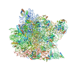 | | Co-crystal Structure of Blasticidin S Bound to the 50S Ribosomal Subunit | | 分子名称: | 23S RRNA, 5S RRNA, BLASTICIDIN S, ... | | 著者 | Hansen, J.L, Ban, N, Nissen, P, Moore, P.B, Steitz, T.A. | | 登録日 | 2001-11-07 | | 公開日 | 2003-07-22 | | 最終更新日 | 2023-08-16 | | 実験手法 | X-RAY DIFFRACTION (3.01 Å) | | 主引用文献 | Structures of Five Antibiotics Bound at the Peptidyl Transferase Center of
the Large Ribosomal Subunit
J.Mol.Biol., 330, 2003
|
|
1B8H
 | | SLIDING CLAMP, DNA POLYMERASE | | 分子名称: | DNA POLYMERASE PROCESSIVITY COMPONENT, DNA POLYMERASE fragment | | 著者 | Shamoo, Y, Steitz, T.A. | | 登録日 | 1999-02-01 | | 公開日 | 1999-02-09 | | 最終更新日 | 2023-08-09 | | 実験手法 | X-RAY DIFFRACTION (3 Å) | | 主引用文献 | Building a replisome from interacting pieces: sliding clamp complexed to a peptide from DNA polymerase and a polymerase editing complex.
Cell(Cambridge,Mass.), 99, 1999
|
|
1BDN
 | |
1B77
 | |
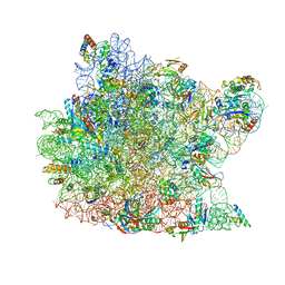
1N8R
 | | Structure of large ribosomal subunit in complex with virginiamycin M | | 分子名称: | 23S ribosomal RNA, 50S ribosomal protein L10e, 50S ribosomal protein L13P, ... | | 著者 | Hansen, J.L, Moore, P.B, Steitz, T.A. | | 登録日 | 2002-11-21 | | 公開日 | 2003-07-22 | | 最終更新日 | 2024-03-13 | | 実験手法 | X-RAY DIFFRACTION (3 Å) | | 主引用文献 | Structures of Five Antibiotics Bound at the Peptidyl Transferase Center of
the Large Ribosomal Subunit
J.Mol.Biol., 330, 2003
|
|


