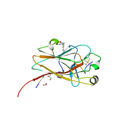
5JR2
 
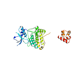
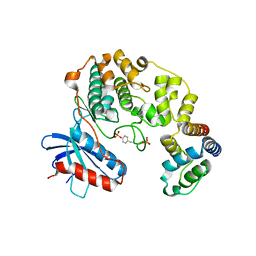
 | | Crystal structure of the EphA4 LBD in complex with APYd3 peptide inhibitor | | 分子名称: | APYd3 peptide, Ephrin type-A receptor 4, GLYCEROL, ... | | 著者 | Lechtenberg, B.C, Olson, E.J, Pasquale, E.B, Dawson, P.E, Riedl, S.J. | | 登録日 | 2016-05-05 | | 公開日 | 2016-07-06 | | 最終更新日 | 2023-09-27 | | 実験手法 | X-RAY DIFFRACTION (1.75 Å) | | 主引用文献 | Modifications of a Nanomolar Cyclic Peptide Antagonist for the EphA4 Receptor To Achieve High Plasma Stability.
Acs Med.Chem.Lett., 7, 2016
|
|
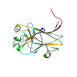
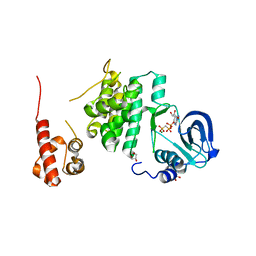
2QBX
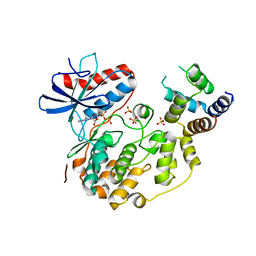
 | | EphB2/SNEW Antagonistic Peptide Complex | | 分子名称: | Ephrin type-B receptor 2, SULFATE ION, antagonistic peptide | | 著者 | Chrencik, J.E, Brooun, A, Recht, M.I, Nicola, G, Pasquale, E.B, Kuhn, P, Accelerated Technologies Center for Gene to 3D Structure (ATCG3D) | | 登録日 | 2007-06-18 | | 公開日 | 2007-11-06 | | 最終更新日 | 2023-08-30 | | 実験手法 | X-RAY DIFFRACTION (2.3 Å) | | 主引用文献 | Three-dimensional structure of the EphB2 receptor in complex with an antagonistic peptide reveals a novel mode of inhibition.
J.Biol.Chem., 282, 2007
|
|
6NK0
 | |
6NKP
 | |
6NJZ
 | |
6NK1
 | |
6NK2
 | |
7KJC
 | |
7KJB
 | |
7KJA
 | |
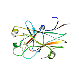
4W4Z
 | | Structure of the EphA4 LBD in complex with peptide | | 分子名称: | APY-bAla8.am peptide, Ephrin type-A receptor 4, GLYCEROL, ... | | 著者 | Lechtenberg, B.C, Mace, P.D, Riedl, S.J. | | 登録日 | 2014-08-15 | | 公開日 | 2014-10-08 | | 最終更新日 | 2023-12-27 | | 実験手法 | X-RAY DIFFRACTION (2.41 Å) | | 主引用文献 | Development and Structural Analysis of a Nanomolar Cyclic Peptide Antagonist for the EphA4 Receptor.
Acs Chem.Biol., 9, 2014
|
|
3GXU
 | |
2BBA
 | |
4W50
 | | Structure of the EphA4 LBD in complex with peptide | | 分子名称: | 1,3-BUTANEDIOL, APY peptide, Ephrin type-A receptor 4, ... | | 著者 | Lechtenberg, B.C, Mace, P.D, Riedl, S.J. | | 登録日 | 2014-08-16 | | 公開日 | 2014-10-08 | | 最終更新日 | 2023-09-27 | | 実験手法 | X-RAY DIFFRACTION (2.42 Å) | | 主引用文献 | Development and Structural Analysis of a Nanomolar Cyclic Peptide Antagonist for the EphA4 Receptor.
Acs Chem.Biol., 9, 2014
|
|
2GJY
 | |
3T6G
 | |
3T6A
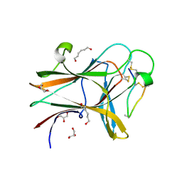
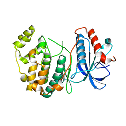
 | | Structure of the C-terminal domain of BCAR3 | | 分子名称: | (20S)-2,5,8,11,14,17-HEXAMETHYL-3,6,9,12,15,18-HEXAOXAHENICOSANE-1,20-DIOL, Breast cancer anti-estrogen resistance protein 3, UNKNOWN ATOM OR ION | | 著者 | Mace, P.D, Robinson, H, Riedl, S.J. | | 登録日 | 2011-07-28 | | 公開日 | 2011-11-23 | | 最終更新日 | 2024-02-28 | | 実験手法 | X-RAY DIFFRACTION (2.4 Å) | | 主引用文献 | NSP-Cas protein structures reveal a promiscuous interaction module in cell signaling.
Nat.Struct.Mol.Biol., 18, 2011
|
|
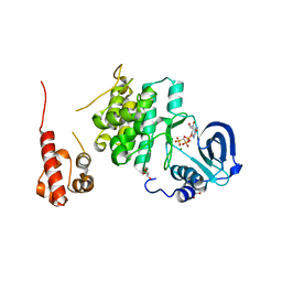
5V60
 | | Phospho-ERK2 bound to AMP-PCP | | 分子名称: | GLYCEROL, MAGNESIUM ION, Mitogen-activated protein kinase 1, ... | | 著者 | Lechtenberg, B.C, Riedl, S.J. | | 登録日 | 2017-03-15 | | 公開日 | 2017-07-26 | | 最終更新日 | 2023-11-15 | | 実験手法 | X-RAY DIFFRACTION (2.18 Å) | | 主引用文献 | Structure-Guided Strategy for the Development of Potent Bivalent ERK Inhibitors.
ACS Med Chem Lett, 8, 2017
|
|
5V61
 | | Phospho-ERK2 bound to bivalent inhibitor SBP2 | | 分子名称: | 2-oxo-6,9,12,15-tetraoxa-3-azaoctadecan-18-oic acid, 5-(2-PHENYLPYRAZOLO[1,5-A]PYRIDIN-3-YL)-1H-PYRAZOLO[3,4-C]PYRIDAZIN-3-AMINE, GLYCEROL, ... | | 著者 | Lechtenberg, B.C, Riedl, S.J. | | 登録日 | 2017-03-15 | | 公開日 | 2017-07-26 | | 最終更新日 | 2023-11-15 | | 実験手法 | X-RAY DIFFRACTION (2.2 Å) | | 主引用文献 | Structure-Guided Strategy for the Development of Potent Bivalent ERK Inhibitors.
ACS Med Chem Lett, 8, 2017
|
|
5V62
 | | Phospho-ERK2 bound to bivalent inhibitor SBP3 | | 分子名称: | 5-(2-PHENYLPYRAZOLO[1,5-A]PYRIDIN-3-YL)-1H-PYRAZOLO[3,4-C]PYRIDAZIN-3-AMINE, GLYCEROL, Mitogen-activated protein kinase 1, ... | | 著者 | Lechtenberg, B.C, Riedl, S.J. | | 登録日 | 2017-03-15 | | 公開日 | 2017-07-26 | | 最終更新日 | 2019-12-04 | | 実験手法 | X-RAY DIFFRACTION (1.9 Å) | | 主引用文献 | Structure-Guided Strategy for the Development of Potent Bivalent ERK Inhibitors.
ACS Med Chem Lett, 8, 2017
|
|
3CKH
 | | Crystal structure of Eph A4 receptor | | 分子名称: | Ephrin type-A receptor 4 | | 著者 | Shi, J.H, Song, J.X. | | 登録日 | 2008-03-15 | | 公開日 | 2008-09-23 | | 最終更新日 | 2023-11-01 | | 実験手法 | X-RAY DIFFRACTION (2.8 Å) | | 主引用文献 | Crystal Structure and NMR Binding Reveal That Two Small Molecule Antagonists Target the High Affinity Ephrin-binding Channel of the EphA4 Receptor.
J.Biol.Chem., 283, 2008
|
|
4IZA
 | |
4IZ5
 | | Structure of the complex between ERK2 phosphomimetic mutant and PEA-15 | | 分子名称: | ADENOSINE-5'-DIPHOSPHATE, Astrocytic phosphoprotein PEA-15, Mitogen-activated protein kinase 1, ... | | 著者 | Mace, P.D, Robinson, H, Riedl, S.J. | | 登録日 | 2013-01-29 | | 公開日 | 2013-04-10 | | 最終更新日 | 2024-02-28 | | 実験手法 | X-RAY DIFFRACTION (3.19 Å) | | 主引用文献 | Structure of ERK2 bound to PEA-15 reveals a mechanism for rapid release of activated MAPK.
Nat Commun, 4, 2013
|
|
4IZ7
 | |
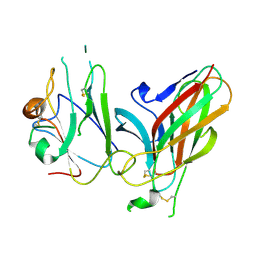
4L0P
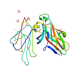 | | Structure of the human EphA3 receptor ligand binding domain complexed with ephrin-A5 | | 分子名称: | 2-acetamido-2-deoxy-beta-D-glucopyranose-(1-4)-2-acetamido-2-deoxy-beta-D-glucopyranose, CALCIUM ION, Ephrin type-A receptor 3, ... | | 著者 | Forse, G.J, Kolatkar, A.R, Kuhn, P. | | 登録日 | 2013-05-31 | | 公開日 | 2014-06-11 | | 最終更新日 | 2023-09-20 | | 実験手法 | X-RAY DIFFRACTION (2.26 Å) | | 主引用文献 | Distinctive Structure of the EphA3/Ephrin-A5 Complex Reveals a Dual Mode of Eph Receptor Interaction for Ephrin-A5.
Plos One, 10, 2015
|
|


