Loading
PDBj
MenuPDBj@FacebookPDBj@TwitterPDBj@YouTubewwPDB FoundationwwPDB
RCSB PDBPDBeBMRBAdv. SearchSearch help
Search by PDB author
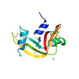
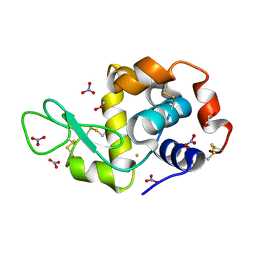
9EO2
DownloadVisualize
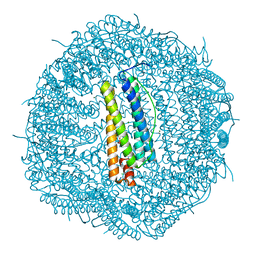
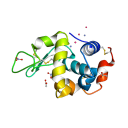
BU of 9eo2 by Molmil
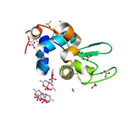
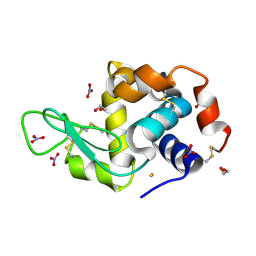
X-ray structure of the adduct formed upon reaction of picoplatin with lysozyme (structure B)
Descriptor: ACETATE ION, GLYCEROL, Lysozyme C, ...
Authors:Ferraro, G, Merlino, A.
Deposit date:2024-03-14
Release date:2024-05-22
Last modified:2024-10-16
Method:X-RAY DIFFRACTION (1.36 Å)
Cite:Picoplatin binding to proteins: X-ray structures and mass spectrometry data on the adducts with lysozyme and ribonuclease A.
Dalton Trans, 53, 2024
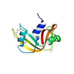
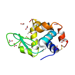
9ENZ
DownloadVisualize
BU of 9enz by Molmil
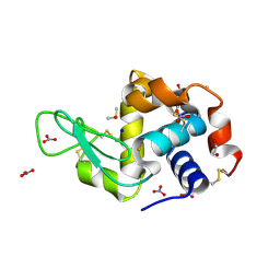
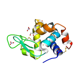
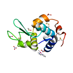
X-ray structure of the adduct formed upon reaction of picoplatin with lysozyme (structure A)
Descriptor: ACETATE ION, AMMONIA, Lysozyme C, ...
Authors:Ferraro, G, Merlino, A.
Deposit date:2024-03-14
Release date:2024-05-22
Last modified:2024-10-16
Method:X-RAY DIFFRACTION (1.6 Å)
Cite:Picoplatin binding to proteins: X-ray structures and mass spectrometry data on the adducts with lysozyme and ribonuclease A.
Dalton Trans, 53, 2024
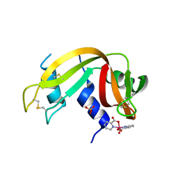
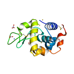
9EO8
DownloadVisualize
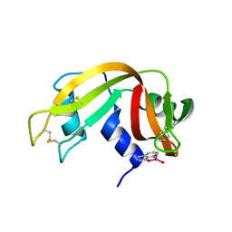
BU of 9eo8 by Molmil
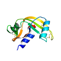
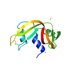
X-ray structure of the adduct formed upon reaction of picoplatin with bovine pancreatic ribonuclease (structure D)
Descriptor: AMMONIA, CHLORIDE ION, PLATINUM (II) ION, ...
Authors:Ferraro, G, Merlino, A.
Deposit date:2024-03-14
Release date:2024-05-22
Last modified:2024-05-29
Method:X-RAY DIFFRACTION (1.76 Å)
Cite:Picoplatin binding to proteins: X-ray structures and mass spectrometry data on the adducts with lysozyme and ribonuclease A.
Dalton Trans, 53, 2024
9EO5
DownloadVisualize
BU of 9eo5 by Molmil
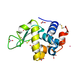
X-ray structure of the adduct formed upon reaction of picoplatin with bovine pancreatic ribonuclease (structure C)
Descriptor: AMMONIA, PLATINUM (II) ION, Ribonuclease pancreatic
Authors:Ferraro, G, Merlino, A.
Deposit date:2024-03-14
Release date:2024-05-22
Last modified:2024-05-29
Method:X-RAY DIFFRACTION (1.99 Å)
Cite:Picoplatin binding to proteins: X-ray structures and mass spectrometry data on the adducts with lysozyme and ribonuclease A.
Dalton Trans, 53, 2024
7P8R
DownloadVisualize
BU of 7p8r by Molmil
X-ray structure of the adduct of a vanadium picolinate complex with RNase A at pH 5.1
Descriptor: Ribonuclease pancreatic, SULFATE ION, hydroxy(oxo)bis(pyridine-2-carboxylato-kappa~2~N,O)vanadium(3+)
Authors:Ferraro, G, Merlino, A.
Deposit date:2021-07-23
Release date:2021-12-22
Last modified:2024-01-31
Method:X-RAY DIFFRACTION (1.27 Å)
Cite:Spectroscopic/Computational Characterization and the X-ray Structure of the Adduct of the V IV O-Picolinato Complex with RNase A.
Inorg.Chem., 60, 2021
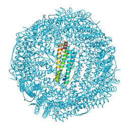
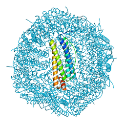
5IX6
DownloadVisualize
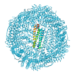
BU of 5ix6 by Molmil
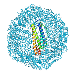
X-ray structure of Auoxo3-encapsulated horse spleen apoferritin
Descriptor: CADMIUM ION, Ferritin light chain, GOLD ION, ...
Authors:Ferraro, G, Pontillo, N, Merlino, A.
Deposit date:2016-03-23
Release date:2016-07-06
Last modified:2024-01-10
Method:X-RAY DIFFRACTION (1.85 Å)
Cite:Gold-based drug encapsulation within a ferritin nanocage: X-ray structure and biological evaluation as a potential anticancer agent of the Auoxo3-loaded protein.
Chem.Commun.(Camb.), 52, 2016
6Y3D
DownloadVisualize
BU of 6y3d by Molmil
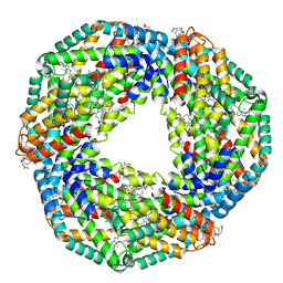
X-ray structure of thermophilic C-phycocyanin from Galdiera phlegrea
Descriptor: ACETATE ION, C-phycocyanin alpha chain, C-phycocyanin beta chain, ...
Authors:Ferraro, G, Lucignano, R, Marseglia, A, Merlino, A.
Deposit date:2020-02-18
Release date:2020-12-30
Last modified:2024-01-24
Method:X-RAY DIFFRACTION (1.8 Å)
Cite:X-ray structure of C-phycocyanin from Galdieria phlegrea: Determinants of thermostability and comparison with a C-phycocyanin in the entire phycobilisome.
Biochim Biophys Acta Bioenerg, 1861, 2020
5JVX
DownloadVisualize
BU of 5jvx by Molmil
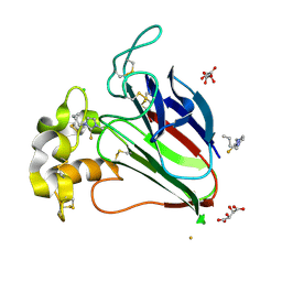
X-ray structure of the adduct formed in the reaction between thaumatin and a gold carbene compound
Descriptor: (1-butyl-3-methyl-1,3-dihydro-2H-imidazol-2-ylidene)(chloro)gold, CHLORIDE ION, L(+)-TARTARIC ACID, ...
Authors:Ferraro, G, Merlino, A.
Deposit date:2016-05-11
Release date:2016-07-13
Last modified:2024-10-16
Method:X-RAY DIFFRACTION (1.7 Å)
Cite:First Crystal Structure for a Gold Carbene-Protein Adduct.
Bioconjug.Chem., 27, 2016
8OO3
DownloadVisualize
BU of 8oo3 by Molmil
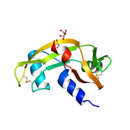
X-ray structure of the adduct formed upon reaction of cisplatin with human angiogenin after 5 days soaking
Descriptor: AMMONIA, Angiogenin, D(-)-TARTARIC ACID, ...
Authors:Ferraro, G, Merlino, A.
Deposit date:2023-04-04
Release date:2023-07-12
Last modified:2024-01-24
Method:X-RAY DIFFRACTION (1.76 Å)
Cite:Cisplatin binding to angiogenin protein: new molecular pathways and targets for the drug's anticancer activity.
Dalton Trans, 52, 2023
8OO4
DownloadVisualize
BU of 8oo4 by Molmil
X-ray structure of the adduct formed upon reaction of cisplatin with human angiogenin after 1 month soaking
Descriptor: AMMONIA, Angiogenin, PLATINUM (II) ION
Authors:Ferraro, G, Merlino, A.
Deposit date:2023-04-04
Release date:2023-07-12
Last modified:2024-01-24
Method:X-RAY DIFFRACTION (1.99 Å)
Cite:Cisplatin binding to angiogenin protein: new molecular pathways and targets for the drug's anticancer activity.
Dalton Trans, 52, 2023
6ZS8
DownloadVisualize
BU of 6zs8 by Molmil
X-ray structure of the adduct formed upon treating lysozyme with an aged solution of arsenoplatin-1
Descriptor: 1,2-ETHANEDIOL, DIMETHYL SULFOXIDE, Lysozyme, ...
Authors:Ferraro, G, Merlino, A.
Deposit date:2020-07-15
Release date:2020-12-23
Last modified:2024-01-31
Method:X-RAY DIFFRACTION (2.153 Å)
Cite:The first step of arsenoplatin-1 aggregation in solution unveiled by solving the crystal structure of its protein adduct.
Dalton Trans, 50, 2021
7BD7
DownloadVisualize
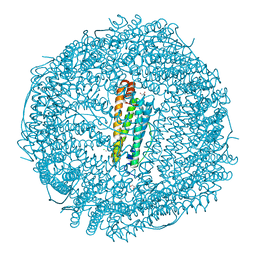
BU of 7bd7 by Molmil
X-ray structure of Arsenoplatin-1-encapsulated horse spleen ferritin
Descriptor: CADMIUM ION, CHLORIDE ION, Ferritin light chain, ...
Authors:Ferraro, G, Merlino, A.
Deposit date:2020-12-21
Release date:2021-03-17
Last modified:2024-01-31
Method:X-RAY DIFFRACTION (1.5 Å)
Cite:Arsenoplatin-Ferritin Nanocage: Structure and Cytotoxicity.
Int J Mol Sci, 22, 2021
6SET
DownloadVisualize
BU of 6set by Molmil
X-ray structure of the gold/lysozyme adduct formed upon 3 days exposure of protein crystals to compound 1
Descriptor: 1,2-ETHANEDIOL, CHLORIDE ION, DIMETHYL SULFOXIDE, ...
Authors:Ferraro, G, Giorgio, A, Merlino, A.
Deposit date:2019-07-30
Release date:2019-09-18
Last modified:2024-01-24
Method:X-RAY DIFFRACTION (1.9 Å)
Cite:Protein-mediated disproportionation of Au(i): insights from the structures of adducts of Au(iii) compounds bearing N,N-pyridylbenzimidazole derivatives with lysozyme.
Dalton Trans, 48, 2019
6SEU
DownloadVisualize
BU of 6seu by Molmil
X-ray structure of the gold/lysozyme adduct formed upon 21h exposure of protein crystals to compound 2
Descriptor: 1,2-ETHANEDIOL, GOLD ION, Lysozyme C, ...
Authors:Ferraro, G, Giorgio, A, Merlino, A.
Deposit date:2019-07-30
Release date:2019-09-18
Last modified:2024-10-16
Method:X-RAY DIFFRACTION (1.95 Å)
Cite:Protein-mediated disproportionation of Au(i): insights from the structures of adducts of Au(iii) compounds bearing N,N-pyridylbenzimidazole derivatives with lysozyme.
Dalton Trans, 48, 2019
6SEW
DownloadVisualize
BU of 6sew by Molmil
X-ray structure of the gold/lysozyme adduct formed upon 24h exposure of protein crystals to compound 2
Descriptor: 1,2-ETHANEDIOL, GOLD ION, Lysozyme C, ...
Authors:Ferraro, G, Giorgio, A, Merlino, A.
Deposit date:2019-07-30
Release date:2019-09-18
Last modified:2024-10-23
Method:X-RAY DIFFRACTION (2.12 Å)
Cite:Protein-mediated disproportionation of Au(i): insights from the structures of adducts of Au(iii) compounds bearing N,N-pyridylbenzimidazole derivatives with lysozyme.
Dalton Trans, 48, 2019
6SEX
DownloadVisualize
BU of 6sex by Molmil
X-ray structure of the gold/lysozyme adduct formed upon 21h exposure of protein crystals to compound 1
Descriptor: 1,2-ETHANEDIOL, ACETATE ION, DIMETHYL SULFOXIDE, ...
Authors:Ferraro, G, Giorgio, A, Merlino, A.
Deposit date:2019-07-30
Release date:2019-09-18
Last modified:2024-01-24
Method:X-RAY DIFFRACTION (1.78 Å)
Cite:Protein-mediated disproportionation of Au(i): insights from the structures of adducts of Au(iii) compounds bearing N,N-pyridylbenzimidazole derivatives with lysozyme.
Dalton Trans, 48, 2019
6SEZ
DownloadVisualize
BU of 6sez by Molmil
X-ray structure of the gold/lysozyme adduct formed upon 24h exposure of protein crystals to compound 1
Descriptor: 1,2-ETHANEDIOL, GOLD ION, Lysozyme C, ...
Authors:Ferraro, G, Giorgio, A, Merlino, A.
Deposit date:2019-07-30
Release date:2019-09-18
Last modified:2024-01-24
Method:X-RAY DIFFRACTION (2.22 Å)
Cite:Protein-mediated disproportionation of Au(i): insights from the structures of adducts of Au(iii) compounds bearing N,N-pyridylbenzimidazole derivatives with lysozyme.
Dalton Trans, 48, 2019
5N27
DownloadVisualize
BU of 5n27 by Molmil
X-ray structure of human heavy chain ferritin (apo form)
Descriptor: CHLORIDE ION, Ferritin heavy chain, MAGNESIUM ION
Authors:Ferraro, G, Merlino, A.
Deposit date:2017-02-07
Release date:2017-08-02
Last modified:2024-01-17
Method:X-RAY DIFFRACTION (1.74 Å)
Cite:Cisplatin Binding Sites in Human H-Chain Ferritin.
Inorg Chem, 56, 2017
5N26
DownloadVisualize
BU of 5n26 by Molmil
X-ray structure of human heavy chain ferritin in complex with cisplatin
Descriptor: CHLORIDE ION, Cisplatin, Ferritin heavy chain, ...
Authors:Ferraro, G, Merlino, A.
Deposit date:2017-02-07
Release date:2017-08-02
Last modified:2024-01-17
Method:X-RAY DIFFRACTION (2.05 Å)
Cite:Cisplatin Binding Sites in Human H-Chain Ferritin.
Inorg Chem, 56, 2017
7QWH
DownloadVisualize
BU of 7qwh by Molmil
X-ray structure of the adduct formed upon reaction of a vanadium hydroxyquinoline complex with RNase A
Descriptor: 2,2-bis($l^{1}-oxidanyl)-3-oxa-1$l^{4}-aza-2$l^{4}-vanadatricyclo[6.3.1.0^{4,12}]dodeca-1(12),4,6,8,10-pentaene, Ribonuclease pancreatic
Authors:Merlino, A, Ferraro, G.
Deposit date:2022-01-25
Release date:2023-02-01
Last modified:2024-10-23
Method:X-RAY DIFFRACTION (1.573 Å)
Cite:Interaction of VIVO-8-hydroxyquinoline species with RNase A: the effect of metal ligands in the protein adduct stabilization
Inorg Chem Front, 2023
6ENV
DownloadVisualize
BU of 6env by Molmil
X-ray structure of Au2phen-encapsulated horse spleen apoferritin
Descriptor: CADMIUM ION, CHLORIDE ION, Ferritin light chain, ...
Authors:Ferraro, G, Merlino, A.
Deposit date:2017-10-06
Release date:2017-12-13
Last modified:2024-01-17
Method:X-RAY DIFFRACTION (1.82 Å)
Cite:Ferritin nanocages loaded with gold ions induce oxidative stress and apoptosis in MCF-7 human breast cancer cells.
Dalton Trans, 46, 2017
6ENW
DownloadVisualize
BU of 6enw by Molmil
X-ray structure of Auoxo4-encapsulated horse spleen apoferritin
Descriptor: CADMIUM ION, CHLORIDE ION, Ferritin light chain, ...
Authors:Ferraro, G, Merlino, A.
Deposit date:2017-10-06
Release date:2017-12-13
Last modified:2024-01-17
Method:X-RAY DIFFRACTION (2.6 Å)
Cite:Ferritin nanocages loaded with gold ions induce oxidative stress and apoptosis in MCF-7 human breast cancer cells.
Dalton Trans, 46, 2017
5OLE
DownloadVisualize
BU of 5ole by Molmil
X-ray structure of the adduct formed upon reaction of hen egg white lysozyme with a tetranuclear Pt-thiosemicarbazone compound
Descriptor: 1,2-ETHANEDIOL, DIMETHYL SULFOXIDE, Lysozyme C, ...
Authors:Ferraro, G, Merlino, A.
Deposit date:2017-07-27
Release date:2018-08-29
Last modified:2024-10-16
Method:X-RAY DIFFRACTION (1.78 Å)
Cite:Reactions of a tetranuclear Pt-thiosemicarbazone complex with model proteins.
J. Inorg. Biochem., 181, 2018
5OLD
DownloadVisualize
BU of 5old by Molmil
X-ray structure of the adduct formed upon reaction of ribonuclease A with a tetranuclear Pt-thiosemicarbazone compound
Descriptor: DIMETHYL SULFOXIDE, PLATINUM (II) ION, Ribonuclease pancreatic
Authors:Ferraro, G, Merlino, A.
Deposit date:2017-07-27
Release date:2018-08-29
Last modified:2024-01-17
Method:X-RAY DIFFRACTION (1.78 Å)
Cite:Reactions of a tetranuclear Pt-thiosemicarbazone complex with model proteins.
J. Inorg. Biochem., 181, 2018
6QEA
DownloadVisualize
BU of 6qea by Molmil
The X-ray structure of the adduct formed in the reaction between hen egg white lysozyme and complex I, a pentacoordinate Pt(II) compound containing 2,9-dimethyl-1,10-phenanthroline, dimethylfumarate, methyl and iodine as ligands
Descriptor: DIMETHYL SULFOXIDE, Lysozyme C, NITRATE ION, ...
Authors:Merlino, A, Ferraro, G.
Deposit date:2019-01-07
Release date:2019-03-27
Last modified:2024-01-24
Method:X-RAY DIFFRACTION (1.96 Å)
Cite:Reaction with Proteins of a Five-Coordinate Platinum(II) Compound.
Int J Mol Sci, 20, 2019

226707

PDB entries from 2024-10-30

PDB statisticsPDBj update infoContact PDBjnumon