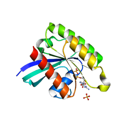
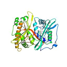
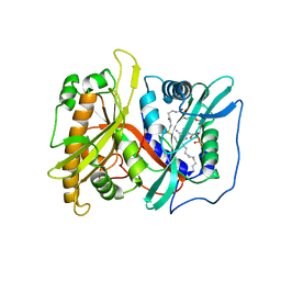
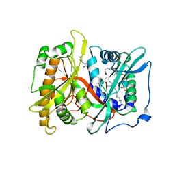
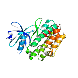
2AJI
 
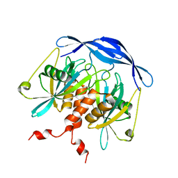
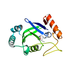 | | Crystal structure of the editing domain of E. coli leucyl-tRNA synthetase complexes with isoleucine | | 分子名称: | ISOLEUCINE, Leucyl-tRNA synthetase | | 著者 | Liu, Y, Liao, J, Zhu, B, Wang, E.D, Ding, J. | | 登録日 | 2005-08-02 | | 公開日 | 2006-01-24 | | 最終更新日 | 2024-03-13 | | 実験手法 | X-RAY DIFFRACTION (3.2 Å) | | 主引用文献 | Crystal structures of the editing domain of Escherichia coli leucyl-tRNA synthetase and its complexes with Met and Ile reveal a lock-and-key mechanism for amino acid discrimination
Biochem.J., 394, 2006
|
|
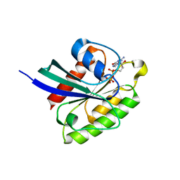
1HQU
 | | HUMAN IMMUNODEFICIENCY VIRUS TYPE 1 | | 分子名称: | (S)-4-ISOPROPOXYCARBONYL-6-METHOXY-3-METHYLTHIOMETHYL-3,4-DIHYDROQUINOXALIN-2(1H)-THIONE, POL POLYPROTEIN | | 著者 | Hsiou, Y, Ding, J, Arnold, E. | | 登録日 | 2000-12-19 | | 公開日 | 2001-05-30 | | 最終更新日 | 2023-08-09 | | 実験手法 | X-RAY DIFFRACTION (2.7 Å) | | 主引用文献 | The Lys103Asn mutation of HIV-1 RT: a novel mechanism of drug resistance.
J.Mol.Biol., 309, 2001
|
|
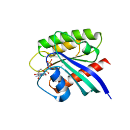
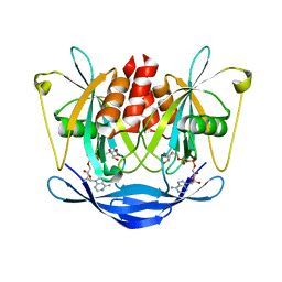
5H4B
 | | Crystal structure of Cbln4 | | 分子名称: | 2-acetamido-2-deoxy-beta-D-glucopyranose, Cerebellin-4 | | 著者 | Zhong, C, Shen, J, Zhang, H, Ding, J. | | 登録日 | 2016-10-31 | | 公開日 | 2017-09-13 | | 最終更新日 | 2020-07-29 | | 実験手法 | X-RAY DIFFRACTION (2.8 Å) | | 主引用文献 | Cbln1 and Cbln4 Are Structurally Similar but Differ in GluD2 Binding Interactions.
Cell Rep, 20, 2017
|
|
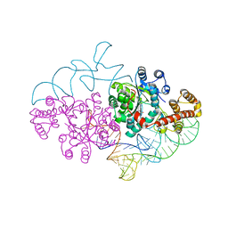
3IU3
 | | Crystal structure of the Fab fragment of therapeutic antibody Basiliximab in complex with IL-2Ra (CD25) ectodomain | | 分子名称: | Heavy chain of Fab fragment of Basiliximab, Interleukin-2 receptor alpha chain, Light chain of Fab fragment of Basiliximab, ... | | 著者 | Du, J, Yang, H, Wang, J, Ding, J. | | 登録日 | 2009-08-29 | | 公開日 | 2010-01-26 | | 最終更新日 | 2023-11-01 | | 実験手法 | X-RAY DIFFRACTION (2.9 Å) | | 主引用文献 | Structural basis for the blockage of IL-2 signaling by therapeutic antibody basiliximab
J.Immunol., 184, 2010
|
|
7BTA
 | |
7BTC
 | |
7BTD
 | |
1HYS
 | | CRYSTAL STRUCTURE OF HIV-1 REVERSE TRANSCRIPTASE IN COMPLEX WITH A POLYPURINE TRACT RNA:DNA | | 分子名称: | 5'-D(*CP*TP*TP*TP*TP*CP*TP*TP*TP*TP*AP*AP*AP*AP*AP*GP*TP*GP*GP*CP*TP*G)-3', 5'-R(*UP*CP*AP*GP*CP*CP*AP*CP*UP*UP*UP*UP*UP*AP*AP*AP*AP*GP*AP*AP*AP*AP*G)-3', FAB-28 MONOCLONAL ANTIBODY FRAGMENT HEAVY CHAIN, ... | | 著者 | Sarafianos, S.G, Das, K, Tantillo, C, Clark Jr, A.D, Ding, J, Whitcomb, J, Boyer, P.L, Hughes, S.H, Arnold, E. | | 登録日 | 2001-01-22 | | 公開日 | 2001-03-26 | | 最終更新日 | 2023-08-09 | | 実験手法 | X-RAY DIFFRACTION (3 Å) | | 主引用文献 | Crystal structure of HIV-1 reverse transcriptase in complex with a polypurine tract RNA:DNA.
EMBO J., 20, 2001
|
|
3NFS
 | | Crystal structure the Fab fragment of therapeutic antibody daclizumab | | 分子名称: | Heavy chain of Fab fragment of daclizumab, Light chain of Fab fragment of daclizumab | | 著者 | Yang, H, Wang, J, Du, J, Zhong, C, Guo, Y, Ding, J. | | 登録日 | 2010-06-10 | | 公開日 | 2010-09-15 | | 最終更新日 | 2023-11-01 | | 実験手法 | X-RAY DIFFRACTION (2.6 Å) | | 主引用文献 | Structural basis of immunosuppression by the therapeutic antibody daclizumab
Cell Res., 20, 2010
|
|
3NFP
 | | Crystal structure of the Fab fragment of therapeutic antibody daclizumab in complex with IL-2Ra (CD25) ectodomain | | 分子名称: | Heavy chain of Fab fragment of daclizumab, Interleukin-2 receptor subunit alpha, Light chain of Fab fragment of daclizumab | | 著者 | Yang, H, Wang, J, Du, J, Zhong, C, Guo, Y, Ding, J. | | 登録日 | 2010-06-10 | | 公開日 | 2010-09-15 | | 最終更新日 | 2023-11-01 | | 実験手法 | X-RAY DIFFRACTION (2.86 Å) | | 主引用文献 | Structural basis of immunosuppression by the therapeutic antibody daclizumab
Cell Res., 20, 2010
|
|
2P6E
 | |
2OSL
 | | Crystal structure of Rituximab Fab in complex with an epitope peptide | | 分子名称: | B-lymphocyte antigen CD20, heavy chain of the Rituximab Fab fragment,heavy chain of the Rituximab Fab fragment, light chain of the Rituximab Fab fragment,light chain of the Rituximab Fab fragment | | 著者 | Du, J, Zhong, C, Ding, J. | | 登録日 | 2007-02-06 | | 公開日 | 2007-04-10 | | 最終更新日 | 2024-04-10 | | 実験手法 | X-RAY DIFFRACTION (2.6 Å) | | 主引用文献 | Structural basis for recognition of CD20 by therapeutic antibody Rituximab
J.Biol.Chem., 282, 2007
|
|
2P6G
 | |
2P6F
 | |
1NFG
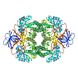 | | Structure of D-hydantoinase | | 分子名称: | D-hydantoinase, ZINC ION | | 著者 | Xu, Z, Yang, Y, Jiang, W, Arnold, E, Ding, J. | | 登録日 | 2002-12-14 | | 公開日 | 2003-07-15 | | 最終更新日 | 2023-11-15 | | 実験手法 | X-RAY DIFFRACTION (2.7 Å) | | 主引用文献 | Crystal Structure of D-Hydantoinase from Burkholderia pickettii at a Resolution of 2.7 Angstroms: Insights into the Molecular Basis of Enzyme Thermostability.
J.Bacteriol., 185, 2003
|
|
3IWP
 | |
3V9R
 | | Crystal structure of Saccharomyces cerevisiae MHF complex | | 分子名称: | SULFATE ION, Uncharacterized protein YDL160C-A, Uncharacterized protein YOL086W-A | | 著者 | Yang, H, Zhang, T, Zhong, C, Li, H, Zhou, J, Ding, J. | | 登録日 | 2011-12-28 | | 公開日 | 2012-02-29 | | 実験手法 | X-RAY DIFFRACTION (2.4 Å) | | 主引用文献 | Saccharomyces Cerevisiae MHF Complex Structurally Resembles the Histones (H3-H4)(2) Heterotetramer and Functions as a Heterotetramer
Structure, 20, 2012
|
|
2DSB
 | |
2DSC
 | |
2DSD
 | |
2DR2
 | | Structure of human tryptophanyl-tRNA synthetase in complex with tRNA(Trp) | | 分子名称: | SULFATE ION, TRYPTOPHAN, Tryptophanyl-tRNA synthetase, ... | | 著者 | Shen, N, Guo, L, Yang, B, Jin, Y, Ding, J. | | 登録日 | 2006-06-05 | | 公開日 | 2006-07-11 | | 最終更新日 | 2023-10-25 | | 実験手法 | X-RAY DIFFRACTION (3 Å) | | 主引用文献 | Structure of human tryptophanyl-tRNA synthetase in complex with tRNA(Trp) reveals the molecular basis of tRNA recognition and specificity
Nucleic Acids Res., 34, 2006
|
|
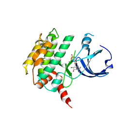
4FGB
 | | Crystal structure of human calcium/calmodulin-dependent protein kinase I apo form | | 分子名称: | Calcium/calmodulin-dependent protein kinase type 1 | | 著者 | Zha, M, Zhong, C, Ou, Y, Wang, J, Han, L, Ding, J. | | 登録日 | 2012-06-04 | | 公開日 | 2013-01-23 | | 最終更新日 | 2023-09-13 | | 実験手法 | X-RAY DIFFRACTION (2.6 Å) | | 主引用文献 | Crystal structures of human CaMKIalpha reveal insights into the regulation mechanism of CaMKI.
Plos One, 7, 2012
|
|
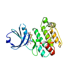
4FG9
 | | Crystal structure of human calcium/calmodulin-dependent protein kinase I 1-320 in complex with ATP | | 分子名称: | ADENOSINE-5'-TRIPHOSPHATE, Calcium/calmodulin-dependent protein kinase type 1 | | 著者 | Zha, M, Zhong, C, Ou, Y, Wang, J, Han, L, Ding, J. | | 登録日 | 2012-06-04 | | 公開日 | 2013-01-23 | | 最終更新日 | 2023-09-13 | | 実験手法 | X-RAY DIFFRACTION (2.4 Å) | | 主引用文献 | Crystal structures of human CaMKIalpha reveal insights into the regulation mechanism of CaMKI.
Plos One, 7, 2012
|
|
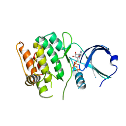
4FG8
 | | Crystal structure of human calcium/calmodulin-dependent protein kinase I 1-315 in complex with ATP | | 分子名称: | ADENOSINE-5'-TRIPHOSPHATE, Calcium/calmodulin-dependent protein kinase type 1 | | 著者 | Zha, M, Zhong, C, Ou, Y, Wang, J, Han, L, Ding, J. | | 登録日 | 2012-06-04 | | 公開日 | 2013-01-23 | | 最終更新日 | 2023-09-13 | | 実験手法 | X-RAY DIFFRACTION (2.2 Å) | | 主引用文献 | Crystal structures of human CaMKIalpha reveal insights into the regulation mechanism of CaMKI.
Plos One, 7, 2012
|
|
4FG7
 | | Crystal structure of human calcium/calmodulin-dependent protein kinase I 1-293 in complex with ATP | | 分子名称: | ADENOSINE-5'-TRIPHOSPHATE, Calcium/calmodulin-dependent protein kinase type 1 | | 著者 | Zha, M, Zhong, C, Ou, Y, Wang, J, Han, L, Ding, J. | | 登録日 | 2012-06-04 | | 公開日 | 2013-01-23 | | 最終更新日 | 2023-09-13 | | 実験手法 | X-RAY DIFFRACTION (2.7 Å) | | 主引用文献 | Crystal structures of human CaMKIalpha reveal insights into the regulation mechanism of CaMKI.
Plos One, 7, 2012
|
|


