Loading
PDBj
MenuPDBj@FacebookPDBj@TwitterPDBj@YouTubewwPDB FoundationwwPDB
RCSB PDBPDBeBMRBAdv. SearchSearch help
Search by PDB author
1N4K
DownloadVisualize
BU of 1n4k by Molmil
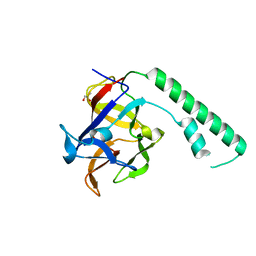
Crystal structure of the inositol 1,4,5-trisphosphate receptor binding core in complex with IP3
Descriptor: D-MYO-INOSITOL-1,4,5-TRIPHOSPHATE, Inositol 1,4,5-trisphosphate receptor type 1
Authors:Bosanac, I, Alattia, J.R, Mal, T.K, Chan, J, Talarico, S, Tong, F.K, Tong, K.I, Yoshikawa, F, Furuichi, T, Iwai, M, Michikawa, T, Mikoshiba, K, Ikura, M.
Deposit date:2002-10-31
Release date:2002-12-25
Last modified:2024-02-14
Method:X-RAY DIFFRACTION (2.2 Å)
Cite:Structure of the inositol 1,4,5-trisphosphate receptor binding core in complex with its ligand.
Nature, 420, 2002
1XZZ
DownloadVisualize
BU of 1xzz by Molmil
Crystal structure of the ligand binding suppressor domain of type 1 inositol 1,4,5-trisphosphate receptor
Descriptor: GLYCEROL, Inositol 1,4,5-trisphosphate receptor type 1
Authors:Bosanac, I, Yamazaki, H, Matsu-ura, T, Michikawa, T, Mikoshiba, K, Ikura, M.
Deposit date:2004-11-13
Release date:2005-01-25
Last modified:2024-02-14
Method:X-RAY DIFFRACTION (1.8 Å)
Cite:Crystal structure of the ligand binding suppressor domain of type 1 inositol 1,4,5-trisphosphate receptor.
Mol.Cell, 17, 2005
3PTF
DownloadVisualize
BU of 3ptf by Molmil
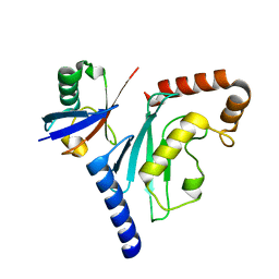
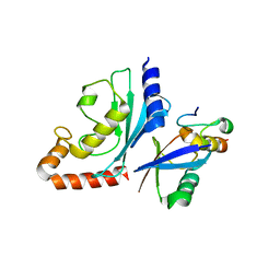
X-ray structure of the non-covalent complex between UbcH5A and Ubiquitin
Descriptor: Polyubiquitin-B, Ubiquitin-conjugating enzyme E2 D1
Authors:Bosanac, I, Hymowitz, S.G.
Deposit date:2010-12-02
Release date:2011-05-11
Last modified:2023-09-06
Method:X-RAY DIFFRACTION (2.7 Å)
Cite:Modulation of K11-Linkage Formation by Variable Loop Residues within UbcH5A.
J.Mol.Biol., 408, 2011
3HO3
DownloadVisualize
BU of 3ho3 by Molmil
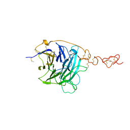
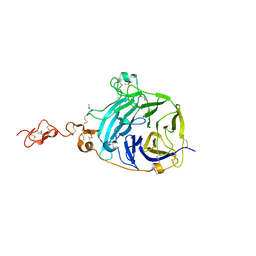
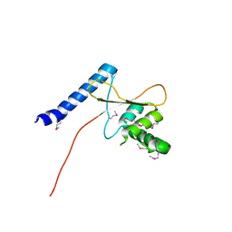
Crystal structure of Hedgehog-interacting protein (HHIP)
Descriptor: Hedgehog-interacting protein
Authors:Bosanac, I, Hymowitz, S.G.
Deposit date:2009-06-01
Release date:2009-06-23
Last modified:2023-09-06
Method:X-RAY DIFFRACTION (2.9 Å)
Cite:The structure of SHH in complex with HHIP reveals a recognition role for the Shh pseudo active site in signaling.
Nat.Struct.Mol.Biol., 16, 2009
3OJ4
DownloadVisualize
BU of 3oj4 by Molmil
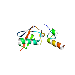
Crystal structure of the A20 ZnF4, ubiquitin and UbcH5A complex
Descriptor: Tumor necrosis factor alpha-induced protein 3, Ubiquitin, Ubiquitin-conjugating enzyme E2 D1, ...
Authors:Bosanac, I, Hymowitz, S.G.
Deposit date:2010-08-20
Release date:2010-12-08
Last modified:2023-09-06
Method:X-RAY DIFFRACTION (3.4 Å)
Cite:Ubiquitin Binding to A20 ZnF4 Is Required for Modulation of NF-κB Signaling
Mol.Cell, 40, 2010
3OJ3
DownloadVisualize
BU of 3oj3 by Molmil
Crystal structure of the A20 ZnF4 and ubiquitin complex
Descriptor: Tumor necrosis factor alpha-induced protein 3, Ubiquitin, ZINC ION
Authors:Bosanac, I, Hymowitz, S.G.
Deposit date:2010-08-20
Release date:2010-12-08
Last modified:2023-09-06
Method:X-RAY DIFFRACTION (2.5 Å)
Cite:Ubiquitin Binding to A20 ZnF4 Is Required for Modulation of NF-KB Signaling
Mol.Cell, 40, 2010
3HO4
DownloadVisualize
BU of 3ho4 by Molmil
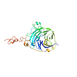
Crystal structure of Hedgehog-interacting protein (HHIP)
Descriptor: Hedgehog-interacting protein
Authors:Hymowitz, S.G, Bosanac, I.
Deposit date:2009-06-01
Release date:2009-06-23
Last modified:2011-07-13
Method:X-RAY DIFFRACTION (3.1 Å)
Cite:The structure of SHH in complex with HHIP reveals a recognition role for the Shh pseudo active site in signaling.
Nat.Struct.Mol.Biol., 16, 2009
3HO5
DownloadVisualize
BU of 3ho5 by Molmil
Crystal structure of Hedgehog-interacting protein (HHIP) and Sonic hedgehog (SHH) complex
Descriptor: CALCIUM ION, Hedgehog-interacting protein, Sonic hedgehog protein, ...
Authors:Hymowitz, S.G, Bosanac, I.
Deposit date:2009-06-01
Release date:2009-06-23
Last modified:2023-09-06
Method:X-RAY DIFFRACTION (3.01 Å)
Cite:The structure of SHH in complex with HHIP reveals a recognition role for the Shh pseudo active site in signaling.
Nat.Struct.Mol.Biol., 16, 2009
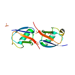
2IB5
DownloadVisualize
BU of 2ib5 by Molmil
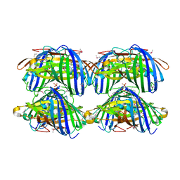
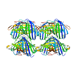
Structural characterization of a blue chromoprotein and its yellow mutant from the sea anemone cnidopus japonicus
Descriptor: Chromo protein, PHOSPHATE ION
Authors:Chan, M.C.Y, Bosanac, I, Ikura, M.
Deposit date:2006-09-10
Release date:2006-10-10
Last modified:2023-11-15
Method:X-RAY DIFFRACTION (1.8 Å)
Cite:Structural Characterization of a Blue Chromoprotein and Its Yellow Mutant from the Sea Anemone Cnidopus Japonicus
J.Biol.Chem., 281, 2006
2IB6
DownloadVisualize
BU of 2ib6 by Molmil
Structural characterization of a blue chromoprotein and its yellow mutant from the sea anemone cnidopus japonicus
Descriptor: PHOSPHATE ION, Yellow mutant chromo protein
Authors:Chan, M.C.Y, Bosanac, I, Ho, D, Prive, G, Ikura, M.
Deposit date:2006-09-10
Release date:2006-10-10
Last modified:2023-11-15
Method:X-RAY DIFFRACTION (2 Å)
Cite:Structural Characterization of a Blue Chromoprotein and Its Yellow Mutant from the Sea Anemone Cnidopus Japonicus
J.Biol.Chem., 281, 2006
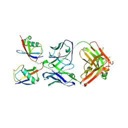
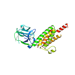
4Z5R
DownloadVisualize
BU of 4z5r by Molmil
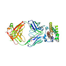
Rontalizumab Fab bound to Interferon-a2
Descriptor: Interferon alpha-2, SULFATE ION, anti-IFN-a antibody rontalizumab heavy chain modules VH and CH1 (Fab), ...
Authors:Eigenbrot, C, Maurer, B, Bosanac, I.
Deposit date:2015-04-02
Release date:2015-07-08
Last modified:2023-09-27
Method:X-RAY DIFFRACTION (3 Å)
Cite:Structural basis of the broadly neutralizing anti-interferon-alpha antibody rontalizumab.
Protein Sci., 24, 2015
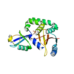
3TMO
DownloadVisualize
BU of 3tmo by Molmil
The catalytic domain of human deubiquitinase DUBA
Descriptor: OTU domain-containing protein 5
Authors:Yin, J, Bosanac, I, Ma, X, Hymowitz, S, Starovasnik, M, Cochran, A.
Deposit date:2011-08-31
Release date:2012-01-11
Last modified:2012-02-29
Method:X-RAY DIFFRACTION (2.2 Å)
Cite:Phosphorylation-dependent activity of the deubiquitinase DUBA.
Nat.Struct.Mol.Biol., 19, 2012
3DVG
DownloadVisualize
BU of 3dvg by Molmil
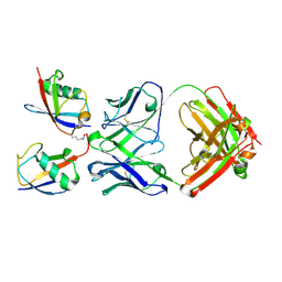
Crystal structure of K63-specific fab Apu.3A8 bound to K63-linked di-ubiquitin
Descriptor: Human IgG1 fab fragment heavy chain, Human IgG1 fab fragment light chain, Ubiquitin, ...
Authors:Hymowitz, S.G.
Deposit date:2008-07-18
Release date:2008-09-30
Last modified:2021-10-20
Method:X-RAY DIFFRACTION (2.6 Å)
Cite:Ubiquitin chain editing revealed by polyubiquitin linkage-specific antibodies.
Cell(Cambridge,Mass.), 134, 2008
3DVN
DownloadVisualize
BU of 3dvn by Molmil
Crystal structure of K63-specific fab Apu2.16 bound to K63-linked di-ubiquitin
Descriptor: Human IgG1 fab fragment heavy chain, Human IgG1 fab fragment light chain, Ubiquitin, ...
Authors:Hymowitz, S.G.
Deposit date:2008-07-18
Release date:2008-09-30
Last modified:2021-10-20
Method:X-RAY DIFFRACTION (2.7 Å)
Cite:Ubiquitin chain editing revealed by polyubiquitin linkage-specific antibodies.
Cell(Cambridge,Mass.), 134, 2008
3TMP
DownloadVisualize
BU of 3tmp by Molmil
The catalytic domain of human deubiquitinase DUBA in complex with ubiquitin aldehyde
Descriptor: OTU domain-containing protein 5, Polyubiquitin-C
Authors:Ma, X, Yin, J, Hymowitz, S, Starovasnik, M, Cochran, A.
Deposit date:2011-08-31
Release date:2012-01-11
Last modified:2012-02-29
Method:X-RAY DIFFRACTION (1.91 Å)
Cite:Phosphorylation-dependent activity of the deubiquitinase DUBA.
Nat.Struct.Mol.Biol., 19, 2012
3NOB
DownloadVisualize
BU of 3nob by Molmil
Structure of K11-linked di-ubiquitin
Descriptor: SULFATE ION, Ubiquitin
Authors:Dong, K.C, Matsumoto, M.L, Hymowitz, S.G.
Deposit date:2010-06-25
Release date:2010-08-11
Last modified:2023-09-06
Method:X-RAY DIFFRACTION (2.19 Å)
Cite:K11-Linked Polyubiquitination in Cell Cycle Control Revealed by a K11 Linkage-Specific Antibody.
Mol.Cell, 39, 2010

222926

PDB entries from 2024-07-24

PDB statisticsPDBj update infoContact PDBjnumon