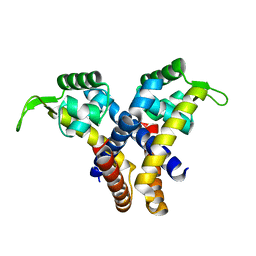
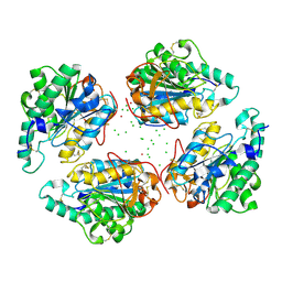
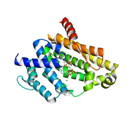
4QID
 
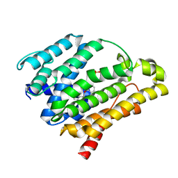
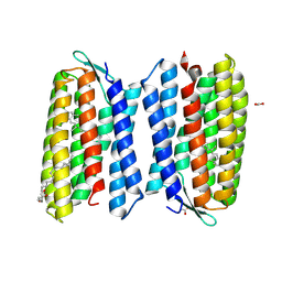 | | Crystal structure of Haloquadratum walsbyi bacteriorhodopsin | | 分子名称: | ACETATE ION, Bacteriorhodopsin-I, RETINAL, ... | | 著者 | Wang, A.H.J, Hsu, M.F, Yang, C.S, Fu, H.Y. | | 登録日 | 2014-05-30 | | 公開日 | 2015-07-29 | | 実験手法 | X-RAY DIFFRACTION (2.57 Å) | | 主引用文献 | An acid-tolerant light-driven proton pump
To be Published
|
|
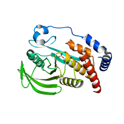
4XZW
 | | Endo-glucanase chimera C10 | | 分子名称: | 1,4,7,10,13,16-HEXAOXACYCLOOCTADECANE, 4-(2-HYDROXYETHYL)-1-PIPERAZINE ETHANESULFONIC ACID, CALCIUM ION, ... | | 著者 | Lee, C.C, Chang, C.J, Ho, T.H.D, Chao, Y.C, Wang, A.H.J. | | 登録日 | 2015-02-05 | | 公開日 | 2016-02-10 | | 最終更新日 | 2023-11-08 | | 実験手法 | X-RAY DIFFRACTION (1.5 Å) | | 主引用文献 | Endo-glucanase chimera C10
To Be Published
|
|
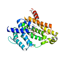
4WAV
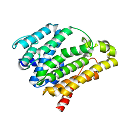
 | | Crystal Structure of Haloquadratum walsbyi bacteriorhodopsin mutant D93N | | 分子名称: | Bacteriorhodopsin-I, RETINAL, [(Z)-octadec-9-enyl] (2R)-2,3-bis(oxidanyl)propanoate | | 著者 | Wang, A.H.J, Hsu, M.F, Yang, C.S, Fu, H.Y. | | 登録日 | 2014-09-02 | | 公開日 | 2015-09-23 | | 最終更新日 | 2023-11-08 | | 実験手法 | X-RAY DIFFRACTION (2.8 Å) | | 主引用文献 | Crystal structure of an acid-tolerant light-driven proton pump at 1.85 Angstroms resolution
To be published
|
|
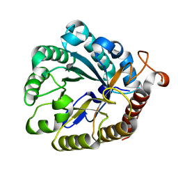
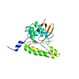
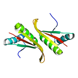
2QN5
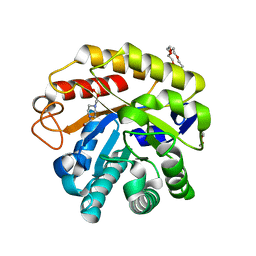
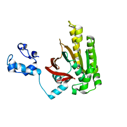
 | | Crystal Structure and Functional Study of the Bowman-Birk Inhibitor from Rice Bran in Complex with Bovine Trypsin | | 分子名称: | Bowman-Birk type bran trypsin inhibitor, Cationic trypsin | | 著者 | Li, H.T, Lin, Y.H, Guan, H.H, Hsieh, Y.C, Wang, A.H.J, Chen, C.J. | | 登録日 | 2007-07-18 | | 公開日 | 2008-07-22 | | 最終更新日 | 2023-10-25 | | 実験手法 | X-RAY DIFFRACTION (3 Å) | | 主引用文献 | Crystal Structure and Functional Study of the Bowman-Birk Inhibitor from Rice Bran in Complex with Bovine Trypsin
To be Published
|
|
4EJW
 | |
4EJU
 | |
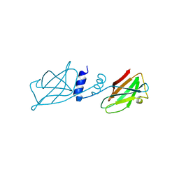
4EJV
 | | Staphylococcus epidermidis TcaR in complex with chloramphenicol | | 分子名称: | 2,2-dichloro-N-[(1S,2S)-1,3-dihydroxy-1-(4-nitrophenyl)propan-2-yl]acetamide, Transcriptional regulator TcaR | | 著者 | Chang, Y.M, Chen, C.K.M, Wang, A.H.J. | | 登録日 | 2012-04-07 | | 公開日 | 2013-05-08 | | 最終更新日 | 2023-11-08 | | 実験手法 | X-RAY DIFFRACTION (2.9 Å) | | 主引用文献 | Structural analysis of the antibiotic recognition mechanism of MarR family proteins
Acta Crystallogr.,Sect.D, 2013
|
|
4XZB
 | |
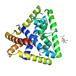
3KCW
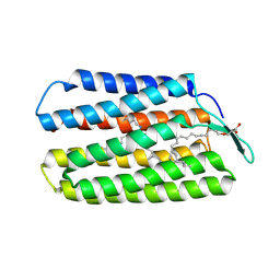
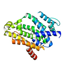
 | | Crystal structure of Ganoderma fungal immunomodulatory protein, GMI | | 分子名称: | immunomodulatory protein | | 著者 | Hsu, M.F, Wang, A.H.J, Yang, C.S, Huang, C.T, Hseu, R.S, Lin, C.W, Wu, M.Y, Huang, C.S, Fu, H.Y. | | 登録日 | 2009-10-22 | | 公開日 | 2010-11-03 | | 最終更新日 | 2023-11-01 | | 実験手法 | X-RAY DIFFRACTION (2 Å) | | 主引用文献 | Single cysteine replacement at Leu6 increase the potent and thermostability in Ganoderma fungal immunomodulatory proteins
To be Published
|
|
2Z43
 | |
2ZUB
 | | Left handed RadA | | 分子名称: | DNA repair and recombination protein radA | | 著者 | Chang, Y.W, Ko, T.P, Wang, T.F, Wang, A.H.J. | | 登録日 | 2008-10-15 | | 公開日 | 2009-04-07 | | 最終更新日 | 2023-11-01 | | 実験手法 | X-RAY DIFFRACTION (2.9 Å) | | 主引用文献 | Three new structures of left-handed RADA helical filaments: structural flexibility of N-terminal domain is critical for recombinase activity
Plos One, 4, 2009
|
|
2ZUD
 | |
2ZUC
 | |
2DFL
 | | Crystal structure of left-handed RadA filament | | 分子名称: | DNA repair and recombination protein radA | | 著者 | Chen, L.T, Ko, T.P, Wang, T.F, Wang, A.H.J. | | 登録日 | 2006-03-02 | | 公開日 | 2007-01-23 | | 最終更新日 | 2023-10-25 | | 実験手法 | X-RAY DIFFRACTION (2.9 Å) | | 主引用文献 | Crystal structure of the left-handed archaeal RadA helical filament: identification of a functional motif for controlling quaternary structures and enzymatic functions of RecA family proteins
Nucleic Acids Res., 35, 2007
|
|
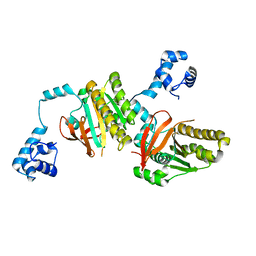
3ACW
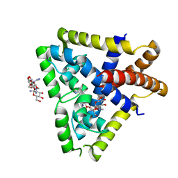
 | | Crystal structure of the C(30) carotenoid dehydrosqualene synthase from Staphylococcus aureus complexed with BPH-651 | | 分子名称: | (3R)-3-biphenyl-4-yl-1-azabicyclo[2.2.2]octan-3-ol, Dehydrosqualene synthase | | 著者 | Liu, C.I, Jeng, W.Y, Wang, A.H.J, Oldfield, E. | | 登録日 | 2010-01-13 | | 公開日 | 2010-11-24 | | 最終更新日 | 2023-11-01 | | 実験手法 | X-RAY DIFFRACTION (1.63 Å) | | 主引用文献 | Mechanism of action and inhibition of dehydrosqualene synthase
Proc.Natl.Acad.Sci.USA, 107, 2010
|
|
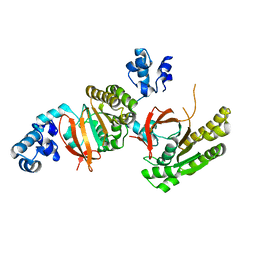
3ACX
 | | Crystal structure of the C(30) carotenoid dehydrosqualene synthase from Staphylococcus aureus complexed with BPH-673 | | 分子名称: | Dehydrosqualene synthase, N-(1-methylethyl)-3-[(3-prop-2-en-1-ylbiphenyl-4-yl)oxy]propan-1-amine | | 著者 | Liu, C.I, Jeng, W.Y, Wang, A.H.J, Oldfield, E. | | 登録日 | 2010-01-13 | | 公開日 | 2010-11-24 | | 最終更新日 | 2023-11-01 | | 実験手法 | X-RAY DIFFRACTION (1.31 Å) | | 主引用文献 | Mechanism of action and inhibition of dehydrosqualene synthase
Proc.Natl.Acad.Sci.USA, 107, 2010
|
|
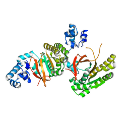
3ACY
 | | Crystal structure of the C(30) carotenoid dehydrosqualene synthase from Staphylococcus aureus complexed with BPH-702 | | 分子名称: | (1R)-4-[3-(2-benzylphenoxy)phenyl]-1-phosphonobutane-1-sulfonic acid, Dehydrosqualene synthase, MAGNESIUM ION | | 著者 | Liu, C.I, Jeng, W.Y, Wang, A.H.J, Oldfield, E. | | 登録日 | 2010-01-13 | | 公開日 | 2010-11-24 | | 最終更新日 | 2023-11-01 | | 実験手法 | X-RAY DIFFRACTION (1.84 Å) | | 主引用文献 | Mechanism of action and inhibition of dehydrosqualene synthase
Proc.Natl.Acad.Sci.USA, 107, 2010
|
|
2ZY1
 | | Crystal structure of the C(30) carotenoid dehydrosqualene synthase from Staphylococcus aureus complexed with bisphosphonate BPH-830 | | 分子名称: | Dehydrosqualene synthase, dipotassium (2-oxo-2-{[3-(3-phenoxyphenyl)propyl]amino}ethyl)phosphonate | | 著者 | Liu, C.I, Jeng, W.Y, Wang, A.H.J, Oldfield, E. | | 登録日 | 2009-01-10 | | 公開日 | 2009-09-01 | | 最終更新日 | 2023-11-01 | | 実験手法 | X-RAY DIFFRACTION (1.78 Å) | | 主引用文献 | Inhibition of staphyloxanthin virulence factor biosynthesis in Staphylococcus aureus: in vitro, in vivo, and crystallographic results.
J.Med.Chem., 52, 2009
|
|
4QNN
 | |
4QUN
 | |
4QUM
 | |
3ADZ
 | | Crystal structure of the C(30) carotenoid dehydrosqualene synthase from Staphylococcus aureus complexed with intermediate PSPP | | 分子名称: | Dehydrosqualene synthase, MAGNESIUM ION, {(1R,2R,3R)-2-[(3E)-4,8-dimethylnona-3,7-dien-1-yl]-2-methyl-3-[(1E,5E)-2,6,10-trimethylundeca-1,5,9-trien-1-yl]cyclopropyl}methyl trihydrogen diphosphate | | 著者 | Liu, C.I, Jeng, W.Y, Wang, A.H.J, Oldfield, E. | | 登録日 | 2010-01-31 | | 公開日 | 2010-11-24 | | 最終更新日 | 2023-11-01 | | 実験手法 | X-RAY DIFFRACTION (1.89 Å) | | 主引用文献 | Mechanism of action and inhibition of dehydrosqualene synthase.
Proc.Natl.Acad.Sci.USA, 107, 2010
|
|
3AE0
 | | Crystal structure of the C(30) carotenoid dehydrosqualene synthase from Staphylococcus aureus complexed with geranylgeranyl thiopyrophosphate | | 分子名称: | Dehydrosqualene synthase, MAGNESIUM ION, phosphonooxy-[(10E)-3,7,11,15-tetramethylhexadeca-2,6,10,14-tetraenyl]sulfanyl-phosphinic acid | | 著者 | Liu, C.I, Jeng, W.Y, Wang, A.H.J, Oldfield, E. | | 登録日 | 2010-01-31 | | 公開日 | 2010-11-24 | | 最終更新日 | 2023-11-01 | | 実験手法 | X-RAY DIFFRACTION (2.37 Å) | | 主引用文献 | Mechanism of action and inhibition of dehydrosqualene synthase.
Proc.Natl.Acad.Sci.USA, 107, 2010
|
|
1UDV
 | | Crystal structure of the hyperthermophilic archaeal dna-binding protein Sso10b2 at 1.85 A | | 分子名称: | DNA binding protein SSO10b, ZINC ION | | 著者 | Chou, C.-C, Lin, T.-W, Chen, C.-Y, Wang, A.H.J. | | 登録日 | 2003-05-07 | | 公開日 | 2003-08-05 | | 最終更新日 | 2023-12-27 | | 実験手法 | X-RAY DIFFRACTION (1.85 Å) | | 主引用文献 | Crystal structure of the hyperthermophilic archaeal DNA-binding protein Sso10b2 at a resolution of 1.85 Angstroms
J.BACTERIOL., 185, 2003
|
|
3VJE
 | | Crystal structure of the Y248A mutant of C(30) carotenoid dehydrosqualene synthase from Staphylococcus aureus in complex with zaragozic acid A | | 分子名称: | Dehydrosqualene synthase, Zaragozic acid A | | 著者 | Liu, C.I, Jeng, W.Y, Chang, W.J, Wang, A.H.J. | | 登録日 | 2011-10-14 | | 公開日 | 2012-04-11 | | 最終更新日 | 2023-11-08 | | 実験手法 | X-RAY DIFFRACTION (2.12 Å) | | 主引用文献 | Binding modes of zaragozic acid A to human squalene synthase and staphylococcal dehydrosqualene synthase
J.Biol.Chem., 287, 2012
|
|


