Loading
PDBj
MenuPDBj@FacebookPDBj@TwitterPDBj@YouTubewwPDB FoundationwwPDB
RCSB PDBPDBeBMRBAdv. SearchSearch help
Search by PDB author
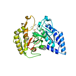
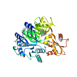
6E97
DownloadVisualize
BU of 6e97 by Molmil
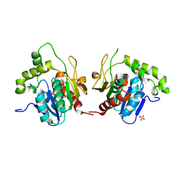
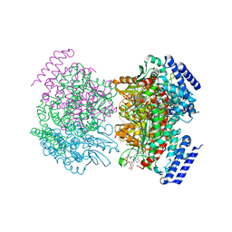
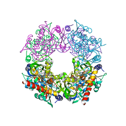
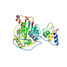
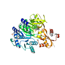
Crystal structure of the aryl acid adenylating enzyme FscC from Fuscachelin NRPS in complex with DHB-adenylate
Descriptor: 2,3-dihydroxybenzoate-AMP ligase, 5'-O-[(S)-[(2,3-dihydroxybenzene-1-carbonyl)oxy](hydroxy)phosphoryl]adenosine, GLYCEROL, ...
Authors:Bruner, S.D, Zagulyaeva, A.A.
Deposit date:2018-07-31
Release date:2019-08-21
Last modified:2023-10-11
Method:X-RAY DIFFRACTION (1.8 Å)
Cite:Implication of MbtH-like proteins in crystallization of the independent NRPS A domains. Crystal structure of FscC: supporting rationale for revised mechanism of freestanding aryl acid adenylating enzymes
To Be Published
6E8O
DownloadVisualize
BU of 6e8o by Molmil
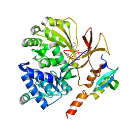
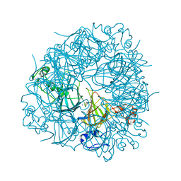
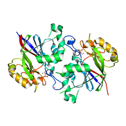
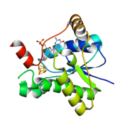
Crystal structure of aryl acid adenylating enzyme FscC from Fuscachelin NRPS in complex with AMP
Descriptor: 2,3-dihydroxybenzoate-AMP ligase, ADENOSINE MONOPHOSPHATE, GLYCEROL, ...
Authors:Bruner, S.D, Zagulyaeva, A.A.
Deposit date:2018-07-30
Release date:2019-08-21
Last modified:2023-10-11
Method:X-RAY DIFFRACTION (1.7 Å)
Cite:Implication of MbtH-like proteins in crystallization of the independent NRPS A domains. Crystal structure of FscC: supporting rationale for revised mechanism of freestanding aryl acid adenylating enzymes
To Be Published
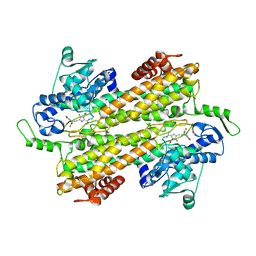
6EBY
DownloadVisualize
BU of 6eby by Molmil
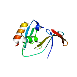
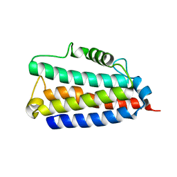
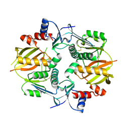
Crystal structure of the MbtH-like protein FscK bound to the interface forming region of FscH adenylation domain from Thermobifida fusca
Descriptor: Amino acid adenylation, Conserved protein MbtH
Authors:Bruner, S.D, Zagulyaeva, A.A.
Deposit date:2018-08-07
Release date:2019-08-21
Last modified:2023-10-11
Method:X-RAY DIFFRACTION (1.85 Å)
Cite:Comprehensive analysis of protein-protein interactions between MbtH-like protein FscK and adenylation domains in nonribosomal biosynthesis of Fuscachelins.
To Be Published
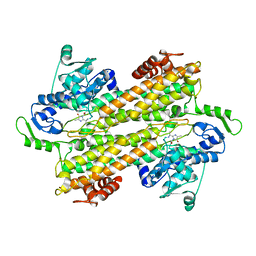
6EA3
DownloadVisualize
BU of 6ea3 by Molmil
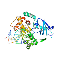
Thermobifida fusca FscH adenylation domain complexed with MbtH-like protein FscK and Ser-AMP
Descriptor: MbtH-like protein, SERYL ADENYLATE, adenylation domain of Fuscachelin synthetase component H
Authors:Bruner, S.D, Zagulyaeva, A.A.
Deposit date:2018-08-02
Release date:2019-08-07
Last modified:2023-10-11
Method:X-RAY DIFFRACTION (1.65 Å)
Cite:Comprehensive analysis of protein-protein interactions between MbtH-like protein FscK and adenylation domains in nonribosomal biosynthesis of Fuscachelins.
To Be Published
1JMK
DownloadVisualize
BU of 1jmk by Molmil
Structural Basis for the Cyclization of the Lipopeptide Antibiotic Surfactin by the Thioesterase Domain SrfTE
Descriptor: SULFATE ION, Surfactin Synthetase
Authors:Bruner, S.D, Weber, T, Kohli, R.M, Schwarzer, D, Marahiel, M.A, Walsh, C.T, Stubbs, M.T.
Deposit date:2001-07-18
Release date:2002-03-27
Last modified:2024-02-07
Method:X-RAY DIFFRACTION (1.71 Å)
Cite:Structural basis for the cyclization of the lipopeptide antibiotic surfactin by the thioesterase domain SrfTE.
Structure, 10, 2002
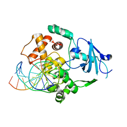
1EBM
DownloadVisualize
BU of 1ebm by Molmil
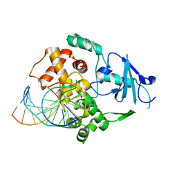
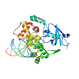
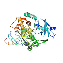
CRYSTAL STRUCTURE OF THE HUMAN 8-OXOGUANINE GLYCOSYLASE (HOGG1) BOUND TO A SUBSTRATE OLIGONUCLEOTIDE
Descriptor: 8-OXOGUANINE DNA GLYCOSYLASE, CALCIUM ION, DNA (5'-D(*GP*CP*GP*TP*CP*CP*AP*(8OG)P*GP*TP*CP*TP*AP*CP*C)-3'), ...
Authors:Bruner, S.D, Norman, D.P, Verdine, G.L.
Deposit date:2000-01-24
Release date:2000-03-20
Last modified:2024-02-07
Method:X-RAY DIFFRACTION (2.1 Å)
Cite:Structural basis for recognition and repair of the endogenous mutagen 8-oxoguanine in DNA.
Nature, 403, 2000
2NP9
DownloadVisualize
BU of 2np9 by Molmil
Crystal structure of a dioxygenase in the Crotonase superfamily
Descriptor: DpgC, OXYGEN MOLECULE, [(2R,3S,4R,5R)-5-(6-AMINO-9H-PURIN-9-YL)-4-HYDROXY-3-(PHOSPHONOOXY)TETRAHYDROFURAN-2-YL]METHYL (3R)-4-({3-[(2-{[(3,5-DIHYDROXYPHENYL)ACETYL]AMINO}ETHYL)AMINO]-3-OXOPROPYL}AMINO)-3-HYDROXY-2,2-DIMETHYL-4-OXOBUTYL DIHYDROGEN DIPHOSPHATE
Authors:Bruner, S.D, Widboom, P.F, Fielding, E.N.
Deposit date:2006-10-26
Release date:2007-05-22
Last modified:2024-04-03
Method:X-RAY DIFFRACTION (2.45 Å)
Cite:Structural basis for cofactor-independent dioxygenation in vancomycin biosynthesis.
Nature, 447, 2007
1HU0
DownloadVisualize
BU of 1hu0 by Molmil
CRYSTAL STRUCTURE OF AN HOGG1-DNA BOROHYDRIDE TRAPPED INTERMEDIATE COMPLEX
Descriptor: 5'-D(*GP*CP*GP*TP*CP*CP*AP*(PED)P*GP*TP*CP*TP*AP*CP*C)-3', 5'-D(*GP*GP*TP*AP*GP*AP*CP*CP*TP*GP*GP*AP*CP*GP*C)-3', 8-OXOGUANINE, ...
Authors:Fromme, J.C, Bruner, S.D, Yang, W, Karplus, M, Verdine, G.L.
Deposit date:2001-01-03
Release date:2003-02-25
Last modified:2024-10-30
Method:X-RAY DIFFRACTION (2.35 Å)
Cite:Product-Assisted Catalysis in base-excision DNA Repair
Nat.Struct.Biol., 10, 2003
7MTT
DownloadVisualize
BU of 7mtt by Molmil
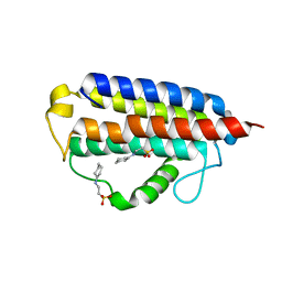
Crystal structure of colibactin self-resistance protein ClbS in complex with two molecules of CHES
Descriptor: 2-[N-CYCLOHEXYLAMINO]ETHANE SULFONIC ACID, Colibactin self-protection protein ClbS
Authors:Tripathi, P, Bruner, S.D.
Deposit date:2021-05-13
Release date:2021-05-26
Last modified:2023-10-18
Method:X-RAY DIFFRACTION (1.85 Å)
Cite:Structural Basis for the Interactions of the Colibactin Resistance Gene Product ClbS with DNA.
Biochemistry, 60, 2021
7MTL
DownloadVisualize
BU of 7mtl by Molmil
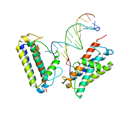
Crystal structure of colibactin self-resistance protein ClbS in complex with a dsDNA
Descriptor: Colibactin self-protection protein ClbS, DNA (5'-D(*AP*AP*TP*TP*CP*CP*CP*AP*CP*TP*TP*CP*CP*AP*G)-3'), DNA (5'-D(*CP*TP*GP*GP*AP*AP*GP*TP*GP*GP*GP*AP*AP*TP*T)-3')
Authors:Tripathi, P, Bruner, S.D.
Deposit date:2021-05-13
Release date:2021-05-26
Last modified:2023-10-18
Method:X-RAY DIFFRACTION (2.446 Å)
Cite:Structural Basis for the Interactions of the Colibactin Resistance Gene Product ClbS with DNA.
Biochemistry, 60, 2021
7RK0
DownloadVisualize
BU of 7rk0 by Molmil
Crystal structure of Thermovibrio ammonificans THI4
Descriptor: 2-[(E)-[(4R)-5-[[[(2R,3S,4R,5R)-5-(6-aminopurin-9-yl)-3,4-bis(oxidanyl)oxolan-2-yl]methoxy-oxidanyl-phosphoryl]oxy-oxidanyl-phosphoryl]oxy-4-oxidanyl-3-oxidanylidene-pentan-2-ylidene]amino]ethanoic acid, FE (III) ION, Thiamine thiazole synthase
Authors:Li, Q, Bruner, S.D.
Deposit date:2021-07-21
Release date:2021-09-01
Last modified:2023-10-18
Method:X-RAY DIFFRACTION (2.28 Å)
Cite:Structure and function of aerotolerant, multiple-turnover THI4 thiazole synthases.
Biochem.J., 478, 2021
6UFI
DownloadVisualize
BU of 6ufi by Molmil
W96Y Oxalate Decarboxylase (Bacillus subtilis)
Descriptor: CHLORIDE ION, Cupin domain-containing protein, GLYCEROL, ...
Authors:Pastore, A.J, Burg, M.J, Twahir, U.T, Bruner, S.D, Angerhofer, A.
Deposit date:2019-09-24
Release date:2020-09-30
Last modified:2023-10-11
Method:X-RAY DIFFRACTION (1.72 Å)
Cite:Oxalate decarboxylase uses electron hole hopping for catalysis.
J.Biol.Chem., 297, 2021
8UTL
DownloadVisualize
BU of 8utl by Molmil
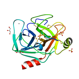
Bovine trypsin in complex with Thr3Dap mutated microviridin J
Descriptor: CALCIUM ION, Cationic trypsin, GLYCEROL, ...
Authors:Chen, W, Bruner, S.D.
Deposit date:2023-10-31
Release date:2024-02-14
Last modified:2024-02-28
Method:X-RAY DIFFRACTION (1.56 Å)
Cite:Alternative Linkage Chemistries in the Chemoenzymatic Synthesis of Microviridin-Based Cyclic Peptides.
Org.Lett., 26, 2024
8UO7
DownloadVisualize
BU of 8uo7 by Molmil
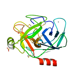
Bovine trypsin in complex with deacetylated wild type microviridin J
Descriptor: CALCIUM ION, Cationic trypsin, Deacetylated wildtype microviridin J, ...
Authors:Chen, W, Bruner, S.D.
Deposit date:2023-10-19
Release date:2024-02-14
Last modified:2024-02-28
Method:X-RAY DIFFRACTION (2.2 Å)
Cite:Alternative Linkage Chemistries in the Chemoenzymatic Synthesis of Microviridin-Based Cyclic Peptides.
Org.Lett., 26, 2024
1FN7
DownloadVisualize
BU of 1fn7 by Molmil
COUPLING OF DAMAGE RECOGNITION AND CATALYSIS BY A HUMAN BASE-EXCISION DNA REPAIR PROTEIN
Descriptor: 8-OXOGUANINE DNA GLYCOSYLASE 1, CALCIUM ION, DNA (5'-D(*GP*CP*GP*TP*CP*CP*AP*(3DR)P*GP*TP*CP*TP*AP*CP*C)-3'), ...
Authors:Norman, D.P.G, Bruner, S.D, Verdine, G.L.
Deposit date:2000-08-21
Release date:2001-04-21
Last modified:2024-02-07
Method:X-RAY DIFFRACTION (2.6 Å)
Cite:Coupling of substrate recognition and catalysis by a human base-excision DNA repair protein.
J.Am.Chem.Soc., 123, 2001
8ES6
DownloadVisualize
BU of 8es6 by Molmil
Crystal structure of an unusual amidase ClbL from colibactin gene cluster
Descriptor: Colibactin biosynthesis amidase ClbL
Authors:Tripathi, P, Bruner, S.D.
Deposit date:2022-10-13
Release date:2023-09-20
Method:X-RAY DIFFRACTION (1.9 Å)
Cite:Structural basis of the amidase ClbL central to the biosynthesis of the genotoxin colibactin.
Acta Crystallogr D Struct Biol, 79, 2023
3KDY
DownloadVisualize
BU of 3kdy by Molmil
X-ray crystal structure of tyrosine aminomutase mutant construct
Descriptor: Histidine ammonia-lyase
Authors:Cooke, H.A, Bruner, S.D.
Deposit date:2009-10-23
Release date:2010-07-07
Last modified:2023-11-15
Method:X-RAY DIFFRACTION (2.2 Å)
Cite:Probing the active site of MIO-dependent aminomutases, key catalysts in the biosynthesis of beta-amino acids incorporated in secondary metabolites
Biopolymers, 93, 2010
3KDZ
DownloadVisualize
BU of 3kdz by Molmil
X-ray crystal structure of a tyrosine aminomutase mutant construct with bound ligand
Descriptor: Histidine ammonia-lyase, TYROSINE
Authors:Cooke, H.A, Bruner, S.D.
Deposit date:2009-10-23
Release date:2010-07-07
Last modified:2024-10-30
Method:X-RAY DIFFRACTION (2.2 Å)
Cite:Probing the active site of MIO-dependent aminomutases, key catalysts in the biosynthesis of beta-amino acids incorporated in secondary metabolites
Biopolymers, 93, 2010
8TYJ
DownloadVisualize
BU of 8tyj by Molmil
Crystal structure of SARS-CoV-2 nsp10/nsp16 complex with bound SAH
Descriptor: 2'-O-methyltransferase, GLYCEROL, Non-structural protein 10, ...
Authors:Patel, K.P, Bruner, S.D.
Deposit date:2023-08-25
Release date:2024-01-24
Method:X-RAY DIFFRACTION (1.9 Å)
Cite:Crystal structure of SARS-CoV-2 nsp10/nsp16 complex with bound SAH
To Be Published
6B9V
DownloadVisualize
BU of 6b9v by Molmil
Crystal Structure of a New Diphosphatase from the PhnP Family
Descriptor: Beta-lactamase-like protein, MANGANESE (II) ION, PHOSPHATE ION, ...
Authors:Li, Q, Bruner, S.D.
Deposit date:2017-10-11
Release date:2018-10-17
Last modified:2023-10-04
Method:X-RAY DIFFRACTION (1.88 Å)
Cite:An unusual diphosphatase from the PhnP family cleaves reactive FAD photoproducts.
Biochem.J., 475, 2018
6ANR
DownloadVisualize
BU of 6anr by Molmil
Crystal structure of a self resistance protein ClbS from colibactin biosynthetic gene cluster
Descriptor: Colibactin self-protection protein ClbS
Authors:Tripathi, P, Bruner, S.D.
Deposit date:2017-08-14
Release date:2018-06-27
Last modified:2023-10-04
Method:X-RAY DIFFRACTION (2.098 Å)
Cite:ClbS Is a Cyclopropane Hydrolase That Confers Colibactin Resistance.
J. Am. Chem. Soc., 139, 2017
1LWW
DownloadVisualize
BU of 1lww by Molmil
Borohydride-trapped hOgg1 Intermediate Structure Co-Crystallized with 8-bromoguanine
Descriptor: 5'-D(*GP*CP*GP*TP*CP*CP*AP*(PED)P*GP*TP*CP*TP*AP*CP*C)-3', 5'-D(*GP*GP*TP*AP*GP*AP*CP*CP*TP*GP*GP*AP*CP*GP*C)-3', 8-BROMOGUANINE, ...
Authors:Fromme, J.C, Bruner, S.D, Yang, W, Karplus, M, Verdine, G.L.
Deposit date:2002-06-03
Release date:2003-02-25
Last modified:2011-07-13
Method:X-RAY DIFFRACTION (2.1 Å)
Cite:Product-Assisted Catalysis in Base Excision DNA Repair
Nat.Struct.Biol., 10, 2003
1LWV
DownloadVisualize
BU of 1lwv by Molmil
Borohydride-trapped hOgg1 Intermediate Structure Co-Crystallized with 8-aminoguanine
Descriptor: 5'-D(*GP*CP*GP*TP*CP*CP*AP*(PED)P*GP*TP*CP*TP*AP*CP*C)-3', 5'-D(*GP*GP*TP*AP*GP*AP*CP*CP*TP*GP*GP*AP*CP*GP*C)-3', 8-AMINOGUANINE, ...
Authors:Fromme, J.C, Bruner, S.D, Yang, W, Karplus, M, Verdine, G.L.
Deposit date:2002-06-03
Release date:2003-02-25
Last modified:2011-07-13
Method:X-RAY DIFFRACTION (2.3 Å)
Cite:Product-Assisted Catalysis in Base Excision DNA Repair
Nat.Struct.Biol., 10, 2003
7LC7
DownloadVisualize
BU of 7lc7 by Molmil
Crystal structure of epoxyqueuosine reductase QueH in complex with GMP from Thermotoga maritima
Descriptor: CHLORIDE ION, Epoxyqueuosine reductase QueH, FE (III) ION, ...
Authors:Li, Q, Bruner, S.D.
Deposit date:2021-01-09
Release date:2022-01-12
Last modified:2023-10-18
Method:X-RAY DIFFRACTION (1.58 Å)
Cite:The epoxyqueuosine reductase QueH in the biosynthesis of tRNA queuosine is a unique metalloenzyme
To Be Published
7M4S
DownloadVisualize
BU of 7m4s by Molmil
Crystal structure of macrocyclase AMdnB from Anabaena sp. PCC 7120
Descriptor: AMdnB protein
Authors:Li, G, Bruner, S.D.
Deposit date:2021-03-22
Release date:2021-11-03
Last modified:2024-10-23
Method:X-RAY DIFFRACTION (2.493 Å)
Cite:Structural and biochemical studies of an iterative ribosomal peptide macrocyclase.
Proteins, 90, 2022


123>

226707

数据于2024-10-30公开中

PDB statisticsPDBj update infoContact PDBjnumon