Loading
PDBj
MenuPDBj@FacebookPDBj@TwitterPDBj@YouTubewwPDB FoundationwwPDB
RCSB PDBPDBeBMRBAdv. SearchSearch help
Search by PDB author
1LT1
DownloadVisualize
BU of 1lt1 by Molmil
SLIDING HELIX INDUCED CHANGE OF COORDINATION GEOMETRY IN A MODEL DI-MN(II) PROTEIN
Descriptor: L13G-DF1, MANGANESE (II) ION
Authors:Di Costanzo, L, Geremia, S.
Deposit date:2002-05-20
Release date:2003-05-20
Last modified:2023-10-25
Method:X-RAY DIFFRACTION (1.91 Å)
Cite:Sliding helix and change of coordination geometry in a model di-MnII protein
Angew.Chem.Int.Ed.Engl., 42, 2003
3MFV
DownloadVisualize
BU of 3mfv by Molmil
Crystal structure of human arginase I in complex with 2-aminohomohistidine
Descriptor: (2S)-2-amino-4-(2-amino-1H-imidazol-5-yl)butanoic acid, Arginase-1, MANGANESE (II) ION
Authors:Di Costanzo, L, Christianson, D.W.
Deposit date:2010-04-04
Release date:2010-05-05
Last modified:2023-09-06
Method:X-RAY DIFFRACTION (1.9 Å)
Cite:2-aminoimidazole amino acids as inhibitors of the binuclear manganese metalloenzyme human arginase I.
J.Med.Chem., 53, 2010
3MFW
DownloadVisualize
BU of 3mfw by Molmil
Crystal structure of human arginase I in complex with L-2-aminohistidine and sulphate
Descriptor: 2-amino-L-histidine, Arginase-1, MANGANESE (II) ION, ...
Authors:Di Costanzo, L, Christianson, D.W.
Deposit date:2010-04-04
Release date:2010-05-05
Last modified:2023-09-06
Method:X-RAY DIFFRACTION (1.47 Å)
Cite:2-aminoimidazole amino acids as inhibitors of the binuclear manganese metalloenzyme human arginase I.
J.Med.Chem., 53, 2010
3MJL
DownloadVisualize
BU of 3mjl by Molmil
Crystal structure of human arginase I in complex with 2-aminoimidazole. Resolution 1.90 A.
Descriptor: 1H-imidazol-2-amine, Arginase-1, MANGANESE (II) ION
Authors:Di Costanzo, L, Christianson, D.W.
Deposit date:2010-04-13
Release date:2010-05-05
Last modified:2023-09-06
Method:X-RAY DIFFRACTION (1.9 Å)
Cite:Crystal structure of human arginase I in complex with 2-aminoimidazole. Resolution 1.90 A.
To be Published
3SJT
DownloadVisualize
BU of 3sjt by Molmil
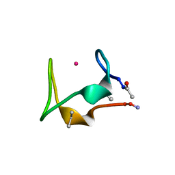
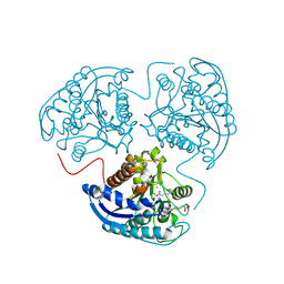
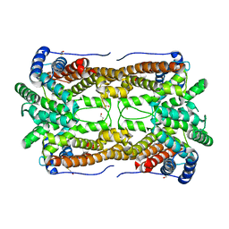
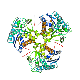
Crystal structure of human arginase I in complex with the inhibitor Me-ABH, Resolution 1.60 A, twinned structure
Descriptor: Arginase-1, MANGANESE (II) ION, [(5S)-5-amino-5-carboxyhexyl](trihydroxy)borate
Authors:Di Costanzo, L, Christianson, D.W.
Deposit date:2011-06-21
Release date:2011-07-20
Last modified:2023-09-13
Method:X-RAY DIFFRACTION (1.597 Å)
Cite:Binding of alpha , alpha-disubstituted amino acids to arginase suggests new avenues for inhibitor design.
J.Med.Chem., 54, 2011
5SBI
DownloadVisualize
BU of 5sbi by Molmil
The crystal structure of METP in complex with Co at a resolution of 1.80A.
Descriptor: COBALT (II) ION, METP, miniaturized rubredoxin
Authors:Di Costanzo, L, La Gatta, S, Leone, L, Chino, M.
Deposit date:2021-08-24
Release date:2023-02-15
Last modified:2024-04-03
Method:X-RAY DIFFRACTION (1.799 Å)
Cite:Miniaturization process reloaded - structural and functional insights from a miniaturized rubredoxin
To be published
5SBJ
DownloadVisualize
BU of 5sbj by Molmil
The crystal structure of METP in complex with Cd at a resolution of 1.29 A.
Descriptor: CADMIUM ION, METP, miniaturized rubredoxin
Authors:Di Costanzo, L, La Gatta, S, Pavone, V.
Deposit date:2021-08-24
Release date:2023-02-15
Last modified:2024-04-03
Method:X-RAY DIFFRACTION (1.28 Å)
Cite:Miniaturization process reloaded - structural and functional insights from a miniaturized rubredoxin
To be published
2Z6M
DownloadVisualize
BU of 2z6m by Molmil
Crystal structure of Human Ferritin H8 as biotemplate for noble metal nanoparticle synthesis
Descriptor: (4S)-2-METHYL-2,4-PENTANEDIOL, CALCIUM ION, Ferritin heavy chain, ...
Authors:Di Costanzo, L, Christianson, D.W.
Deposit date:2007-08-04
Release date:2008-07-22
Last modified:2023-11-01
Method:X-RAY DIFFRACTION (2.72 Å)
Cite:Directing noble metal ion chemistry within a designed ferritin protein
Biochemistry, 47, 2008
3E6K
DownloadVisualize
BU of 3e6k by Molmil
X-ray structure of Human Arginase I: the mutant D183A in complex with ABH
Descriptor: 2(S)-AMINO-6-BORONOHEXANOIC ACID, Arginase-1, MANGANESE (II) ION
Authors:Di Costanzo, L, Christianson, D.W.
Deposit date:2008-08-15
Release date:2008-12-02
Last modified:2023-08-30
Method:X-RAY DIFFRACTION (2.1 Å)
Cite:Probing the specificity determinants of amino acid recognition by arginase.
Biochemistry, 48, 2009
3E6V
DownloadVisualize
BU of 3e6v by Molmil
X-ray structure of human arginase I-D183N mutant: the complex with ABH
Descriptor: 2(S)-AMINO-6-BORONOHEXANOIC ACID, Arginase-1, MANGANESE (II) ION
Authors:Di Costanzo, L, Christianson, D.W.
Deposit date:2008-08-16
Release date:2008-12-02
Last modified:2023-08-30
Method:X-RAY DIFFRACTION (1.72 Å)
Cite:Probing the specificity determinants of amino acid recognition by arginase.
Biochemistry, 48, 2009
3ES3
DownloadVisualize
BU of 3es3 by Molmil
Directing Noble Metal Ion Chemistry within a Designed Ferritin Protein. The Complex with Gold ions. Ferritin H8-H9x Mutant
Descriptor: CALCIUM ION, Ferritin heavy chain, GOLD ION
Authors:Di Costanzo, L, Christianson, D.W.
Deposit date:2008-10-03
Release date:2008-10-21
Last modified:2023-09-06
Method:X-RAY DIFFRACTION (2.795 Å)
Cite:Directing noble metal ion chemistry within a designed ferritin protein.
Biochemistry, 47, 2008
3ERZ
DownloadVisualize
BU of 3erz by Molmil
Directing Noble Metal Ion Chemistry within a Designed Ferritin Protein. Mercury Ions on the Three-Fold Channel
Descriptor: (4S)-2-METHYL-2,4-PENTANEDIOL, CALCIUM ION, Ferritin heavy chain, ...
Authors:Di Costanzo, L, Christianson, D.W.
Deposit date:2008-10-03
Release date:2008-10-21
Last modified:2023-12-27
Method:X-RAY DIFFRACTION (3.056 Å)
Cite:Directing noble metal ion chemistry within a designed ferritin protein.
Biochemistry, 47, 2008
3G1R
DownloadVisualize
BU of 3g1r by Molmil
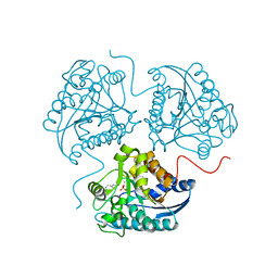
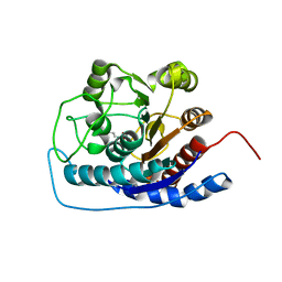
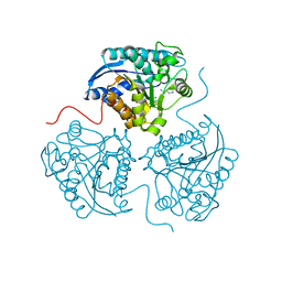
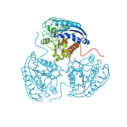
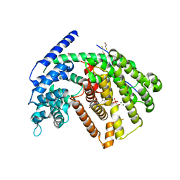
Crystal structure of human liver 5beta-reductase (AKR1D1) in complex with NADP and Finasteride. Resolution 1.70 A
Descriptor: (4aR,4bS,6aS,7S,9aS,9bS,11aR)-N-tert-butyl-4a,6a-dimethyl-2-oxo-2,4a,4b,5,6,6a,7,8,9,9a,9b,10,11,11a-tetradecahydro-1H-indeno[5,4-f]quinoline-7-carboxamide, 3-oxo-5-beta-steroid 4-dehydrogenase, NADP NICOTINAMIDE-ADENINE-DINUCLEOTIDE PHOSPHATE
Authors:Di Costanzo, L, Drury, J.E, Penning, T.M, Christianson, D.W.
Deposit date:2009-01-30
Release date:2009-06-09
Last modified:2023-09-06
Method:X-RAY DIFFRACTION (1.701 Å)
Cite:Inhibition of human steroid 5beta-reductase (AKR1D1) by finasteride and structure of the enzyme-inhibitor complex.
J.Biol.Chem., 284, 2009
3DJ8
DownloadVisualize
BU of 3dj8 by Molmil
Synthesis of (2S)-2-amino-7,8-epoxyoctanoic acid and structure of its metal-bridging complex with human arginase I
Descriptor: 6-[(2R)-oxiran-2-yl]-L-norleucine, Arginase-1, MANGANESE (II) ION
Authors:Di Costanzo, L, Christianson, D.W.
Deposit date:2008-06-22
Release date:2008-07-08
Last modified:2023-11-01
Method:X-RAY DIFFRACTION (1.51 Å)
Cite:Synthesis of (2S)-2-amino-7,8-epoxyoctanoic acid and structure of its metal-bridging complex with human arginase I
Org.Biomol.Chem., 6, 2008
3F80
DownloadVisualize
BU of 3f80 by Molmil
(S)-2-amino-6-nitrohexanoic acid binds to human arginase I through multiple nitro-metal coordination interactions in the binuclear manganese cluster. Resolution 1.60 A.
Descriptor: 6-nitro-L-norleucine, Arginase-1, MANGANESE (II) ION
Authors:Di Costanzo, L, Christianson, D.W.
Deposit date:2008-11-10
Release date:2008-12-02
Last modified:2023-09-06
Method:X-RAY DIFFRACTION (1.6 Å)
Cite:(S)-2-Amino-6-nitrohexanoic Acid Binds to Human Arginase I through Multiple Nitro-Metal Coordination Interactions in the Binuclear Manganese Cluster.
J.Am.Chem.Soc., 130, 2008
3GMZ
DownloadVisualize
BU of 3gmz by Molmil
Crystal of human arginase in complex with L-ornithine. Resolution 1.43 A.
Descriptor: Arginase-1, L-ornithine, MANGANESE (II) ION
Authors:Di Costanzo, L, Christianson, D.W.
Deposit date:2009-03-15
Release date:2010-01-26
Last modified:2023-09-06
Method:X-RAY DIFFRACTION (1.43 Å)
Cite:Binding of alpha,alpha-Disubstituted Amino Acids to Arginase Suggests New Avenues for Inhibitor Design
J.Med.Chem., 54, 2011
3GN0
DownloadVisualize
BU of 3gn0 by Molmil
Crystal structure of human arginase I in complex with difluoromethylornithine (DFMO)
Descriptor: ALPHA-DIFLUOROMETHYLORNITHINE, Arginase-1, MANGANESE (II) ION
Authors:Di Costanzo, L, Christianson, D.W.
Deposit date:2009-03-15
Release date:2010-02-23
Last modified:2023-09-06
Method:X-RAY DIFFRACTION (1.7 Å)
Cite:Binding of alpha,alpha-Disubstituted Amino Acids to Arginase Suggests New Avenues for Inhibitor Design
J.Med.Chem., 54, 2011
3M1S
DownloadVisualize
BU of 3m1s by Molmil
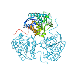
Structure of Ruthenium Half-Sandwich Complex Bound to Glycogen Synthase Kinase 3
Descriptor: Glycogen synthase kinase-3 beta, Ruthenium pyridocarbazole
Authors:Atilla-Gokcumen, G.E, Di Costanzo, L, Zimmermann, G, Meggers, E.
Deposit date:2010-03-05
Release date:2010-12-22
Last modified:2023-09-06
Method:X-RAY DIFFRACTION (3.134 Å)
Cite:Structure of anticancer ruthenium half-sandwich complex bound to glycogen synthase kinase 3beta
J.Biol.Inorg.Chem., 16, 2011
2E4O
DownloadVisualize
BU of 2e4o by Molmil
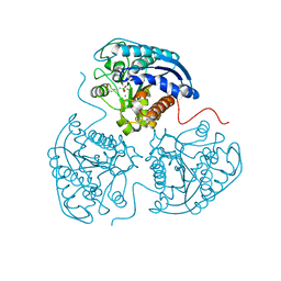
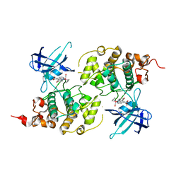
X-ray Crystal Structure of Aristolochene Synthase from Aspergillus terreus and the Evolution of Templates for the Cyclization of Farnesyl Diphosphate
Descriptor: 2-(N-MORPHOLINO)-ETHANESULFONIC ACID, Aristolochene synthase, BETA-MERCAPTOETHANOL, ...
Authors:Shishova, E.Y, Di Costanzo, L, Cane, D.E, Christianson, D.W.
Deposit date:2006-12-15
Release date:2007-01-23
Last modified:2023-10-25
Method:X-RAY DIFFRACTION (2.2 Å)
Cite:X-ray crystal structure of aristolochene synthase from Aspergillus terreus and evolution of templates for the cyclization of farnesyl diphosphate.
Biochemistry, 46, 2007
2OA6
DownloadVisualize
BU of 2oa6 by Molmil
Aristolochene synthase from Aspergillus terreus complexed with pyrophosphate
Descriptor: Aristolochene synthase, BETA-MERCAPTOETHANOL, GLYCEROL, ...
Authors:Shishova, E.Y, Di Costanzo, L, Cane, D.E, Christianson, D.W.
Deposit date:2006-12-15
Release date:2007-01-30
Last modified:2023-11-15
Method:X-RAY DIFFRACTION (2.15 Å)
Cite:X-ray crystal structure of aristolochene synthase from Aspergillus terreus and evolution of templates for the cyclization of farnesyl diphosphate
Biochemistry, 46, 2007
3SKK
DownloadVisualize
BU of 3skk by Molmil
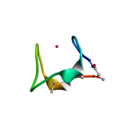
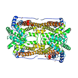
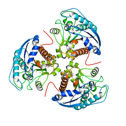
Crystal structure of human arginase I in complex with the inhibitor FABH, Resolution 1.70 A, twinned structure
Descriptor: Arginase-1, MANGANESE (II) ION, [(5S)-5-amino-5-carboxy-6,6-difluorohexyl](trihydroxy)borate(1-)
Authors:Thorn, K.J, Di Costanzo, L, Christianson, D.W.
Deposit date:2011-06-22
Release date:2011-07-20
Last modified:2023-09-13
Method:X-RAY DIFFRACTION (1.701 Å)
Cite:Binding of alpha , alpha-disubstituted amino acids to arginase suggests new avenues for inhibitor design.
J.Med.Chem., 54, 2011
3CYU
DownloadVisualize
BU of 3cyu by Molmil
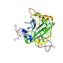
Human Carbonic Anhydrase II complexed with Cryptophane biosensor and xenon
Descriptor: Carbonic anhydrase 2, MoMo-2-[4-(2-(4-(methoxy)-1H-1,2,3-triazol-1-yl)ethyl)benzenesulfonamide]-7,12-bis-[3-(4-(methoxy)-1H-1,2,3-triazol-1-y l)propanoic acid]-cryptophane-A, PoPo-2-[4-(2-(4-(methoxy)-1H-1,2,3-triazol-1-yl)ethyl)benzenesulfonamide]-7,12-bis-[3-(4-(methoxy)-1H-1,2,3-triazol-1-yl)propanoic acid]-cryptophane-A, ...
Authors:Aaron, J.A, Jude, K.M, Di Costanzo, L, Christianson, D.W.
Deposit date:2008-04-26
Release date:2008-05-27
Last modified:2023-08-30
Method:X-RAY DIFFRACTION (1.7 Å)
Cite:Structure of a 129Xe-cryptophane biosensor complexed with human carbonic anhydrase II.
J.Am.Chem.Soc., 130, 2008
3E8Z
DownloadVisualize
BU of 3e8z by Molmil
X-ray structure of rat arginase I-N130A mutant: the unliganded complex
Descriptor: Arginase-1, MANGANESE (II) ION
Authors:Shishova, E.Y, Di Costanzo, L, Christianson, D.W.
Deposit date:2008-08-20
Release date:2008-12-02
Last modified:2023-11-29
Method:X-RAY DIFFRACTION (2 Å)
Cite:Probing the specificity determinants of amino acid recognition by arginase.
Biochemistry, 48, 2009
3E8Q
DownloadVisualize
BU of 3e8q by Molmil
X-ray structure of rat arginase I-T135A: the unliganded complex
Descriptor: Arginase-1, MANGANESE (II) ION
Authors:Shishova, E.Y, Di Costanzo, L, Emig, F.A, Ash, D.E, Christianson, D.W.
Deposit date:2008-08-20
Release date:2008-12-02
Last modified:2023-08-30
Method:X-RAY DIFFRACTION (2.9 Å)
Cite:Probing the specificity determinants of amino acid recognition by arginase.
Biochemistry, 48, 2009
3G4F
DownloadVisualize
BU of 3g4f by Molmil
Crystal Structure of (+)- -Cadinene Synthase from Gossypium arboreum in complex with 2-fluorofarnesyl diphosphate
Descriptor: (+)-delta-cadinene synthase isozyme XC1, (2Z,6E)-2-fluoro-3,7,11-trimethyldodeca-2,6,10-trien-1-yl trihydrogen diphosphate, BETA-MERCAPTOETHANOL, ...
Authors:Gennadios, H.A, Di Costanzo, L, Christianson, D.W.
Deposit date:2009-02-03
Release date:2009-06-23
Last modified:2024-04-03
Method:X-RAY DIFFRACTION (2.651 Å)
Cite:Crystal structure of (+)-delta-cadinene synthase from Gossypium arboreum and evolutionary divergence of metal binding motifs for catalysis.
Biochemistry, 48, 2009

226262

数据于2024-10-16公开中

PDB statisticsPDBj update infoContact PDBjnumon