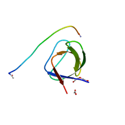
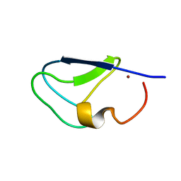
1IO0
 
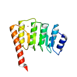 | | CRYSTAL STRUCTURE OF TROPOMODULIN C-TERMINAL HALF | | 分子名称: | TROPOMODULIN, ZINC ION | | 著者 | Krieger, I, Kostyukova, A, Yamashita, A, Maeda, Y. | | 登録日 | 2000-12-14 | | 公開日 | 2002-11-27 | | 最終更新日 | 2023-12-27 | | 実験手法 | X-RAY DIFFRACTION (1.45 Å) | | 主引用文献 | Crystal structure of the C-terminal half of tropomodulin and structural basis of actin filament pointed-end capping.
Biophys.J., 83, 2002
|
|
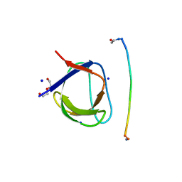
5KHT
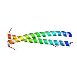 | | Crystal structure of the N-terminal fragment of tropomyosin isoform Tpm1.1 at 1.5 A resolution | | 分子名称: | DI(HYDROXYETHYL)ETHER, Tropomyosin alpha-1 chain,General control protein GCN4 | | 著者 | Kostyukova, A.S, Krieger, I, Yoon, Y.-H, Tolkatchev, D, Samatey, F.A. | | 登録日 | 2016-06-15 | | 公開日 | 2017-06-21 | | 最終更新日 | 2024-04-03 | | 実験手法 | X-RAY DIFFRACTION (1.4964 Å) | | 主引用文献 | Structural destabilization of tropomyosin induced by the cardiomyopathy-linked mutation R21H.
Protein Sci., 27, 2018
|
|
3AZD
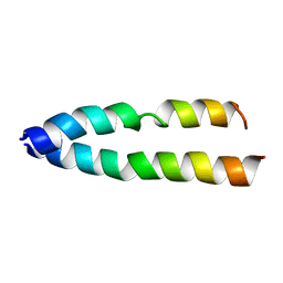 | | Crystal structure of tropomyosin N-terminal fragment at 0.98A resolution | | 分子名称: | short alpha-tropomyosin,transcription factor GCN4 | | 著者 | Meshcheryakov, V.A, Krieger, I, Kostyukova, A.S, Samatey, F.A. | | 登録日 | 2011-05-23 | | 公開日 | 2011-10-19 | | 最終更新日 | 2023-11-01 | | 実験手法 | X-RAY DIFFRACTION (0.98 Å) | | 主引用文献 | Structure of a tropomyosin N-terminal fragment at 0.98 A resolution
Acta Crystallogr.,Sect.D, 67, 2011
|
|
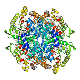
6W32
 | | Crystal structure of Sfh5 | | 分子名称: | PROTOPORPHYRIN IX CONTAINING FE, Phosphatidylinositol transfer protein SFH5 | | 著者 | Gulten, G, Khan, D, Aggarwal, A, Krieger, I, Sacchettini, J.C, Bankaitis, V.A. | | 登録日 | 2020-03-08 | | 公開日 | 2020-11-25 | | 最終更新日 | 2024-03-06 | | 実験手法 | X-RAY DIFFRACTION (2.9 Å) | | 主引用文献 | A Sec14-like phosphatidylinositol transfer protein paralog defines a novel class of heme-binding proteins.
Elife, 9, 2020
|
|
5L23
 | |
5IH2
 | | Structure, thermodynamics, and the role of conformational dynamics in the interactions between the N-terminal SH3 domain of CrkII and proline-rich motifs in cAbl | | 分子名称: | 1-ETHOXY-2-(2-ETHOXYETHOXY)ETHANE, Adapter molecule crk, DI(HYDROXYETHYL)ETHER, ... | | 著者 | Bhatt, V.S, Zeng, D, Krieger, I, Sacchettini, J.C, Cho, J.-H. | | 登録日 | 2016-02-28 | | 公開日 | 2016-06-29 | | 最終更新日 | 2023-09-27 | | 実験手法 | X-RAY DIFFRACTION (1.8 Å) | | 主引用文献 | Binding Mechanism of the N-Terminal SH3 Domain of CrkII and Proline-Rich Motifs in cAbl.
Biophys.J., 110, 2016
|
|
4P0M
 | | Crystal structure of an evolved putative penicillin-binding protein homolog, Rv2911, from Mycobacterium tuberculosis | | 分子名称: | D-alanyl-D-alanine carboxypeptidase | | 著者 | Krieger, I, Yu, M, Bursey, E, Hung, L.-W, Terwilliger, T.C, TB Structural Genomics Consortium (TBSGC) | | 登録日 | 2014-02-21 | | 公開日 | 2014-03-12 | | 最終更新日 | 2023-12-27 | | 実験手法 | X-RAY DIFFRACTION (2 Å) | | 主引用文献 | Subfamily-Specific Adaptations in the Structures of Two Penicillin-Binding Proteins from Mycobacterium tuberculosis.
Plos One, 9, 2014
|
|
8GKF
 | |
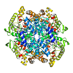
6VB9
 | | Covalent adduct of cis-2,3-epoxysuccinic acid with Isocitrate Lyase-1 from Mycobacterium tuberculosis | | 分子名称: | ACETIC ACID, DI(HYDROXYETHYL)ETHER, Isocitrate lyase, ... | | 著者 | Krieger, I.V, Pham, T.V, Meek, T.D, Sacchettini, J.C, TB Structural Genomics Consortium (TBSGC) | | 登録日 | 2019-12-18 | | 公開日 | 2020-12-30 | | 最終更新日 | 2023-10-11 | | 実験手法 | X-RAY DIFFRACTION (1.881 Å) | | 主引用文献 | Covalent Inactivation of Mycobacterium tuberculosis Isocitrate Lyase by cis -2,3-Epoxy-Succinic Acid.
Acs Chem.Biol., 16, 2021
|
|
6WSI
 | | Intact cis-2,3-epoxysuccinic acid bound to Isocitrate Lyase-1 from Mycobacterium tuberculosis | | 分子名称: | (2R,3S)-oxirane-2,3-dicarboxylic acid, DI(HYDROXYETHYL)ETHER, GLYCEROL, ... | | 著者 | Krieger, I.V, Mellott, D, Meek, T.D, Sacchettini, J.C, TB Structural Genomics Consortium (TBSGC) | | 登録日 | 2020-05-01 | | 公開日 | 2021-03-24 | | 最終更新日 | 2023-10-18 | | 実験手法 | X-RAY DIFFRACTION (1.749 Å) | | 主引用文献 | Covalent Inactivation of Mycobacterium tuberculosis Isocitrate Lyase by cis -2,3-Epoxy-Succinic Acid.
Acs Chem.Biol., 16, 2021
|
|
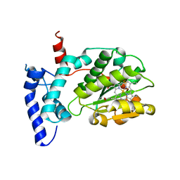
7KND
 | |
7KNJ
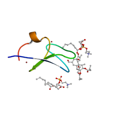 | | C1B domain of Protein kinase C in complex with Phorbol ester and Phosphatidylcholine | | 分子名称: | (4S,7R)-7-(heptanoyloxy)-4-hydroxy-N,N,N-trimethyl-10-oxo-3,5,9-trioxa-4-phosphahexadecan-1-aminium 4-oxide, Phorbol 12,13-dibutyrate, Protein kinase C delta type, ... | | 著者 | Katti, S.S, Krieger, I. | | 登録日 | 2020-11-04 | | 公開日 | 2022-05-04 | | 最終更新日 | 2023-10-18 | | 実験手法 | X-RAY DIFFRACTION (1.57 Å) | | 主引用文献 | Structural anatomy of Protein Kinase C C1 domain interactions with diacylglycerol and other agonists.
Nat Commun, 13, 2022
|
|
7LEO
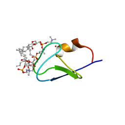 | | C1B domain of Protein kinase C in complex with diacylglycerol-lactone (AJH-836) and 1,2-diheptanoyl-sn-glycero-3-phosphocholine | | 分子名称: | (4S,7R)-7-(heptanoyloxy)-4-hydroxy-N,N,N-trimethyl-10-oxo-3,5,9-trioxa-4-phosphahexadecan-1-aminium 4-oxide, Protein kinase C delta type, ZINC ION, ... | | 著者 | Katti, S.S, Krieger, I. | | 登録日 | 2021-01-14 | | 公開日 | 2022-05-04 | | 最終更新日 | 2023-10-18 | | 実験手法 | X-RAY DIFFRACTION (1.65 Å) | | 主引用文献 | Structural anatomy of Protein Kinase C C1 domain interactions with diacylglycerol and other agonists.
Nat Commun, 13, 2022
|
|
1PGV
 | | Structural Genomics of Caenorhabditis elegans: tropomodulin C-terminal domain | | 分子名称: | tropomodulin TMD-1 | | 著者 | Symersky, J, Lu, S, Li, S, Chen, L, Meehan, E, Luo, M, Qiu, S, Bunzel, R.J, Luo, D, Arabashi, A, Nagy, L.A, Lin, G, Luan, W.C.-H, Carson, M, Gray, R, Huang, W, Southeast Collaboratory for Structural Genomics (SECSG) | | 登録日 | 2003-05-28 | | 公開日 | 2003-06-10 | | 最終更新日 | 2023-08-16 | | 実験手法 | X-RAY DIFFRACTION (1.8 Å) | | 主引用文献 | Structural genomics of Caenorhabditis elegans: crystal structure of the tropomodulin C-terminal domain
Proteins, 56, 2004
|
|


